Cytokine, Antibody and Proliferative Cellular
Responses Elicited by
Taenia solium
Calreticulin upon Experimental Infection in
Hamsters
Fela Mendlovic1,2, Mayra Cruz-Rivera1, Guillermina Ávila1, Gilberto Vaughan3, Ana Flisser1
*
1Departamento de Microbiología y Parasitología, Facultad de Medicina, Universidad Nacional Autónoma de México, Av. Universidad 3000, Coyoacán, México D.F. 04510, México,2Facultad de Ciencias de la Salud, Universidad Anáhuac, México Norte, Av. Universidad Anáhuac 46, Huixquilucan, 52786 Edo. de México, México,3AV BioSciences, Mexico City, Mexico
*flisser@unam.mx
Abstract
Taenia soliumcauses two diseases in humans, cysticercosis and taeniosis. Tapeworm
car-riers are the main risk factor for neurocysticercosis. Limited information is available about the immune response elicited by the adult parasite, particularly the induction of Th2 re-sponses, frequently associated to helminth infections. Calreticulin is a ubiquitous, multifunc-tional protein involved in cellular calcium homeostasis, which has been suggested to play a role in the regulation of immune responses. In this work, we assessed the effect of recombi-nantT.soliumcalreticulin (rTsCRT) on the cytokine, humoral and cellular responses upon experimental infection in Syrian Golden hamsters (Mesocricetus auratus). Animals were in-fected withT.soliumcysticerci and euthanized at different times after infection. Specific serum antibodies, proliferative responses in mesenteric lymph nodes and spleen cells, as well as cytokines messenger RNA (mRNA) were analyzed. The results showed that one third of the infected animals elicited anti-rTsCRT IgG antibodies. Interestingly, mesenteric lymph node (MLN) cells from either infected or non-infected animals did not proliferate upon in vitrostimulation with rTsCRT. Additionally, stimulation with a tapeworm crude extract
re-sulted in increased expression of IL-4 and IL-5 mRNA. Upon stimulation, rTsCRT increased the expression levels of IL-10 in spleen and MLN cells from uninfected and infected ham-sters. The results showed that rTsCRT favors a Th2-biased immune response character-ized by the induction of IL-10 in mucosal and systemic lymphoid organs. Here we provide the first data on the cytokine, antibody and cellular responses to rTsCRT uponin vitro stimu-lation during taeniasis.
a11111
OPEN ACCESS
Citation:Mendlovic F, Cruz-Rivera M, Ávila G, Vaughan G, Flisser A (2015) Cytokine, Antibody and Proliferative Cellular Responses Elicited byTaenia soliumCalreticulin upon Experimental Infection in Hamsters. PLoS ONE 10(3): e0121321. doi:10.1371/ journal.pone.0121321
Academic Editor:Alejandro Escobar-Gutiérrez, Instituto de Diagnostico y Referencia Epidemiologicos, MEXICO
Received:December 10, 2014
Accepted:January 30, 2015
Published:March 26, 2015
Copyright:© 2015 Mendlovic et al. This is an open access article distributed under the terms of the
Creative Commons Attribution License, which permits unrestricted use, distribution, and reproduction in any medium, provided the original author and source are credited.
Data Availability Statement:All relevant data are within the paper. The accession number for the cytokine sequences used to design the qRTPCR primers are listed inTable 1.
Introduction
Taenia soliumis responsible for two diseases in humans, i.e. taeniosis and neurocysticercosis, which are caused by the adult tapeworm and the larval stage (cysticercus), respectively. Tape-worms lodge in the small intestine of human beings after ingestion of live cysticerci in contami-nated, undercooked pork meat; develop into adult tapeworms and expel gravid proglottids full of eggs in feces. Accidental intake of eggs by humans results in the development of neurocysti-cercosis, due to the establishment of cysticerci in the central nervous system [1]. Neurocysticer-cosis is a public health problem in many developing countries [2]. Taeniosis is usually
asymptomatic but epidemiological studies have shown that tapeworm carriers are the main risk factor for developing neurocysticercosis [3]. Human beings are the only definitive hosts forT.solium. Thus, the use of experimental models, such as the Syrian golden hamster (Meso-cricetus auratus), is necessary to study the mechanisms involved in the immune response elic-ited by the tapeworm [4].
Helminth infections generally induce Th2 responses. Molecules derived from helminths that stimulate Th2 responses have been subject of current research for their potential regulato-ry immune functions [5,6]. However, only few molecules involved in triggering such responses have been recognized [7]. Calreticulin (CRT) is a ubiquitous and well-conserved protein found in all living cells except prokaryotes and fungi. CRT is a multifunctional protein with a house-keeping role in cellular calcium homeostasis and glycoprotein synthesis [8,9]. Additionally, there is growing evidence supporting its role in the induction and regulation of immune re-sponses in different parasitic diseases [10]. For example,Schistosoma mansoniCRT is a strong T-cell immunogen capable of inducing IL-4 synthesis [11], while nativeHeligmosomoides poly-gyrusCRT has been shown to induce production of IL-4 and IL-10 by T cells from infected mice [12], suggesting that CRT is able to induce a Th2 response during helminth infections.
We have previously identified, cloned and expressedT.soliumCRT as a recombinant pro-tein (rTsCRT) with calcium binding functions and analyzed its expression pattern during T.soliumdevelopment [12,13]. We have also characterized the immune response elicited by rTsCRT as an oral vaccine [14–16]. Nonetheless, the immune response to rTsCRT upon tape-worm infection has not been investigated. The aim of the present study was to characterize the cytokine, humoral and cellular immune responses against rTsCRT during experimental infec-tion in the hamster model.
Methods
Ethics Statement
The Institutional Research and Ethics Committee of the Faculty of Medicine, National Univer-sity of Mexico (UNAM) in accordance with the Mexican Official Guidelines (NOM-062-ZOO-1999) approved all animal procedures and the experimental protocol (Approval numbers for immune response experiments: 004–2010 and quantitative RT-PCR: 020–2011).
T
.
solium
infection
Six-month-old outbreed female Syrian hamsters (Mesocricetus auratus) were supplied with water and foodad libitum. Five animals were keptpercage and light/darkness was on a 12:12 hour cycle. Two weeks prior to infection, all hamsters were treated for intestinal parasites with three daily doses of albendazole (30mg/Kg) and a single dose of praziquantel (30 mg/kg). Two experiments were performed and hamsters were infected orally with 4T.soliumcysticerci ob-tained from the skeletal muscle of one naturally infected pig per experiment. Prior to infection, cysticerci were assayed for viability byin vitroevagination in the presence of 25% porcine bile
and only cysticerci from pigs that had parasites with>90% evagination were used. All hamsters
in each experiment were infected at day 1 and were euthanized at 10, 20, and 30 days post in-fection (DPI) by inhalation of sevofluorane (Svofast, Baxter Int., Deerfield, IL) Two control groups were left uninfected. Blood, spleen and mesenteric lymph node (MLN) cells were col-lected for ELISA, lymphocyte proliferation assays and RT-PCR analyses.
Preparation of tapeworm crude extract and excretion/secretion products
Excretion and secretion (E/S) products and crude extract (TsCE) from tapeworms were pre-pared as follows: Hamsters were orally infected with 8 cysticerci at day 1 and immunosup-pressed with methyl prednisolone acetate (2 mg, Depo-medrol, Pfizer, Mexico) at days 1,15 and 30 [17].T.soliummature tapeworms were obtained at 35 DPI, rinsed and incubated in sterile RPMI supplemented with antibiotics (Gibco, Grand Island, NY) for 24h at 37°C. For E/S products preparation culture medium was centrifuged at 3300xg for 15 min and E/S products were concentrated 100 fold using a Millipore concentrator (Millipore Corp, Bedford, MA) with a 10kDa molecular weight cut-off. For TsCE preparation, tapeworms were recovered from the culture medium, washed with phosphate buffer saline (PBS) and homogenized in 6.7 mM phosphate buffer plus 0.4 mM KCl and 1 mM MgCl2, pH 7.4 using a homogenizer (Power-Gen125, Fisher Scientific, UK). Homogenates were then sonicated 3 times (Omniruptor 250, Omni Int. Inc., GA), centrifuged at 12000xg for 30 min to eliminate any particulate material and filtered using a 0.2μm membrane. All working solutions contained complete proteasein-hibitors (Roche Applied Science, Indianapolis, IN). Bradford protein assay (Bio-Rad, Hercules, CA) was used to measure protein concentration. Aliquots were kept at -70°C until use.
Expression and purification of recombinant TsCRT
The full coding region of the mature rTsCRT was cloned, expressed and the resulting protein was purified as described previously [13,14]. Briefly,Escherichia colibacterial cultures were in-duced to express rTsCRT, treated with 20% sucrose in Tris buffer, centrifuged and sonicated. rTsCRT was submitted to 10% SDS-PAGE, the 50kDa band was electro-eluted. Residual LPS was eliminated by Detoxi-gel endotoxin removing columns (Pierce Biotechnology, IL, USA) and endotoxins were measured as reported elsewhere [14]. Purified rTsCRT was filtered using a 0.2μm filter and kept at -70°C until use.
Electrophoresis and western blot analysis of E/S products
To evaluate if TsCRT was secreted/excreted by the tapeworms, 25μg of E/S products were
dilut-ed in loading buffer under rdilut-educing conditions, boildilut-ed at 100°C for 5 min, and subjectdilut-ed to SDS-PAGE using a 10% polyacrylamide gel. Proteins were blotted to PVDF membranes (Milli-pore, Billerica, MA) and incubated with either anti rTsCRT or control sera at a 1:5000 dilution [12]. HRP-labeled goat anti mouse IgG was used as secondary antibody (Zymed, CA). Blots were developed using 3–3’diaminobenzidine.
Analysis of antibody detection by enzyme-linked immunosorbent assay
Serum samples were analyzed by ELISA for the specific detection of rTSCRT and anti-TsCE IgG antibodies as described previously [18]. Biotin conjugated anti-hamster IgGLymphocyte proliferation assay
Spleens and mesenteric lymph nodes (MLN) were dissected from uninfected and infected ham-sters at 10, 20 and 30 DPI. Cell suspensions were obtained by disaggregation through a 70μm
cell strainer (BD Biosciences, San Jose, CA). 0.15 M ammonium-chloride-potassium buffer was used to lyse erythrocytes. The resulting cell suspensions were washed three times with RPMI culture medium with antibiotics (Gibco) by centrifugation at 1200 rpm at 4°C. Cells were counted, suspended in 10% FCS-supplemented RPMI and plated in 96-well flat-bottom plates (Nalge Nunc Int., Rochester, NY) in presence of medium alone, 10μg/ml rTsCRT, 25μg/ml
TsCE or 2μg/ml concanavalin A (Con A, Sigma-Aldrich, Saint Louis, MO). Triplicate cultures
consisting of 2x10⁵cells were maintained at 37°C in a CO2incubator for 5 days. Eight hours before harvesting,3H thymidine (PerkinElmer, Wellesley, MA) was added to each well and the radioactive label was measured using a cell harvester and counter (MicroBeta Trilux,
PerkinElmer).
RNA isolation and cytokine quantification by quantitative RT-PCR
Spleen and MLN cells were isolated and stimulated as described above. Triplicate cultures con-sisting of 3x10⁵were maintained at 37°C in a CO2incubator for 5 days, and plated in 96-well flat-bottom-plates. RNA was isolated using Trizol reagent (Invitrogen, Carlsbad, CA) following the manufacturer’s recommendations. RNA quality was assessed by agarose gel electrophoresis and quantified at 260 nm using a Genequant spectrophotometer (BioRad). RNA samples were treated with DNase (Invitrogen) following manufacturer’s instructions. cDNA was prepared from 1.2μg RNA by reverse transcription using a Superscript First Strand Synthesis System
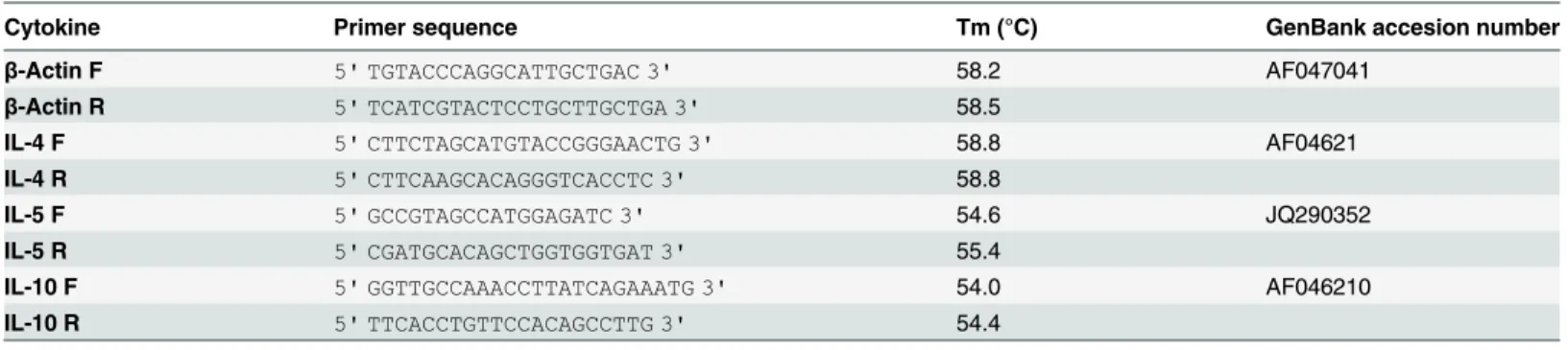
(Invitrogen). The cDNA samples were kept at -20°C until use. TibMolbiol LLC (Adelphia, NJ) designed the primers forβ-actin, IL-4 and IL-5; the primers for IL-10 were synthesized accord-ing to Espitiaet al. [19]. Nucleotide sequences, Tm values, product size and Genebank acces-sion numbers are shown inTable 1.
For each primer combination, optimal primer and MgCl2concentrations, as well as anneal-ing temperatures were experimentally determined. cDNA amplification was performed usanneal-ing the LightCycler 2.0 instrument (Roche Applied Science). The reaction mixture consisted of 1μl
cDNA, 3mM MgCl2, 0.5μM of each primer, and 1μl LightCycler FastStart DNA Master SYBR I
Green mix (Roche Applied Science) in a final volume of 10μl. For the non-template control,
water was added to the reaction instead of cDNA. The PCR cycling conditions were as follows: initial denaturation at 95°C for 10 min, followed by 45 cycles each consisting of denaturation at 95°C for 10s, annealing at 60°C for 5s, and extension at 72°C for 9s. Fluorescence was acquired
Table 1. Nucleotide sequences of primers for hamster genes.
Cytokine Primer sequence Tm (°C) GenBank accesion number
β-Actin F 5' TGTACCCAGGCATTGCTGAC 3' 58.2 AF047041
β-Actin R 5' TCATCGTACTCCTGCTTGCTGA 3' 58.5
IL-4 F 5' CTTCTAGCATGTACCGGGAACTG 3' 58.8 AF04621
IL-4 R 5' CTTCAAGCACAGGGTCACCTC 3' 58.8
IL-5 F 5' GCCGTAGCCATGGAGATC 3' 54.6 JQ290352
IL-5 R 5' CGATGCACAGCTGGTGGTGAT 3' 55.4
IL-10 F 5' GGTTGCCAAACCTTATCAGAAATG 3' 54.0 AF046210
IL-10 R 5' TTCACCTGTTCCACAGCCTTG 3' 54.4
Tm: Melting temperature
after each extension phase. After amplification, a melting curve analysis was performed to as-sess the specificity of the product. Standard curves for each primer set were generated from ten-fold serial dilutions of cDNA starting at a 1:10 and used to calculate the PCR efficiencies using the LightCycler software. Relative quantification calculations were performed by the Pfaffl method [20]. Non-stimulated cells were used as controls in all experiment.
Statistical Analysis
Data are shown as mean ± standard error of the mean (SEM). The Kruskal-Wallis test for non-parametric data with Dunn post-test was used.Pvalues<0.05 were considered significant.
Sta-tistical analysis was performed using the Prism 6.0 software (Graphpad Prism, San Diego, CA).
Results
Presence of TsCRT in E/S of cultured tapeworms
The identification of TsCRT in E/S products was performed in supernatants ofin vitrocultured tapeworms recovered from immunosuppressed hamsters and analyzed by western blot using specific anti-rTsCRT polyclonal antibodies. Among the proteins present in theT.soliumE/S products separated by SDS-PAGE, a band of ~50 kDa reacted with the anti-rTsCRT serum (Fig. 1). This molecular weight corresponds to the expected size of TsCRT.
TsCRT specific humoral immune response
The capacity of TsCRT to induce a specific IgG response in infected animals at different DPI by ELISA was investigated.Fig. 2shows the presence of specific IgG antibodies reacting to TsCE (Fig. 2A) and the purified rTsCRT (Fig. 2B). Between 28 and 33% of hamsters showed anti-TsCRT specific antibodies at 20 and 30 DPI respectively. Anti-TsCE OD values increased along the experiment starting at 10 DPI, and at 20 and 30 DPI all infected hamsters were posi-tive (p<0.05).
Local and systemic lymphoproliferative responses
The ability of TsCE and rTsCRT to inducein vitroresponses duringT.soliuminfection was as-sessed by lymphoproliferative assays in MLN (local) and spleen (systemic) cells. Con A was used as positive control for all proliferation assays (data not shown).Fig. 3shows the prolifer-ative response to TsCE and rTsCRT before infection and at different DPI. MLN cells did not show significant levels of proliferation in response to rTsCRT or TsCE (Fig. 3A). In contrast, TsCE induced a significant proliferative response in spleen cells at 20 and 30 DPI (P<0.05).
rTsCRT showed an increase in the proliferation levels of spleen cells at 20 DPI that were not statistical significant (Fig. 3B).
Cytokine profile induced during
T
.
solium
taeniosis
To analyze if TsCE and rTsCRT induce a Th2 immune response duringT.soliuminfection, the relative expression of IL-4, IL-5 and IL-10 in spleen and MLN cells from uninfected and in-fected hamsters in response toin vitrostimulation at different DPI was evaluated.Fig. 4shows the expression of mRNA for IL-4, IL-5 and IL-10 relative toβ-actin assessed by quantitative RT-PCR. Stimulation with TsCE resulted in an increased expression of IL-4 and IL-5 tran-scripts in both MLN and spleen cells at 20 and 30 DPI compared to non-stimulated cells (Fig. 4A, B, C, and D) while rTsCRT induced a significant increase of the IL-4 mRNA levels in MLN cells from uninfected hamsters (p<0.05) but not from spleen cells (Fig. 4A and B). Only
rTsCRT stimulation (P<0.05,Fig. 4D). Interestingly, cells from uninfected and infected
ham-sters significantly induced IL-10 mRNA expression in response to rTsCRT stimulation, with the exception of MLN cells from 20 DPI. Stimulation with TsCE did not result in a significant increase of transcript levels for the IL-10 immunoregulatory cytokine neither in MLN nor spleen cells (Fig. 4E and F).
Fig 1. TsCRT identification in E/S products obtained fromin vitrocultured tapeworms.E/S products were separated by SDS-PAGE and stained with Coomassie Brilliant Blue (lane 1). Samples were blotted to PVDF membranes after SDS PAGE and analyzed by western blot with anti-rTsCRT serum (lane 2). Molecular weight markers (kDa) are shown in the center lane.
Discussion
Here we have described the immune response elicited byT.soliumTsCRT upon infection in the hamster model. Interestingly,T.soliumTsCRT induced a robust IL-10 response in both MLN and spleen cells from infected and uninfected animals within a prevalent Th2 microenvi-ronment characterized by increased levels of IL-4 and IL-5. These findings are of importance owed to the fact that Th2 responses prevail during helminth infections. Additionally, the pres-ence of native TsCRT in tapeworms E/S products was confirmed.
CRT is involved in cellular Ca2+ homeostasis; however, there is growing evidence of its role in immune regulation [10]. IL-10 expression induced by rTsCRT in local and systemic lym-phoid organs uponin vitrostimulation is in agreement with previous reports [21]. This is im-portant for the parasite life cycle as a mechanism to modulate the host immune response [22,23]. IL-10 induction is a common feature shared by different helminths, presumably, asso-ciated with parasite survival [24,25]. In human neurocysticercosis, asymptomatic patients usu-ally present elevated levels of IL-10, suggesting that IL-10 might play a role in disease outcome [26]. Increased IL-10 production has been associated to immunoregulation in helminth chron-ic infection in humans and animal models [23], and also contribute to the maintenance of the Th2 response by inhibiting Th1 responses [27]. Additionally, IL-10 acts as an anti-inflammato-ry cytokine preventing the pathology associated with parasite infections, down-regulating the host immune response [6,28]. Thus, TsCRT might contribute to the overall masking strategy
Fig 2. Presence of IgG antibodies after oral infection withT.soliumcysticerci.ELISA testing was used to detect specific antibodies. A) Anti-TsCE IgG and B) anti-rTsCRT antibodies present in the sera obtained from uninfected and infected hamsters. Each dot refers to an individual hamster and the discontinuous lines represent the cutoff value that was calculated as the mean OD of the uninfected group plus 2 standard deviations. Kruskal-Wallis: **p<0.01 and****p<0.0001 as compared to the uninfected animals.
developed byT.soliumto escape the host response in an unfavorable microenvironment, char-acterized by mediators and cytokines that influence parasite expulsion.
As observed in other helminths [29–31], Th2 type responses are primarily developed during T.soliuminfection. Several reports have suggested the pivotal role of IL-4 in host protective re-sponses against parasite infections [32]. Interestingly, stimulation with TsCE, but not rTsCRT,
Fig 3. Proliferative responses of MLN (A) and spleen (B) cells from uninfected and infected hamsters to purified rTsCRT and TsCE.White bars represent data from cells incubated in RPMI alone, grey bars from cells incubated with rTsCRT, and hatched bars indicate the response to TsCE. Bars represent mean±SEM of3H thymidine incorporation from triplicate cultures of cells obtained from 6 animals. Kruskal-Wallis: ***P<0.001 as compared to the RPMI control.
resulted in increased synthesis of both IL-4 and IL-5 mRNA upon infection. Our data are in agreement with previous studies where IL-4 and IL-5 positive cells were detected at the tape-worm anchor site [33]. IL-4 is an important driver in T helper cell development leading to Th2 responses guided by antigen presenting cells [34]. Additionally, IL-5 is required for eosinophils
Fig 4. Cytokine mRNA expression by MLN and spleen cells afterin vitrostimulation with TsCE and rTsCRT.cDNA was prepared from mRNA obtained from MLN (A, C, D) or spleen (B, D, E). mRNA levels of IL-4 (A, B) IL-5 (C, D) and IL-10 (E, F) were determined by quantitative RT-PCR. Data are expressed as the mean of the ratio of each cytokine relative toβ-actin (housekeeping gene). White bars represent data from cells incubated in RPMI alone, grey bars from cells incubated with rTsCRT and hatched bars with TsCE. Bars represent mean±SEM of relative expression from triplicate cultures of cells obtained from 6 animals. Kruskal-Wallis:*P<0.05,**P<0.01,***P<0.001,****P<0.0001 rTsCRT-induced expression as
compared to the RPMI control.#P<0.05,##P<0.01 rTsCRT-induced expression as compared to TsCE. ND,
not detected.
maturation and is frequently produced along other Th2 type cytokines such as IL-4. The fact that rTsCRT did not stimulate expression of these two Th2 cytokines suggests that other para-site factors present in the TsCE besides TsCRT, contribute to the Th2 response prevailing dur-ingT.soliuminfection.
The golden hamster model of taeniosis reflects to some extent the early stages of infection produced by theT.soliumtapeworm in humans. In this study, no proliferation in response to antigen stimuli was observed in MLN cells. Conversely, spleen cells showed a rather strong cell proliferationin vitroupon stimulation. During experimental taeniosis, parasite antigens are present in circulation, and this could explain, at least partially, the priming of spleen cells [18]. The lack of response in MLN, might depend on“local”dynamics involved in the immune re-sponse being elicited and may vary based on the participating cell subpopulations, as well as the prevailing cytokine microenvironment. MLN display unique immunologic properties and are programmed for tolerance induction [35]. For instance,Trypanosoma cruziinfection leads to lymphocyte depletion in the thymus and MLN due to apoptosis, while subcutaneous lymph nodes and spleen cells undergo vigorous lymphocyte proliferation [36,37]. Other mechanisms, such as cytokine deprivation by subsets of T cells or tolerogenic dendritic cells or macrophages might promote cell anergy and/or hyporesponsiveness, impairing local cell immunity [38,39]. Our results are in agreement with the notion that parasite infections commonly lead to differ-ent degrees of immunosuppresion [31,40]. Nonetheless, the relative unresponsiveness of im-mune cell in specific lymphoid organs duringT.soiluminfection warrants further research.
Our western blot analysis suggests that TsCRT is secreted byT.soliumadult parasites. TsCRT is also present in crude extracts of cysticerci and tapeworms and its localization sug-gests a role in parasite development [12]. Likewise, CRT has been identified as a secreted pro-tein in different nematodes [41]. The identification of anti-TsCRT IgG antibodies observed in one third of infected hamsters suggests that TsCRT is secretedin vivo.
In conclusion, this is the first study characterizing the Th2 immune response elicited by rTsCRT uponT.soliuminfection. The identification of helminth-derived molecules playing a role in the host-parasite interface during experimental taeniosis is an area of current study that may help unveil and better understand the host anti-parasite response. The ability of rTsCRT to induce IL-4 and IL-10 expression in cells from uninfected hamsters might be explained by its interaction with immature dendritic cells or tolerogenic macrophages. Our results suggest that the host response against rTsCRT features a robust IL-10 response. Future studies should aim to analyze the interaction of rTsCRT with different types of immune cells and elucidate the receptors and signaling pathways involved, as well as the resulting phenotypic changes of the responding cells.
Acknowledgments
We thank Dr. Libia Vega Loyo and Elizabet Estrada Muñiz, MSc, from the Department of Tox-icology at CINVESTAV IPN, for their technical assistance in the3H thymidine incorporation experiments. We are thankful to Ulises Gonzalez for obtaining the partial sequence of hamster IL-5 and to Lizeth Hernandez and Bernardo Oldak for technical assistance.
Author Contributions
References
1. Flisser A, Sarti E, Lightowlers M, Schantz P. Neurocysticercosis: regional status, epidemiology, impact and control measures in the Americas. Acta tropica. 2003; 87(1):43–51. Epub 2003/06/05. PMID: 12781377
2. Sorvillo FJ, DeGiorgio C, Waterman SH. Deaths from cysticercosis, United States. Emerging infectious diseases. 2007; 13(2):230–5. Epub 2007/05/08. doi:10.3201/eid1302.060527PMID:17479884
3. Sarti-Gutierrez EJ, Schantz PM, Lara-Aguilera R, Gomez Dandoy H, Flisser A. Taenia solium taeniasis and cysticercosis in a Mexican village. Trop Med Parasitol. 1988; 39(3):194–8. Epub 1988/09/01.
PMID:3194663
4. Flisser A, Avila G, Maravilla P, Mendlovic F, Leon-Cabrera S, Cruz-Rivera M, et al. Taenia solium: cur-rent understanding of laboratory animal models of taeniosis. Parasitology. 2010; 137(3):347–57. Epub
2010/03/02. doi:10.1017/S0031182010000272PMID:20188011
5. McSorley HJ, Maizels RM. Helminth infections and host immune regulation. Clinical microbiology re-views. 2012; 25(4):585–608. Epub 2012/10/05. doi:10.1128/CMR.05040-11PMID:23034321
6. Elliott DE, Weinstock JV. Helminth-host immunological interactions: prevention and control of immune-mediated diseases. Annals of the New York Academy of Sciences. 2012; 1247:83–96. Epub 2012/01/
14. doi:10.1111/j.1749-6632.2011.06292.xPMID:22239614
7. Harnett W, Harnett MM. Helminth-derived immunomodulators: can understanding the worm produce the pill? Nature reviews Immunology. 2010; 10(4):278–84. Epub 2010/03/13. doi:10.1038/nri2730
PMID:20224568
8. Michalak M, Corbett EF, Mesaeli N, Nakamura K, Opas M. Calreticulin: one protein, one gene, many functions. The Biochemical journal. 1999; 344 Pt 2:281–92. Epub 1999/11/24. PMID:10567207
9. Wang WA, Groenendyk J, Michalak M. Calreticulin signaling in health and disease. The international journal of biochemistry & cell biology. 2012; 44(6):842–6. Epub 2012/03/01. doi:10.1016/j.biocel.2012. 02.009
10. Rzepecka J, Rausch S, Klotz C, Schnoller C, Kornprobst T, Hagen J, et al. Calreticulin from the intesti-nal nematode Heligmosomoides polygyrus is a Th2-skewing protein and interacts with murine scaven-ger receptor-A. Molecular immunology. 2009; 46(6):1109–19. Epub 2008/12/26. doi:10.1016/j. molimm.2008.10.032PMID:19108896
11. El Gengehi N, El Ridi R, Tawab NA, El Demellawy M, Mangold BL. A Schistosoma mansoni 62-kDa band is identified as an irradiated vaccine T-cell antigen and characterized as calreticulin. The Journal of parasitology. 2000; 86(5):993–1000. Epub 2000/12/29. doi:10.1645/0022-3395(2000)086[0993: ASMKBI]2.0.CO;2PMID:11128523
12. Mendlovic F, Carrillo-Farga J, Torres J, Laclette JP, Flisser A. Differential expression of calreticulin in developmental stages of Taenia solium. The Journal of parasitology. 2006; 92(4):789–95. Epub 2006/
09/26. doi:10.1645/GE-724R1.1PMID:16995397
13. Mendlovic F, Ostoa-Saloma P, Solis CF, Martinez-Ocana J, Flisser A, Laclette JP. Cloning, characteri-zation, and functional expression of Taenia solium calreticulin. The Journal of parasitology. 2004; 90-(4):891–3. Epub 2004/09/11. doi:10.1645/GE-3325RNPMID:15357095
14. Fonseca-Coronado S, Ruiz-Tovar K, Perez-Tapia M, Mendlovic F, Flisser A. Taenia solium: immune response against oral or systemic immunization with purified recombinant calreticulin in mice. Experi-mental parasitology. 2011; 127(1):313–7. Epub 2010/08/10. doi:10.1016/j.exppara.2010.07.017
PMID:20691181
15. Leon-Cabrera S, Cruz-Rivera M, Mendlovic F, Avila-Ramirez G, Carrero JC, Laclette JP, et al. Stan-dardization of an experimental model of human taeniosis for oral vaccination. Methods. 2009; 49-(4):346–50. Epub 2009/08/05. doi:10.1016/j.ymeth.2009.07.007PMID:19651215
16. Leon-Cabrera S, Cruz-Rivera M, Mendlovic F, Romero-Valdovinos M, Vaughan G, Salazar AM, et al. Immunological mechanisms involved in the protection against intestinal taeniosis elicited by oral immu-nization with Taenia solium calreticulin. Experimental parasitology. 2012; 132(3):334–40. Epub 2012/
08/28. doi:10.1016/j.exppara.2012.08.006PMID:22921496
17. Avila G, Aguilar L, Benitez S, Yepez-Mulia L, Lavenat I, Flisser A. Inflammatory responses in the intesti-nal mucosa of gerbils and hamsters experimentally infected with the adult stage of Taenia solium. Inter-national journal for parasitology. 2002; 32(10):1301–8. Epub 2002/09/03. PMID:12204230
18. Avila G, Benitez M, Aguilar-Vega L, Flisser A. Kinetics of Taenia solium antibodies and antigens in ex-perimental taeniosis. Parasitology research. 2003; 89(4):284–9. Epub 2003/03/13. doi:10.1007/ s00436-002-0605-8PMID:12632165
cutaneous leishmaniasis. BMC immunology. 2010; 11:31. Epub 2010/06/24. doi: 10.1186/1471-2172-11-31PMID:20569429
20. Pfaffl MW. A new mathematical model for relative quantification in real-time RT-PCR. Nucleic acids re-search. 2001; 29(9):e45. Epub 2001/05/09. PMID:11328886
21. Figueiredo CA, Barreto ML, Rodrigues LC, Cooper PJ, Silva NB, Amorim LD, et al. Chronic intestinal helminth infections are associated with immune hyporesponsiveness and induction of a regulatory net-work. Infection and immunity. 2010; 78(7):3160–7. Epub 2010/04/21. doi:10.1128/IAI.01228-09PMID: 20404082
22. Allen JE, Maizels RM. Diversity and dialogue in immunity to helminths. Nature reviews Immunology. 2011; 11(6):375–88. Epub 2011/05/26. doi:10.1038/nri2992PMID:21610741
23. McSorley HJ, Hewitson JP, Maizels RM. Immunomodulation by helminth parasites: defining mecha-nisms and mediators. International journal for parasitology. 2013; 43(3–4):301–10. Epub 2013/01/08.
doi:10.1016/j.ijpara.2012.11.011PMID:23911309
24. Hernandez JL, Leung G, McKay DM. Cestode regulation of inflammation and inflammatory diseases. International journal for parasitology. 2013; 43(3–4):233–43. Epub 2012/10/13. doi:10.1016/j.ijpara. 2012.09.005PMID:23911309
25. Persaud R, Wang A, Reardon C, McKay DM. Characterization of the immuno-regulatory response to the tapeworm Hymenolepis diminuta in the non-permissive mouse host. International journal for parasi-tology. 2007; 37(3–4):393–403. Epub 2006/11/10. doi:10.1016/j.ijpara.2006.09.012
26. Verma A, Prasad KN, Cheekatla SS, Nyati KK, Paliwal VK, Gupta RK. Immune response in symptomat-ic and asymptomatsymptomat-ic neurocystsymptomat-icercosis. Medsymptomat-ical msymptomat-icrobiology and immunology. 2011; 200(4):255–61.
Epub 2011/05/03. doi:10.1007/s00430-011-0198-xPMID:21533784
27. Balic A, Harcus YM, Taylor MD, Brombacher F, Maizels RM. IL-4R signaling is required to induce IL-10 for the establishment of T(h)2 dominance. International immunology. 2006; 18(10):1421–31. Epub
2006/08/31. doi:10.1093/intimm/dxl075PMID:16940042
28. Yazdanbakhsh M, Wahyuni S. The role of helminth infections in protection from atopic disorders. Cur-rent opinion in allergy and clinical immunology. 2005; 5(5):386–91. Epub 2005/09/01. PMID:16131911
29. Allen JE, Maizels RM. Immunology of human helminth infection. International archives of allergy and immunology. 1996; 109(1):3–10. Epub 1996/01/01. PMID:8527948
30. Bancroft AJ, Grencis RK. Th1 and Th2 cells and immunity to intestinal helminths. Chemical immunolo-gy. 1998; 71:192–208. Epub 1998/10/08. PMID:9761955
31. Loukas A, Prociv P. Immune responses in hookworm infections. Clinical microbiology reviews. 2001; 14(4):689–703, table of contents. Epub 2001/10/05. doi:10.1128/CMR.14.4.689-703.2001PMID: 11585781
32. Anthony RM, Rutitzky LI, Urban JF Jr., Stadecker MJ, Gause WC. Protective immune mechanisms in helminth infection. Nature reviews Immunology. 2007; 7(12):975–87. Epub 2007/11/17. doi:10.1038/ nri2199PMID:18007680
33. Avila G, Aguilar L, Romero-Valdovinos M, Garcia-Vazquez F, Flisser A. Cytokine response in the intes-tinal mucosa of hamsters infected with Taenia solium. Annals of the New York Academy of Sciences. 2008; 1149:170–3. Epub 2009/01/06. doi:10.1196/annals.1428.079PMID:19120202
34. Liu Q, Liu Z, Rozo CT, Hamed HA, Alem F, Urban JF Jr., et al. The role of B cells in the development of CD4 effector T cells during a polarized Th2 immune response. J Immunol. 2007; 179(6):3821–30. Epub
2007/09/06. PMID:17785819
35. Pabst O, Wahl B, Bernhardt G, Hammerschmidt SI. Mesenteric lymph node stroma cells in the genera-tion of intestinal immune responses. J Mol Med (Berl). 2009; 87(10):945–51. Epub 2009/08/04. doi:10. 1007/s00109-009-0502-zPMID:19649572
36. de Meis J, Morrot A, Farias-de-Oliveira DA, Villa-Verde DM, Savino W. Differential regional immune re-sponse in Chagas disease. PLoS Negl Trop Dis. 2009; 3(7):e417. Epub 2009/07/08. doi:10.1371/ journal.pntd.0000417PMID:19582140
37. de Meis J, Barreto de Albuquerque J, Silva Dos Santos D, Farias-de-Oliveira DA, Berbert LR, Cotta-de-Almeida V, et al. Trypanosoma cruzi Entrance through Systemic or Mucosal Infection Sites Differential-ly Modulates Regional Immune Response Following Acute Infection in Mice. Frontiers in immunology. 2013; 4:216. Epub 2013/07/31. doi:10.3389/fimmu.2013.00216PMID:23898334
38. Pandiyan P, Zheng L, Ishihara S, Reed J, Lenardo MJ. CD4+CD25+Foxp3+ regulatory T cells induce cytokine deprivation-mediated apoptosis of effector CD4+ T cells. Nature immunology. 2007; 8-(12):1353–62. Epub 2007/11/06. doi:10.1038/ni1536PMID:17982458
40. Olatunde BO, Onyemelukwe GC. Immunosuppression in Nigerians with hookworm infection. African journal of medicine and medical sciences. 1994; 23(3):221–5. Epub 1994/09/01. PMID:7604745
41. Nagaraj SH, Gasser RB, Ranganathan S. Needles in the EST haystack: large-scale identification and analysis of excretory-secretory (ES) proteins in parasitic nematodes using expressed sequence tags (ESTs). PLoS Negl Trop Dis. 2008; 2(9):e301. Epub 2008/09/30. doi:10.1371/journal.pntd.0000301