Case 7530
Broad Ligament Leiomyoma
J Raposo, TM Cunha.
Section: Genital (Female) Imaging Published: 2009, Jun. 1
Patient: 22 year(s), female
Clinical Summary
A 22 year-old female patient presenting with pelvic pain.
Clinical History and Imaging Procedures
A 22 year old woman presented with a 3 months history of persistent pelvic pain. Menstrual cycles were
regular and there was no abnormal bleeding. She had menarche at 11, gravida 1, para 1 and using oral
contraceptives for 3 years. Physical examination evidenced a firm pelvic mass in the right lateral vaginal
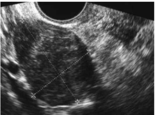
recess. Transvaginal ultrasound revealed a solid right adnexal lesion with a diameter of 6 cm,
hypoechoic and well circumscribed (Fig 1). To better characterize the lesion and to determine the
relationship between the nodule and the surrounding structures, magnetic resonance imaging was
performed. MRI demonstrated a low signal intensity lesion on T1- and T2-weighted images (Fig 2-3,
respectively), with no claw of myometrium surrounding tumour and no interface vessels, that after
endovenous contrast administration enhanced less than myometrium (Fig 4). The findings were
consistent with a fibrotic leiomyoma with extrauterine and extraovaric origin. The patient underwent
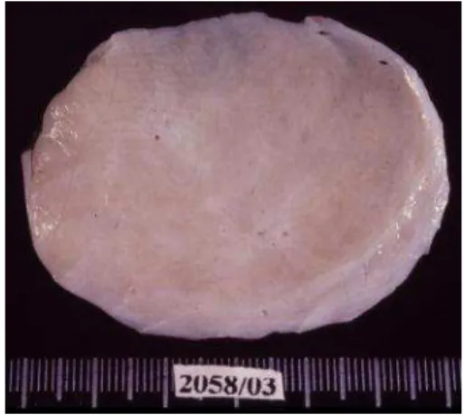
surgical resection of this lesion and a tumour of 6.5x5x5cm attached by a stalk to right broad ligament
was found. Pathological examination confirmed the diagnosis of a right broad ligament fibrotic
leiomyoma weighing 99gr (Fig 5), and showed very good correlation with imaging findings.
Discussion
Extrauterine fibroids are not as common as uterine fibroids, accounting for less than 3% of all pelvic
leiomyomas. These arise from the supporting structures of the uterus, such as the broad ligament, or are
tumours of a uterine ligament and is benign. As a diagnostic criterion, broad ligament leiomyoma must
be completely separated from and in no way connected with either the uterus or the ovary. These
tumours usually are attached by a stalk to the broad ligament, which is also responsible for their blood
supply. Although in most cases broad ligament leiomyomas are asymptomatic, they may present pelvic
pain or a palpable pelvic/ abdominal mass. Pelvic pain may be due to pressure effects on adjacent
organs, such as bladder or rectum, or torsion.
On US, broad ligament leiomyomas have the same appearance as any other uterine fibroma and
present as an hypoechoic solid mass, well-circumscribed, that can be heterogeneous when large.
Usually there is no interface between tumour and uterus and no straight relation to the homolateral
ovary. As sonograms have false negatives and eventually pedunculated leiomyomas may be mistaken
for solid ovarian masses, MRI is indicated whenever ultrasound results are limited, to demonstrate the
exact location of the lesion and to characterize it for differential diagnosis. Broad ligament leiomyomas
have the typical appearance of leiomyomas on MRI and are depicted as sharply emarginated lesions of
low signal intensity on both T1W and T2W sequences due to their high fibrotic content. They are not
surrounded by a pseudocapsule of compressed neighbouring tissue and do not show interface vessels
imaged as perilesional rim enhancement. Most enhance similarly to the myometrium, whereas larger
leiomyomas tend to enhance less and heterogeneously. Postgadolinium images may evidence the
presence of a stalk. In addition to standard MR sagittal and axial views, coronal or oblique views may be
indicated for accurate localization and for establishing the most probable origin of a lesion. The
differential diagnosis is with parasitic leiomyoma of the broad ligament, subserosal leiomyomas, fibrotic
adnexal lesions and other solid tumours from the broad ligament. Parasitic leiomyoma of the broad
ligament originates from the uterus and invades the broad ligament, additionally recruiting arterial supply,
maintaining or losing its original uterine attachment. Subserosal leiomyomas, if pedunculated, may be
hard to distinguish from broad ligament fibroid; identification of the site of attachment is crucial to
establish the uterine origin. Ovarian neoplasms include fibroma, adenofibroma, fibrothecoma, Brenner's
tumour and metastatic ovarian tumours with highly fibrous component, particularly those from the
gastrointestinal tract; usually they originate, and are inseparable, from the ovary, may appear as a
complex mass and may have enhancement of solid portions tumour and septa. Other ligamentous
mesenchymal tumours are lipoma, neurofibroma, schwannoma and leiomyosarcoma. Leiomyosarcoma
is differentiated from leiomyoma microscopically.
Surgical removal is needed for symptomatic relief and for impingement on nearby structures, and
because differential diagnosis may include malignancy and pedunculated tumours with risk of torsion.
Final Diagnosis
Right broad ligament leiomyoma.
Figure 1 Ultrasonography
Ultrasonography shows a hypoechoic right adnexal
mass, well circumscribed, slightly heterogeneous, with
6.5 x 5 cm.
Figure 2 Axial T1-weighted
Axial T1-weighted TSE image shows a right adnexal
mass that is hypointense and slightly heterogeneous.
Figure 3 Axial T2-weighted
Axial T2-weighted TSE image shows a right adnexal
mass that is markedly hypointense.
Figure 4 Gad-enhanced FS T1-W
Figure 5 Surgical specimen
Gross specimen of the broad ligament fibroid.
MeSH
Genital Neoplasms, Female [C13.371.820.800.418]
References
[1] Yuel VI, Kaur V (2006) Broad ligament fibroid - an unusual presentation. JK Science 4:217-8
[2] Meniru GI, Wasdahl D, Onuora CO, Hecht BR, Hopkins MP (2001) Vaginal leiomyoma co-existing
with broad ligament and multiple uterine leiomyomas. Arch Gynecol Obstet J 265: 105-107
[3] Ng Lung Kit HK, Collins RE (1986) Leiomyoma of the broad ligament in an obturator hernia
presenting as a lump in the groin. J R Soc Med 79(3):174-5.
[4] Nappi L, Bettocchi S, Carriero C, Ceci O, Vimercati A, Resta L. Large parasitic leiomyoma of the
broad ligament. Journal of Gynecologic Surgery. 2004, 20(3): 97-102
[5] Pelsang RE, Sorosky J, Woods T (1999) Sonographic evaluation of a leiomyoma of the broad
ligament of the uterus. J Clin Ultrasound 27:402-404
Citation
J Raposo, TM Cunha. (2009, Jun. 1)
Broad Ligament Leiomyoma {Online}