Case 7601
Pedunculated Subserosal Leiomyoma
Ferreira AT, Cunha TM.
Section: Genital (Female) Imaging Published: 2009, Sep. 3
Patient: 45 year(s), female
Clinical Summary
The patient presented with an abdominopelvic mass and recent complaints of abdominal pain and
urinary frequency. Ultrasound and CT were performed. She was then transferred to our institution with
the presumed diagnosis of malignant ovarian tumour.
Clinical History and Imaging Procedures
A 45 year old female presented with abdominal volume enlargement over the previous month and
abdominal pain and urinary frequency of two weeks' duration. On physical examination, an
abdominopelvic mass was palpated.
Imaging work-up (ultrasound and CT) revealed a heterogeneous abdominopelvic mass, with cystic and
solid components, irregular contour and parietal vegetations, measuring approximately 20cm in diameter
and compressing the surrounding structures; mild pielo-caliceal distension and discrete pelvic ascites.
The exams were interpreted as a malignant ovarian mass and she was transferred to our institution.
Blood test results only revealed anaemia (Hb 7,5), with tumoral markers (CA125, CEA, alpha-fetoprotein,
beta-HCG) within normal limits.
MRI findings included a huge, sharply marginated pelvic lesion, reaching the renal hila, hypointense on
T1-weighted images (Fig. 1) and hyperintense on T2-weighted images (Fig. 2), with thick wall that
strongly enhanced, isointense to the well perfused myometrium, with central absence of enhancement
(Fig. 3); the lesion apparently communicated with the uterus by a fundic stalk (Fig. 4).
Laparotomy was performed and revealed a fundic uterine mass with 25cm in larger dimension, adherent
to the small bowel. On gross pathology the tumour had firm, irregular wall, with fasciculated aspect and a
necrotic centre. Microscopic examination revealed subserosal leiomyoma, partially necrotic, probably
Discussion
Subserosal leiomyomas are benign smooth muscle tumors of the uterus, originating beneath serosa.
They are not completely surrounded by normal myometrium and can be sessile or pedunculated. This
location is less common than intramural.
Ultrasonography is the primary diagnostic modality, but accurate assessment of pedunculated
subserosal leiomyomas is not always possible, even with both transvaginal and transabdominal
approach. CT with its poor soft tissue contrast is of limited value. MR is currently considered the most
accurate imaging technique for detection and localization of leiomyomas [1]. They are well-circumscribed
masses, that typically demonstrate low signal intensity relative to that of the myometrium on T2-weighted
images and intermediate signal intensity on T1-weighted images [2]. In pedunculated subserosal
leiomyomas, a low signal intensity pedicle can be identified on T2-weighted images.
Subserosal leiomyomas are usually asymptomatic; however, pedunculated ones may undergo torsion,
which results in infarction and necrosis accompanied by pain. Necrotic leiomyomas that have not
liquefied (ie, coagulative necrosis) have variable signal intensity on T1-weighted images and low signal
intensity on T2-weighted images [1]. In this case, there is a large central area with signal intensity
characteristic of fluid: low on T1-weighted images and high on T2-weighted images with no
enhancement, suggesting liquefaction.
Some torsed leiomyomas may loose their connection with the uterus, become attached to adjacent
structures and be supplied by parasitic vessels [2], which can be traced on contrast enhanced MR
images.
In patients with a markedly enlarged uterus, pedunculated leiomyomas are often difficult to differentiate
from extrauterine or ovarian lesions. The ability of MRI to demonstrate normal ovaries may aid in
determining the origin of pelvic masses by excluding a diagnosis of ovarian neoplasm [1].
Treatment is indicated in symptomatic uterine leiomyomas and surgical removal is the most widely used
therapeutic approach. In pedunculated subserosal leiomyomas with a thin stalk, uterine artery
embolization is relatively contraindicated.
Final Diagnosis
Torsion of pedunculated subserosal leiomyoma
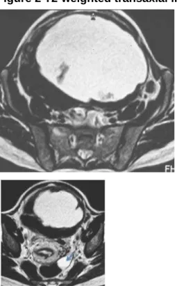
Figure 1 T1-weighted transaxial image.
Large, spherical, sharply marginated lesion, with
hypointense fluid content and hypointense walls.
Figure 2 T2-weighted transaxial image.
The mass has a hyperintense fluid content, and irregular
walls with hypointense signal.
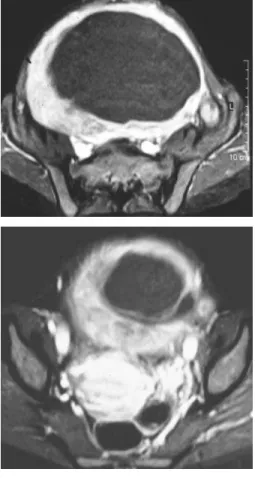
Figure 3 Contrast-enhanced T1-weighted fat-suppressed transaxial images.
Lesion enhances perypherally, isointense to the well
perfused myometrium, with central absence of
enhancement (confirms infarction of the leiomyoma).
Lesion enhances perypherally, isointense to the well
perfused myometrium, with central absence of
enhancement (confirms infarction of the leiomyoma).
Figure 4 T2- weighted para-sagittal image.
The lesion lies anterior to the uterine corpus, displacing
it posteriorly and compressing the bladder inferiorly. The
stalk is detected in the uterine fundus (arrow).
MeSH
Uterine Diseases [C13.371.852]
References
[1] Murase E, Siegelman ES, Outwater EK, Perez-Jaffe LA, Tureck RW (1999) Uterine Leiomyomas:
19:1179-1197
[2] Ueda H, Togashi K, Konishi I, Kataoka ML, Koyama T, Fujiwara T, Kobayashi H, Fujii S, Konishi J
(1999) Unusual Appearances of Uterine Leiomyomas: MR Imaging Findings and Their Histopathologic
Backgrounds. Radiographics 19:S131-S145
[3] Yeh HC, Kaplan M, Deligdisch L (1999) Parasitic and pedunculated leiomyomas: ultrasonographic
features. J Ultrasound Med 18 (11): 789-794
Citation
Ferreira AT, Cunha TM. (2009, Sep. 3)