INSTITUTO SUPERIOR DE CIÊNCIAS DA SAÚDE
EGAS MONIZ
MESTRADO INTEGRADO EM MEDICINA DENTÁRIA
FATORES DE RISCO PARA A PERDA DE IMPLANTES E
PATOLOGIA PERI-IMPLANTAR NUMA POPULAÇÃO
SISTEMICAMENTE COMPROMETIDA
Trabalho submetido por
Joana Leite Neves
para a obtenção do grau de Mestre em Medicina Dentária
INSTITUTO SUPERIOR DE CIÊNCIAS DA SAÚDE
EGAS MONIZ
MESTRADO INTEGRADO EM MEDICINA DENTÁRIA
FATORES DE RISCO PARA A PERDA DE IMPLANTES E
PATOLOGIA PERI-IMPLANTAR NUMA POPULAÇÃO
SISTEMICAMENTE COMPROMETIDA
Trabalho submetido por
Joana Leite Neves
para a obtenção do grau de Mestre em Medicina Dentária
Trabalho orientado por
Doutor Pedro Oliveira
e coorientado por
Professor Doutor José Martins dos Santos
3 Agradecimentos
Gostaria de agradecer a todos que contribuíram para que este trabalho fosse possível de concretizar:
Ao meu orientador Prof. Doutor Pedro Oliveira, pela orientação, disponibilidade e confiança. Pela iniciativa em propor a realização de um projeto desafiante tornando viável a sua elaboração.
Ao meu coorientador Prof. Doutor José Martins dos Santos, pelo apoio prestado, confiança e disponibilidade.
Ao Mestre Miguel de Araújo Nobre, diretor do Departamento de Investigação e Desenvolvimento da Malo Clinic Lisboa, por toda a colaboração e apoio prestado.
Ao meu irmão David Neves, pela colaboração e disponibilidade.
4
RESUMO
Objetivos: Identificar os possíveis fatores de risco para a perda de implantes e patologia peri-implantar numa população de pacientes sistemicamente comprometidos.
Materiais e métodos: Trata-se de estudo clínico retrospetivoque inclui um total de 721 pacientes sistemicamente comprometidos, submetidos a reabilitações com implantes dentários (422 mulheres, 299 homens) e com idade média de 51 anos ( entre 20-87). O tempo médio de seguimento clínico é de 7.3 anos. Foram registadas as variáveis demográficas (idade e género) e as variáveis clínicas (localização dos implantes, tipo de superfície dos implantes e condição sistémica). As variáveis em estudo são a perda de implantes e a patologia peri-implantar. Foram efetuados modelos de regressão logística binários para investigar os efeitos das características demográficas e clínicas dos pacientes para as variáveis dependentes: perda de implantes e patologia peri-implantar. Foi efetuada uma regressão linear para relacionar as características dos pacientes com o número de implantes perdidos. Foram estimados para cada variável o rácio de changes (odds ratio) com intervalos de confiança de 95% e níveis de significância correspondentes.
Resultados: A regressão logística multivariada identificou o aumento da idade (pacientes com mais de 40 anos) como fator de risco para a perda de implantes (OR=2.63); e a hepatite como fator de risco para a patologia peri-implantar (OR=3.74). A regressão linear multivariada identificou as doenças reumatológicas e cardiovasculares como estando relacionadas com um número elevado de implantes perdidos.
Conclusão: Tendo em conta as limitações deste estudo, os resultados sugerem que não existem contraindicações absolutas para a reabilitação com implantes numa população de pacientes sistemicamente comprometidos. No entanto, este estudo sugere que o aumento da idade, as condições reumatológicas, as condições cardiovasculares e a hepatite poderão ser tidos em consideração quando se verifica uma influência negativa nos resultados das reabilitações suportadas por implantes.
5
ABSTRACT
Purpose: The purpose of this study was to identify the possible risk factors for implant failure and peri-implant pathology in a population of systemically compromised patients.
Materials and Methods: This retrospective clinical study included a total of 721 systemically compromised patients (422 women, 299 men), with an average age of 51 years (range: 20-87) rehabilitated with dental implants. The average follow-up time was
7.3 years. The patient’s demographic variables (age and gender), and clinical variables (implant location, type of implant surface and systemic conditions) were recorded. Outcome measures were implant failure and peri-implant pathology. Binary logistic regression models were performed to investigate the effect of the patients’ demographic and clinical characteristics on the dependent variables implant failure and peri-implant
pathology. A linear regression model was performed to correlate the patient’s
characteristics with the number of failed implants. Odds ratios with 95% confidence intervals and corresponding levels of significance were estimated for each variable.
Results: Multivariate logistic regression disclosed increased age (patients over 40 years of age) as a risk factor for implant failure (OR = 2.63); and hepatitis as a risk factor for peri-implant pathology (OR = 3.74). Multivariate linear regression disclosed rheumatologic and cardiac diseases to be correlated with a higher number of failed implants.
Conclusions: Within the limitations of this study, the results suggest no absolute contraindications for implant rehabilitation in a population of systemically compromised patients. Nevertheless, this study suggests that increasing age, rheumatological condition, cardiovascular condition and hepatitis should be taken into consideration when performing implant supported rehabilitations due to their negative influence on the outcome.
6
Índice Geral
I. Introdução ... 8
II. Desenvolvimento ... 13
Paper: Risk factors for implant failure and peri-implant pathology in a systemic compromised population. ... 13
i. Abstract ... 15
ii. Introduction ... 16
iii. Materials and Methods ... 18
Inclusion and Exclusion Criteria ... 18
Surgical Protocol ... 18
Immediate and Final Prosthetic Protocol ... 19
Follow-up ... 20
Outcome Measures ... 20
Statistical Analysis ... 20
iv. Results ... 22
Patient characteristics ... 22
Dental implants and implant failure ... 22
Peri-implant pathology ... 23
v. Discussion ... 26
vi. Conclusions ... 29
vii. Acknowledgments ... 30
viii. References ... 31
III. Conclusões ... 36
7
Índice de tabelas
Tabela 1 Binary logistic regression model for the effect of the independent variables on implant failure. ... 23
Tabela 2 Number of failed implants as a function of patient characteristics using multivariable linear regression. ... 24
8
I.
Introdução
Os implantes dentários têm vindo a ser utilizados ao longo do tempo como opção de tratamento para a substituição de dentes ausentes. A sua eficácia e previsibilidade está demostrada através de elevadas taxas de sucesso, revelando-se em geral a melhor opção quando se pretende reabilitar áreas edentulas (Astrand et al.,2008; Malo et al.,2014; Andersson et al., 2013). Tendo em conta a satisfação por parte dos pacientes, é reportado um aumento do conforto e bem-estar induzido pela melhoria estética e funcional proporcionadas pela reabilitação com implantes, em particular no caso das reabilitações com implantes suportadas por próteses fixas (Annibali et al.,2010; Martín-Ares et al.,2015).
As reabilitações com implantes tem tido cada vez mais sucesso quer no processo de osteointegração como na satisfação por parte dos pacientes. Contudo, existem um conjunto de fatores que podem levar à perda biológica de implantes, precoce ou tardia. Por sua vez, a perda biológica caracteriza-se pela incapacidade dos tecidos estabelecerem ou manterem a osteointegração, ocorrendo precocemente durante o período de osteointegração ou tardiamente após o mesmo (Esposito et al.,1998; Palma-Carrió et al.,2011).
Vários autores identificaram um conjunto de potenciais causas para a ocorrência precoce da perda de implantes. Em particular, a condição sistémica, os hábitos tabágicos, a qualidade óssea, o número de implantes colocados e a distribuição dos mesmos (Esposito et al.,1998; Palma-Carrió et al.,2011; Malo et al.,2011; Alsaadi et al.,2007). Adicionalmente, Esposito et al. (1998) identificou também o trauma cirúrgico associado a um processo de cicatrização debilitado, a contaminação bacteriana e a carga permatura como potenciais causas para a perda precoce de implantes.
Por outro lado, a perda tardia de implantes foi identificada como estando etiologicamente associada ao excesso de carga, à patologia peri-implantar e às características do hospedeiro (Esposito et al.,1998; Malo et al.,2011; Courts et al.,2004; Manor et al.,2007; Moy et al., 2005).
Introdução
9
Manor et al.,2007), género masculino (Manor et al.,2007), tabagismo e condição sistémica (Esposito et al.,1998; Malo et al.,2011).
Segundo Manor et al. (2007), o excesso de carga foi identificado como o fator proporcionalmente mais relevante para ocorrência de perda de implantes tardia. Decorrendo de um equilíbrio funcional impróprio, em que na presença de patologia peri-implantar poderá promover o aumento desta mesma doença. Esposito et al. (1998), salienta que para além do excesso de carga, a persistência de estímulos inflamatórios, provocados por acumulação e maturação de placa bacteriana poderão induzir uma progressiva desintegração da interface implante-osso.
A patologia peri-implantar é classificada como um grupo de situações de origem multifatorial, tais como: a história de periodontite, o nível ósseo localizado no terço médio do implante, a presença de placa bacteriana, hemorragia, o desajuste protético e tipo de material restaurador utilizado. A proximidade com outros implantes ou dentes foi considerado fator de risco para a doença, apenas na presença de placa bacteriana (De Araújo Nobre et al.,2015).
Esta patologia está descrita na literatura como “um termo para reações inflamatórias
com perda de suporte ósseo que rodeiam um implante em função” (Alberktsson et al, 1994). Na literatura, a patologia peri-implantar é definida clinicamente de diferentes formas (Zitzmann et al.,2008). O modo como a incidência da doença é medida também condiciona os resultados. Em particular, varia de 10% a 56% quando o paciente é utilizado como unidade de análise, e de 4% a 43% quando o implante é a unidade de análise ( Zitmann et al., 2008; Froum et al., 2012; Klinge et al.,2012; Cecchinato et al., 2014; Atieh et al.,2013; Daubert et al., 2015). A falta de consenso relativamente à classificação da patologia peri-implantar e aos seus diferentes graus de severidade, levou a que se propusesse uma classificação segundo o grau de severidade (inicial,
moderada ou avançada) tendo em conta “a combinação de hemorragia à sondagem e/ou
supuração, profundidade de sondagem e extensão da perda óssea em torno do implante” (Froum et al., 2012). Desta forma, classificou-se a patologia peri-implantar “inicial” mediante a presença de profundidade à sondagem (PS) ≥ 4 mm e com perda óssea <
25% relativamente ao comprimento do implante. “Moderada”, tendo em conta PS ≥ 6 mm e perda óssea de 25% a 50%. Por último, “avançada” quando se verifica PS ≥ 8 mm
10
classificação permite quer por parte dos clínicos como dos investigadores, uma identificação clara e precisa dos sinais e graus de patologia peri-implantar. Possibilitando assim, um melhor controlo e prevenção da incidência e prevalência da doença, o que aumenta a probabilidade de sucesso e previne a perda de implantes.
Segundo Alsaadi (2008), a localização dos implantes na arcada dentária (maxila ou mandibula e região anterior ou posterior) tem influência na perda de implantes. Moy (2005) documentou uma maior perda de implantes na maxila relativamente à mandibula. De Araújo Nobre (2014a) ao estudar a influência de características tais como a localização e posição dos implantes na patologia peri-implantar, verificou que tanto a localização anterior ou posterior, como a localização na arcada (maxila ou mandibula) não afetam a ocorrência de patologia peri-implantar (Alsaadi et al., 2008; Moy et al., 2005; de Araújo Nobre et al., 2014). Ao avaliar o tipo de superfície dos implantes, Alsaadi (2008) identificou uma tendência para existência de maior perda nos implantes com superfície maquinada relativamente a implantes de superfície tratada através de oxidação anódica (TiUnite). Por outro lado, para Renvert (2011), não há evidência de que o tipo de superfície dos implantes possa afetar a ocorrência de patologia peri-implantar (Alsaadi et al., 2008; Renvert et al., 2011)
A perda de implantes e a incidência de patologia peri-implantar, podem ser influenciadas por diversos fatores, em particular a condição sistémica dos pacientes, que se tem vindo a demostrar como uma área de investigação de grande potencial (Palma-Carrió et al.,2011; Hwang et al.,2007). As condições sistémicas que podem afetar o resultado do tratamento das reabilitações suportadas por implantes são: hepatite, doenças cardiovasculares, doenças da tiroide, diabetes, doenças reumáticas, HIV, doenças oncológicas e hábitos tabágicos (Moy et al., 2005; Coates et al.,2000; Elsubeihi et al., 2002; Hwang et al.,2007; Attard et al., 2002; Ship et al.,2003; Valero et al.,2007; Oates et al., 2009; Weinlander et al., 2010; Krennmair et al., 2010; Balaji et al., 2008; Sham et al., 2003; Baig et al., 2007; Sgolastra et al., 2015).
Os diferentes mecanismos de cada condição sistémica ou de fatores com efeitos nefastos, podem exercer impactos distintos no resultado do tratamento com implantes (Malo et al., 2015).
Introdução
11
tóxicos dos cigarros no organismo, induzem a ocorrência de respostas biológicas tais como a redução da cicatrização dos tecidos, provocada pela vasoconstrição e consequente diminuição da perfusão, resultante dos efeitos nefastos do monóxido de carbono e nicotina nas células sanguíneas e nos mecanismos de transporte de oxigénio e celulares (Hwang et al.,2007). Através dos mecanismos anteriormente descritos bem como de um aumento da expressão de mediadores inflamatórios, observa-se nos pacientes fumadores um menor sucesso nos procedimentos cirúrgicos, nas terapias periodontais e tratamento com implantes (Hwang et al.,2007; Balaji et al., 2008; Sham et al., 2003; Baig et al., 2007). Por sua vez, existe um largo consenso no que diz respeito ao impacto negativo do tabagismo nas reabilitações suportadas por implantes (Esposito et al.,1998; Palma-Carrió et al.,2011; Malo et al.,2011; Alsaadi el al., 2007; Moy et al., 2005; Elsubeihi et al., 2002; Hwang et al.,2007; Balaji et al., 2008; Sham et al., 2003; Baig et al., 2007; Sgolastra et al., 2015; Malo et al., 2015; de Araújo Nobre et al., 2014b).
Por outro lado, o impacto de cada doença sistémica nas reabilitações suportadas por implantes, varia de acordo com o seu tipo, sendo sugerido pela literatura que o grau de controlo da doença poderá ser mais importante que a doença per se (Diz et al., 2013; Scully et al., 2007).
Assim, são necessários mais estudos que avaliem o impacto das condições sistémicas nas reabilitações suportadas por implantes, de modo a aumentar a probabilidade de sucesso neste tipo de tratamentos.
Este trabalho tem como objetivo principal identificar as possíveis causas para ocorrência de patologia peri-implantar e para a perda de implantes, numa população de pacientes sistemicamente comprometidos ou fumadores.
12
Este estudo abrange uma amostra de 721 indivíduos (com doenças sistémicas ou hábitos tabágicos) reabilitados com 3998 implantes (com uma média 5.5 implantes por paciente) e seguidos longitudinalmente. Em que 422 indivíduos são do género feminino e 299 do género masculino. Apresentam uma média de idade de 51 anos de idade, que varia dos 20 aos 87 anos de idade.
A amostra de pacientes presentes neste estudo permitiu estudar de forma estatisticamente significativa as características desta população, levando à concretização de um artigo científico, submetido na revista “Journal of Prosthodontics”.
Desenvolvimento
13
II.
Desenvolvimento
14
Risk factors for implant failure and peri-implant pathology in a
systemic compromised population
Joana Neves, RDH1,2; Miguel de Araújo Nobre RDH, MSc, Epi3; Pedro Oliveira, DDS, MD, PhD1; José Martins dos Santos, MD, PhD1; Paulo Maló DDS, PhD4
Corresponding author:
Miguel de Araújo Nobre, RDH, MSc Epi
Maló Clinic,
Avenida dos Combatentes, 43, 8 floor, Edificio Green Park,
1600-042 Lisbon, Portugal
Telephone: +351 217 228 100
Fax: +351 217 266 965
e-mail: [email protected]
1
Center for Interdisciplinar Research Egas Moniz, Campus Universitário, Quinta da Granja, Monte da Caparica, 2829-511, Caparica, Portugal
2
Oral Hygiene Department, Maló Clinic, Avenida dos Combatentes, 43, 9th floor, Edificio Green Park, 1600-042, Lisbon, Portugal.
3
Research and Development Department, Maló Clinic, Avenida dos Combatentes, 43, 8th floor, Edificio Green Park, 1600-042, Lisbon, Portugal
4
Desenvolvimento
15
i. Abstract
Purpose: The purpose of this study was to identify the possible risk factors for implant failure and peri-implant pathology in a population of systemically compromised patients.
Materials and Methods: This retrospective clinical study included a total of 721 systemically compromised patients (422 women, 299 men), with an average age of 51 years (range: 20-87) rehabilitated with dental implants. The average follow-up time was 7.3 years. The patient’s demographic variables (age and gender), and clinical variables (implant location, type of implant surface and systemic conditions) were recorded. Outcome measures were implant failure and peri-implant pathology. Binary logistic regression models were performed to investigate the effect of the patients’ demographic and clinical characteristics on the dependent variables implant failure and peri-implant
pathology. A linear regression model was performed to correlate the patient’s
characteristics with the number of failed implants. Odds ratios with 95% confidence intervals and corresponding levels of significance were estimated for each variable.
Results: Multivariate logistic regression disclosed increased age (patients over 40 years of age) as a risk factor for implant failure (OR = 2.63); and hepatitis as a risk factor for peri-implant pathology (OR = 3.74). Multivariate linear regression disclosed rheumatologic and cardiac diseases to be correlated with a higher number of failed implants.
Conclusions: Within the limitations of this study, the results suggest no absolute contraindications for implant rehabilitation in a population of systemically compromised patients. Nevertheless, this study suggests that increasing age, rheumatological condition, cardiovascular condition and hepatitis should be taken into consideration when performing implant supported rehabilitations due to their negative influence on the outcome.
16
ii. Introduction
Dental implants are scientifically documented with a high long-term success rate and often represent the best treatment option to replace missing teeth due to their effectiveness and predictability.1-3 Although implant rehabilitations achieve high success rates, there are several situations that can lead to biological implant failure, both early failures (occurring during the osseointegration period) and late failures (after the occurrence of osseointegration.4-5
Previous investigations identified several potential causes for premature implant failure, including smoking habits, systemic condition, bone quality, number of implants placed, and implant distribution.4-7 In addition, factors such as surgical trauma, bacterial contamination and premature loading were previously indicated as potential reasons for early implant failure.4 Late implant failures were previously associated with overloading, peri-implant pathology and host characteristics.4,6,8-10
Peri-implant pathology is classified as a group of multifactorial situations,11 being
described as “the term for inflammatory reactions with loss of supporting bone tissues surrounding the implant in function”.12
Peri-implant pathology is clinically defined in different forms by several authors.13 Due to those different definitions, the prevalence of the condition is reported in several studies with a wide range, varying between 10% and 56% using the patient as unit of analysis, and between 4% and 43% using the implant as unit of analysis.13-18 The lack of consensus on the classification of the different degrees of peri-implant pathology led to the proposal of a classification taking into account the disease severity,14classifying the condition as early, moderate or advanced, based on “a combination of bleeding on probing and/ or suppuration, probing depth and extent of
radiographic bone loss around the implant”.14
Consequently, this could enable clinicians and researchers to clearly identify the signs and degrees of peri-implant pathology, in order to prevent/control its incidence/prevalence and therefore maximize the probability of a successful outcome by avoiding implant loss.
Among the possible causes that can influence implant failure and the incidence of
peri-implant pathology, the patients’ health status is one of the most recently highlighted
Desenvolvimento
17
oncologic disease, and smoking habits can affect implant supported rehabilitations in their management and/or outcome.10, 19-31 The different mechanisms of each systemic condition or deleterious effect factor may exert distinct impact on the implant outcome:32 One of the most reported risk factor for implant failure and peri-implant pathology consists in a deleterious effect factor (smoking habits), with a large consensus on the negative impact on implant supported rehabilitations.4-7,10,20,21,28-33 On the other hand, the extent regarding the impact that different systemic disorders might have on the outcome of implant-supported rehabilitations varies according to the type of systemic disorder, with several authors suggesting that the degree of control of the condition could be more important than the condition itself.34,35 Therefore, more studies are needed to evaluate the impact of systemic disorders on the outcome of implant-supported rehabilitations in order to increase the probability of the success of such treatments.
18
iii. Materials and Methods
This retrospective clinical study was performed in a private practice (Maló Clinic Lisbon, Portugal). The treatments were performed between May 1995 and August 2012 (last follow-up appointment). This study was approved by an ethical committee (Ethical Committee for Health; authorization 003/2010). This manuscript was written according to the Strengthening the Reporting of Observational Studies in Epidemiology (STROBE) guidelines.36
The sample analyzed was retrieved from a list of patients submitted to implant supported rehabilitations and followed longitudinally: The study included 721 consecutively treated patients (299 males and 422 females), with an average age of 51 years (range: 20-87 years).
Inclusion and Exclusion Criteria
The patients who met the inclusion criteria were identified from the medical records.
Patients were included, provided they were rehabilitated with dental implants for at least one year, and had one or more systemic disease or smoking habits present at the time of implant surgery.
The systemic disorders present in the sample were: hepatitis (A,B, and C), cardiovascular conditions, thyroid conditions, diabetes, rheumatological conditions, smoking habits, HIV+ and oncological conditions. The degree of control for each systemic disorder was not known.
Healthy patients and non-smokers were excluded from the study.
Surgical Protocol
Desenvolvimento
19
for the insertion of the implants (Brånemark System® Mk II and Mk III, Mk IV; NobelReplace; and NobelSpeedy; Nobel Biocare AB). Generally, under-preparation was used when a final torque of at least 30Ncm was required before the final seating of the implant. The preparation was performed by full drill depth with a 2.0 or 2.5mm twist drill (depending on bone density), followed by a widening of the cortical bone with a 3-mm twist drill. Countersinking was used only when required to create space for the head of the tilted implants and/or to secure both buccal and lingual cortical bone contact at the implant head in thin bone crests. The implant neck was positioned at bone level, and bicortical anchorage was established whenever possible. After closing and suturing the flap with 3–0 non-resorbable sutures, the access to the abutments was opened by a punch and impression copings were placed.
In case of full-arch rehabilitation (All-on-4 mandible or maxilla), the anterior implants were placed in lateral or central incisor positions and were either 4 or 3.75 mm in diameter, while the posterior implants typically emerging at the second premolar/first molar position were 4 mm in diameter. The posterior implants were inserted just anterior to the foramina and tilted distally about 30 degrees relative to the occlusal plane in the mandible and with 45 degrees of inclination following the anterior sinus wall in the maxilla.
Immediate and Final Prosthetic Protocol
For single teeth or fixed partial prostheses, the intended final abutment was inserted at the time of surgery. The occlusion was adjusted to eliminate direct contact to the prosthesis, and the patients were instructed to avoid biting or chewing directly on the implant-supported crown/bridge. Typically after 6 months, the patients received their permanent prosthetic reconstruction.
all-20
ceramic crowns (Procera forte scanner, Procera and NobelProcera crowns, NobelRondo ceramics; Nobel Biocare AB), were delivered, at the earliest, 6 months post-surgery.
Follow-up
Follow-up clinical examinations were performed for all patients at 10 days, 2, 4, and 6 months after the surgery, and every 6 months thereafter. Data collected in these follow-up appointments were used in this study to evaluate implant failure and peri-implant
pathology. The patients’ medical records were considered for data collection.
Outcome Measures
Outcome measures were implant failure and peri-implant pathology. Implant failure was defined as the loss of an implant in any patient. Peri-implant pathology was defined as presence of peri-implant pockets >4 mm assessed with a 0.25-Ncm calibrated plastic periodontal probe (Click-probe; Hawe Neos, Bioggio, Switzerland)37with concurrent presence of bleeding on probing and marginal bone loss with presence or absence of suppuration.
Statistical Analysis
In order to compute the sample of this study, data was summarized in frequencies for
patient’s baseline characteristics taken in consideration in this study. Frequencies
summarized per patient were the number of implants placed, the number of failed implants, and the number of implants with peri-implant pathology. The patient’s baseline characteristics taken into account were gender, age and systemic condition. The
variable “age” was divided in three categories (less than 40 years, 40 to 59 years, and
older than 59 years).
Multivariable binary logistic regression and linear regressions were performed to
evaluate the relationships between the patient’s characteristics and the occurrence of
Desenvolvimento
21
To identify the factors affecting implant failure, peri-implant pathology, and the number of failed implants per patient, the independent variables included in the models were age, gender and coexisting conditions (hepatitis, cardiovascular condition, thyroid condition, diabetes, rheumatologic condition, HIV+ and smoking habits).
22
iv. Results
Patient characteristics
The systemic disorders included patients with: hepatitis (n=15 patients); cardiovascular diseases (n=222 patients); thyroid diseases (n=37 patients); diabetes (n=56 patients); rheumatic diseases (n=36 patients); smokers (n=476 patients); HIV (n=5 patients); oncological condition (n=7 patients).
Dental implants and implant failure
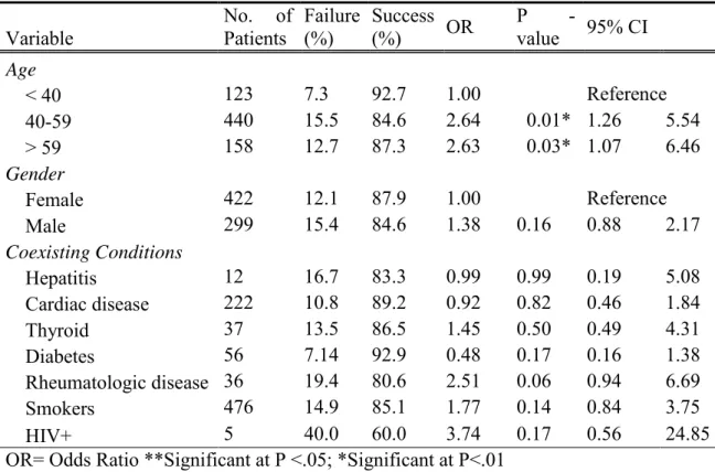
From the 721 patients included in the study, 97 presented at least an implant failure, rendering an implant failure rate of 13.5%. A total of 3998 implants were considered in this study (average of 5.5 implants per patient) with a global failure rate of 4.3% (n=173 implant failures). From the 3998 implants used in the study, 2262 were placed at the maxilla with a failure rate of 5.3% (n=119 implant failures) and the 1736 implants placed at the mandible had a loss rate of 3.1% (n=54 implant failures). Regarding the type of implant surface, there were 820 implants with machined surface and 3178 implants with anodically oxidized surface, with a failure rate of 10.9% (n=89 implant failures) and 2.6% (n=84 implant failures), respectively.
Desenvolvimento
23 Variable
No. of Patients
Failure (%)
Success (%) OR
P -
value 95% CI Age
< 40 123 7.3 92.7 1.00 Reference 40-59 440 15.5 84.6 2.64 0.01* 1.26 5.54 > 59 158 12.7 87.3 2.63 0.03* 1.07 6.46 Gender
Female 422 12.1 87.9 1.00 Reference Male 299 15.4 84.6 1.38 0.16 0.88 2.17 Coexisting Conditions
Hepatitis 12 16.7 83.3 0.99 0.99 0.19 5.08 Cardiac disease 222 10.8 89.2 0.92 0.82 0.46 1.84 Thyroid 37 13.5 86.5 1.45 0.50 0.49 4.31 Diabetes 56 7.14 92.9 0.48 0.17 0.16 1.38 Rheumatologic disease 36 19.4 80.6 2.51 0.06 0.94 6.69 Smokers 476 14.9 85.1 1.77 0.14 0.84 3.75 HIV+ 5 40.0 60.0 3.74 0.17 0.56 24.85 OR= Odds Ratio **Significant at P <.05; *Significant at P<.01
Table 1 Binary logistic regression model for the effect of the independent variables on implant failure.
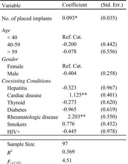
Results obtained from the multiple linear regression (Table 2) yielded rheumatologic and cardiovascular disorders, as well as the number of implants placed per patient as significant predictors (P<0.01) for the number of implants lost.
Peri-implant pathology
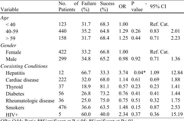
A total of 244 patients (33.8%) and 506 implants (12.7%) registered peri-implant pathology. The peri-implant pathology rate according to implant position was 10.1% (n=176) in the mandible and 14.6% (n=330) in the maxillae. According to the type of implant surface, peri-implant pathology was registered in 13.2% (n=108) and 12.5% (n=398) of the machined surface and anodically oxidized surface implants, respectively.
24
Based on the logistic regression model, hepatitis was the only risk factor for peri-implant pathology (OR= 3.74, p<0.05) when controlled for the presence of the other variables of interest (Table 3).
Variable Coefficient (Std. Err.)
No. of placed implants 0.093* (0.035)
Age
< 40 Ref. Cat.
40-59 -0.200 (0.442) > 59 -0.078 (0.556) Gender
Female Ref. Cat.
Male -0.404 (0.258) Coexisting Conditions
Hepatitis -0.323 (0.967) Cardiac disease 1.125** (0.401) Thyroid -0.273 (0.620) Diabetes -0.965 (0.619) Rheumatologic disease 2.203** (0.550) Smokers 0.776 (0.452) HIV+ -0.445 (0.978)
Sample Size 97 R2 0.369
F(11,85) 4.51
Desenvolvimento
25 Variable
No. of Patients
Failure (%)
Sucess (%) OR
P -
value 95% CI Age
< 40 123 31.7 68.3 1.00 Ref. Cat. 40-59 440 35.2 64.8 1.29 0.26 0.83 2.01 > 59 158 31.7 68.4 1.25 0.44 0.71 2.23 Gender
Female 422 33.2 66.8 1.00 Ref. Cat. Male 299 34.8 65.2 0.98 0.92 0.71 1.36 Coexisting Conditions
Hepatitis 12 66.7 33.3 3.74 0.04* 1.09 12.84 Cardiac disease 222 32.0 68.0 1.14 0.61 0.69 1.88 Thyroid 37 18.9 81.1 0.57 0.23 0.23 1.41 Diabetes 56 26.8 73.2 0.76 0.41 0.41 1.44 Rheumatologic disease 36 25.0 75.0 0.75 0.51 0.32 1.75 Smokers 476 36.6 63.5 1.48 0.15 0.87 2.53 HIV+ 5 60.0 40.0 2.34 0.37 0.36 15.19 OR= Odds Ratio **Significant at P <.05; *Significant at P<.01
26
v. Discussion
In this study, the authors proposed three models with risk factors for implant failure, higher number of failed implants and peri-implant pathology, in patients systemically compromised. The results suggest the impact of some systemic health factors and demographic factors on implant failure and peri-implant pathology.
Regarding the factors associated with implant failure, the demographic variable
“age” revealed a significant association, judging by the higher odds for implant failure in patients over 40 years with patients between 40-59 years and patients with more than 60 years, exhibiting more than 2 times the odds foe implant failure when compared to patients with less than 40 years of age. This result registered in our study using a large sample of systemically compromised patients was outlined in previous research using smaller samples:9,10 Moy et al.10 in a retrospective cohort study analyzed dental implant failure rates and associated risk factors, and observed that increasing age was strongly associated with the risk of dental implant failure, with patients between 60-79 years registering a higher relative risk for implant failure (RR=2.24). Another retrospective cohort study by Manor et al.9 to evaluate the characteristics of early and late implant failure conducted in 194 patients who presented implant failures during a 6-year period, reported old age as one of the factors for late dental implant failure.
Desenvolvimento
27
patients with rheumatoid arthritis and connective tissue diseases had increased bone resorption and more vulnerable soft-tissue conditions than patients with isolated rheumatoid arthritis (p<0.01). Weinlander et al.26 in a retrospective study identified patients with connective tissue diseases present an increased bone resorption (mean: 3.1 mm +/- 0.7 mm) and more peri-implant soft tissue alterations (bleeding index) than patients suffering from rheumatoid arthritis. Moreover, Zahid et al.39 in a retrospective study concluded that patients taking bisphosphonates could be at higher risk for implant thread exposure (OR= 3.25). Furthermore, Alsaadi et al.7 in a retrospective study identified osteoporosis as a significant factor for implant failure on a multivariate logistic regression (OR=2.88). The analysis of the literature and the results of our study suggest at least caution when managing dental implant rehabilitation in these patients.
On the other hand, cardiovascular diseases are a group of disorders that include hypertension, atherosclerosis, vascular stenosis, coronary artery disease, and congestive heart failure. The mechanism of osseointegration and healing process compromising situations can be described by a compromised blood flow which may restrict oxygen supply and nutrients to the tissues.20,21 Nevertheless, several studies registered no evidence that controlled cardiovascular diseases increased the risk of implant failure.7,20,21,34 These potential contradictory results may be explained by the degree of systemic disease control. In fact, this factor has been previously proposed to be of higher significance than the systemic condition itself.34,35 However, the degree of systemic disease control was not known in our study and therefore this question remains open.
Concerning peri-implant pathology, the only risk factor registered in this study was the presence of hepatitis. Despite the low number of studies in the literature, a previous report registered a significant alteration in salivary flow in a group of patients with hepatitis C, which had a significant impact on oral disease, namely the increase of decays, gingival bleeding and deep pockets.19 Marrone et al.38 in a cross-sectional study, evaluated the possible risk indicators for peri-implant pathology assessing implant and patient characteristics, registering a stronger association between hepatitis and peri-implant pathology (OR=2.9).
28
report the strong influence of smoking habits on the implant failure and peri-implant pathology,5,7,20,21,28-33,43 affecting the oral tissues’ healing process, periodontal procedures and implants therapies.21,28-30 Nevertheless, Alssadi et al.42 in a retrospective study reported that smoking did not seem to be relevant player in the etiology of late implant loss; while Renvert et al.44 in a systematic review, suggested smoking should be accounted for as a confounding variable in the etiology of peri-implant infections. Our study accounted with the presence of that possible confounding effect of smoking, by including it in the logistic regression analysis, a statistical procedure that could explain the non-significant result.
The limitations of this study include a single center, the retrospective analysis and the lack of a healthy patients’ control group, which can influence the external validity of the study and the extrapolation to other population. A further limitation is the
lack of analysis of potential risk factors described in the literature such as “history of periodontitis”,33, 43-44
the level of oral hygiene,33, 43-44 and the degree of systemic disease control which could have provided sub-group analysis within this population of systemic compromised patients.
Desenvolvimento
29
vi. Conclusions
30
vii. Acknowledgments
Desenvolvimento
31
viii. References
1. Astrand P, Ahlqvist J, Gunne J, Nilson H. Implant treatment of patients with edentulous jaws: a 20-year follow-up. Clin Implant Dent Relat Res 2008; 10: 207-17.
2. Malo P, de Araújo Nobre M, Lopes A, Ferro A, Gravito I. Single-Tooth Rehabilitations Supported by Dental Implants Used in an Immediate-Provisionalization Protocol: Report on Long-Term Outcome with Retrospective Follow-up. Clin Implant Dent Relat Res 2014; doi: 10.1111/cid.12278.
3. Andersson B, Bergenblock S, Fürst B, Jemt T. Long-term function of single-implant restorations: a 17- to 19-year follow-up study on single-implant infraposition related to the shape of the face and patients' satisfaction. Clin Implant Dent Relat Res 2013; 15: 471-80.
4. Esposito M, Hirsch J-M, Lekholm U, Thomsen P. Biological factors contributing to failures of osseointegrated oral implants (II).Etiopathogenesis. Eur J Oral Sci 1998; 106: 721–764.
5. Palma-Carrió C, Maestre-Ferrín L, Peñarrocha-Oltra D, Peñarrocha-Dia-go MA, Peñarrocha-Diago M. Risk factors associated with early failure of dental implants. A literature review. Med Oral Patol Oral Cir Bucal 2011; 16:e514-7.
6. Malo P, de Araújo Nobre M, Lopes A, Moss SM, Molina GJ. A longitudinal study of the survival of all-on-4 implants in the mandible with up to 10 years of follow-up. J Am Dent Assoc 2011; 142:310–320.
7. Alsaadi G, Quirynen M, Komárek A, van Steenberghe D. Impact of local and systemic factors on the incidence of oral implant failures, up to abutment connection. J Clin Periodontol 2007; 34: 610-617.
32
9. Manor Y, Oubaid S, Mardinger O, Chaushu G, Nissan J. Characteristics of early versus late implant failure: a retrospespective study. J Oral Maxillofac Surg 2009; 67: 2649–2652.
10. Moy PK, Medina D, Shetty V, Aghaloo TL. Dental implant failure rates and associated risk factors. Int J Oral Maxillofac Implants 2005; 20:569-77.
11. de Araújo Nobre M, Mano Azul A, Rocha E, Maló P, Risk factors of peri-implant pathology. Euro J Oral Sci 2015; 123:131-139.
12. Albrektsson T, I F. Consensus report of session IV. In: Lang NP, K T, eds. Proceedings of the First European Workshop on Periodontology. London, United Kingdom: Quintessence; 1994: 365-369.
13. Zitzmann NU, Berglundh T. Definition an prevalence of peri-implant diseases. J Clin Periodontol 2008; 35: 286-291.
14. Froum SJ, Rosen PS. A Proposed Classification for Peri-Implantitis. Int J Periodont Restor Dent 2012; 32:533-540.
15. Klinge B, Meyle J; Working Group 2. Peri-implant tissue destruction. The Third EAO Consensus Conference 2012. Clin Oral Implant Res 2012; 23: 108-110.
16. Cecchinato D, Parpaiola A, Lindhe J. Mucosal inflammation and incidence of crestal bone loss among implant patients: a 10-year study. Clin Oral Implants Res 2014; 25: 791-796.
17. Atieh MA, Alsabeeha NH, Faggion CM Jr, Duncan WJ. The Frequency of Peri-Implant Diseases: A Systematic Review and Meta-Analysis. J Periodontol 2013; 84: 1586-1598.
18. Daubert DM, Weinstein BF, Bordin S, Leroux BG, Flemmig TF. Prevalence and Predictive Factors for Peri-Implant Disease and Implant Failure: A Cross-Sectional Analysis. J Periodontol 2015; 86: 337-347.
Desenvolvimento
33
20. Elsubeihi ES, Zarb GA. Implant Prosthodontics in Medically Challenged Patients: The University of Toronto Experience. J Can Dent Assoc 2002; 68:103-8.
21. Hwang D, Wang H. Medical Contraindications to Implant Therapy: Part II: Relative Contraindications. Implant Dent 2007; 16:13–23.
22. Attard NJ, Zarb GA. A study of dental implants in medically treated hypothyroid patients. Clin Implant Dent Relat Res 2002; 4:220-31.
23. Ship JA. Diabetes and oral health: an overview. J Am Dent Assoc 2003; 134:4S-10S.
24. Valero AM, García JCF, Ballester AH, Rueda CL. Effects of diabetes on the osseointegration of dental implants. Med Oral Patol Oral Cir Bucal 2007; 12:E38-43.
25. Oates TW, Dowell S, Robinson M, McMahan CA. Glycemic Control and Implant Stabilization in Type 2 Diabetes Mellitus. J Dent Res 2009; 88:367-371.
26. Weinlander M, Krennmair G, Piehslinger E. Implant prosthodontic rehabilitation of patients with rheumatic disorders: a case series report. Int J Prosthodont 2010; 23:22-28.
27. Krennmair G, Seemann R, Piehslinger E. Dental implants in patients with rheumatoid arthritis: clinical outcome and peri-implant findings. J Clin Periodontol 2010; 37: 928–936.
28. Balaji SM. Tobacco smoking and surgical healing of oral tissues: A review. Indian J Dent Res 2008; 19:344-348.
29. Sham A, Cheung LK, Jin LJ, Corbet EF. The effects of tobacco use on oral health. Hong Kong Med J 2003; 9:271-277.
30. Baig MR, Rajan M. Effects of smoking on the outcome of implant treatment: A literature review. Indian J Dent Res 2007; 18:190-195.
34
32.Malo P, de Araújo Nobre M, Gonçalves Y, Lopes A. Long-Term Outcome of Implant Rehabilitations in Patients with Systemic Disorders and Smoking Habits: A Retrospective Clinical Study. 2015 Clin Implant Dent Relat Re doi: 10.1111/cid.12346.
33. de Araújo Nobre M, Maló P, Antunes E. Influence of Systemic Conditions on the Incidence of Periimplant Pathology. Implant Dent, 2014; 23: 305-310.
34. Diz P, Scully C, Sanz M. Dental implants in the medically compromised patient. J
dent.2013; 41: 195-206.
35. Scully C, Hobkirk J, Dios PD. Dental endosseous implants in the medically compromised patient. J Oral Rehabil. 2007; 34:590-599
36. von Elm E, Altman DG, Egger M, et al. The Strengthening the Reporting of Observational Studies in Epidemiology (STROBE) statement: guidelines for reporting observational studies. Epidemiology 2007; 18:800–804.
37. de Araújo Nobre M, Cintra N, Maló P. Peri-implant maintenance of immediate function implants: a pilot study comparing hyaluronic acid and chlorhexidine. Int J Dent Hyg 2007; 5:87–94.
38. Marrone A, Lasserre J, Bercy P, Brecx MC. Prevalence and risk factors for peri-implant disease in Belgian adults. Clinical Oral Implants Research. 2013; 24:934-940.
39. Zahid TM, Wang BY, Cohen RE. Influence of bisphosphonates on alveolar bone loss around osseointegrated implants. J Oral Implantol 2011; 37:335-46.
40. World Health Organization. 2012. Chronic rheumatic conditions
http://www.who.int/chp/topics/rheumatic/en (Accessed March 26, 2015)
41. Tsolaki IN, Madianos PN, Vrotsos JA. Outcomes of Dental Implants in Osteoporotic Patients. A Literature Review. J Prosthodont2009; 18: 309–323.
Desenvolvimento
35
43. Mombelli A, Müller N, Ciona N. The epidemiology of peri-implantitis. Clin Oral Implants Res 2012; 23: 67-76.
36
III.
Conclusões
Tendo em conta os resultados estatísticos, o estudo sugere o impacto das seguintes condições sistémicas e fatores demográficos na perda de implantes:
1. Aumento da idade, em que pacientes dos 40-59 anos e acima dos 60 anos de idade, apresentam o dobro da probabilidade para a perda de implantes quando comparados com pacientes a baixo dos 40 anos de idade.
2. A presença de doenças reumatológicas e doenças cardíacas estão associadas a um aumento do número de implantes perdidos ao estudar o grupo de pacientes que perderam implantes. No entanto verifica-se o dobro dos implantes perdidos nos pacientes com doenças reumatológicas.
Relativamente à ocorrência de patologia peri-implantar, o estudo indica:
3. Uma maior incidência da doença em pacientes com hepatite (A, B ou C).
Os resultados obtidos sugerem que se tenham algumas precauções ao reabilitar com implantes o grupo de pacientes acima descritos. Por sua vez, o estudo não identifica qualquer tipo de contraindicações absolutas para a reabilitação com implantes, uma vez que a taxa de perda de implantes absoluta sugere um risco/benefício favorável para os pacientes nestas condições.
As limitações inerentes a este estudo compreendem: (1) o facto de incluir apenas um único centro dentário; (2) o facto de ser um estudo retrospetivo sem um grupo controlo de pacientes saudáveis, que por sua vez poderá influenciar a validade externa do estudo bem como a sua extrapolação para a outra população; (3) a ausência do estudo de potenciais fatores de risco descritos na literatura tais como a “história deperiodontite” e
Conclusões
37
Em investigações futuras, ao estudar os resultados a longo-termo de pacientes sistemicamente comprometidos que foram reabilitados com implantes é importante que se tenha em conta o grau de controlo de cada doença. Assim como a identificação do subtipo de doença de cada grupo. É importante que se utilizem estudos prospetivos, grupos controlo de pacientes saudáveis e que se controle a presença de outros potenciais
fatores de risco, tais como a “história de periodontite”, “nível de higiene oral” ou a “existência de problemas mecânicos”, uma vez que tanto a perda de implantes como a
38
IV.
Bibliografia
Astrand P, Ahlqvist J, Gunne J, Nilson H (2008). Implant treatment of patients with edentulous jaws: a 20-year follow-up. Clinical Implant Dentistry and Related Research, 10(4), 207-17.
Albrektsson T, I F (1994). Consensus report of session IV. In: Lang NP, K T, eds. Proceedings of the First European Workshop on Periodontology. London, United Kingdom: Quintessence; 365-369.
Alsaadi G, Quirynen M, Komárek A, van Steenberghe D (2007). Impact of local and systemic factors on the incidence of oral implant failures, up to abutment connection. Journal of Clinical Periodontology, 34(7), 610-617.
Alsaadi G. Quirynen M, Komárec A, van Steenbergh D (2008). Impact of local and systemic factors on the incidence of late oral implant loss. Clinical Oral Implants Research, 19(7), 670–676.
Andersson B, Bergenblock S, Fürst B, Jemt T (2013). Long-term function of single-implant restorations: a 17- to 19-year follow-up study on single-implant infraposition related to the shape of the face and patients' satisfaction. . Clinical Implant Dentistry and Related Research,15(4), 471-80.
Annibali S, Vestri AN, Pilotto A, La Monaca G, Di Carlo S, Cristalli MP (2010). Patient satisfaction with oral implant rehabilitation: evaluation of responses to a questionnaire. Annali di Stomatologia, 1(3-4) ,2-8.
Atieh MA, Alsabeeha NH, Faggion CM Jr, Duncan WJ (2013). The Frequency of Peri-Implant Diseases: A Systematic Review and Meta-Analysis. Journal of Periodontology, 84(11), 1586-1598.
Attard NJ, Zarb GA (2002). A study of dental implants in medically treated hypothyroid patients. Clinical Implant Dentistry and Related Research, 4(4),220-31.
Bibliografia
39
Balaji SM (2008). Tobacco smoking and surgical healing of oral tissues: A review. Indian Journal of Dental Research, 19(4), 344-348.
Cecchinato D, Parpaiola A, Lindhe J (2014). Mucosal inflammation and incidence of crestal bone loss among implant patients: a 10-year study. Clinical Oral Implants Research, 25(7), 791-796.
Coates EA, Brennan D, Logan RM, Goss AN, Scopacasa B, Spencer AJ, Gorkic E (2000). Hepatitis C infection and associated oral health Problems. Australian Dental Journal, 45(2),108-114.
Daubert DM, Weinstein BF, Bordin S, Leroux BG, Flemmig TF (2015). Prevalence and Predictive Factors for Peri-Implant Disease and Implant Failure: A Cross-Sectional Analysis. Journal of Periodontology, 86(3), 337-347.
De Araújo Nobre M, Cintra N, Maló P (2007). Peri-implant maintenance of immediate function implants: a pilot study comparing hyaluronic acid and chlorhexidine. International Journal of Dental Hygiene, 5(2), 87–94.
De Araújo Nobre M, Maló P, Antunes E (2014b). Influence of Systemic Conditions on the Incidence of Periimplant Pathology. Implant Dentistry, 23(3), 305-310
De Araújo Nobre M, Mano Azul A, Rocha E, Maló P (2015), Risk factors of peri-implant pathology. European Journal of Oral Sciences, 123(3), 131-139.
De Araújo Nobre MA, Malo P (2014c). The influence of Rehabilitation Characteristics in the Incidence of Peri-Implant Pathology: A Case-Control Study. Journal of. Prosthodontics, 23(1): 21-30
De Araújo Nobre MA, Malo PS, Oliveira SH (2014a). The influence of implant location and position characteristics on peri-implant pathology. European. Journal of Prosthodontics and Restorative Dentistry, 22(3): 125-129
Diz P, Scully C, Sanz M (2013). Dental implants in the medically compromised patient. Journal of Dentistry; 41(3): 195-206.
40
Esposito M, Hirsch J-M, Lekholm U, Thomsen P (1998). Biological factors contributing to failures of osseointegrated oral implants (II).Etiopathogenesis. European Journal of Oral Sciences, 106(3), 721–764.
Froum SJ, Rosen PS (2012). A Proposed Classification for Peri-Implantitis. International Journal of Periodontics & Restorative Dentistry, 32(5), 533-540.
Hwang D, Wang H (2007). Medical Contraindications to Implant Therapy: Part II: Relative Contraindications. Implant Dentistry, 16(1),13–23.
Klinge B, Meyle J (2012); Working Group 2. Peri-implant tissue destruction. The Third EAO Consensus Conference 2012. Clinical Oral Implants Research, 23(6), 108-110.
Kourtis SG, Sotiriadou S, Voliotis S, Challas A (2004). Private practice results of dental implants. Part I: survival and evaluation of risk factores- Part II: surgical and prosthetic complication. Implant Dentistry, 13(4), 373-385.
Krennmair G, Seemann R, Piehslinger E (2010). Dental implants in patients with rheumatoid arthritis: clinical outcome and peri-implant findings. Journal of Clinical Periodontology, 37, 928–936.
Malo P, de Araújo Nobre M, Gonçalves Y, Lopes A (2015). Long-Term Outcome of Implant Rehabilitations in Patients with Systemic Disorders and Smoking Habits: A Retrospective Clinical Study. Clinical Implant Dentistry and Related Research, doi: 10.1111/cid.12346.
Malo P, de Araújo Nobre M, Lopes A, Ferro A, Gravito I (2014). Single-Tooth Rehabilitations Supported by Dental Implants Used in an Immediate-Provisionalization Protocol: Report on Long-Term Outcome with Retrospective Follow-up. Clinical Implant Dentistry and Related Research, doi: 10.1111/cid.12278.
Malo P, de Araújo Nobre M, Lopes A, Moss SM, Molina GJ (2011). A longitudinal study of the survival of all-on-4 implants in the mandible with up to 10 years of follow-up. Journal of the American Dental Association, 142(3):310–320.
Bibliografia
41
Marrone A, Lasserre J, Bercy P, Brecx MC (2013). Prevalence and risk factors for peri-implant disease in Belgian adults. Clinical Oral Implants Research., 24(8), 934-940.
Martín-Ares M, Barona‐Dorado C, Guisado‐Moya B, Martínez‐Rodríguez N, Cortés‐Bretón‐Brinkmann J, Martínez‐González JM (2015). Prosthetic hygiene and functional efficacy in completely edentulous patients: satisfaction and quality of life during a 5-year follow-up. Clinical Oral Implants Research, doi: 10.1111/clr.12604
Mombelli A, Müller N, Ciona N (2012). The epidemiology of peri-implantitis. . Clinical Oral Implants Research , 23(6), 67-76.
Moy PK, Medina D, Shetty V, Aghaloo TL (2005). Dental implant failure rates and associated risk factors. International Journal of Oral and Maxillofacial implants, 20(4), 569-77.
Oates TW, Dowell S, Robinson M, McMahan CA (2009). Glycemic Control and Implant Stabilization in Type 2 Diabetes Mellitus. Journal of Dental Research, 88(4),367-371.
Palma-Carrió C, Maestre-Ferrín L, Peñarrocha-Oltra D, Peñarrocha-Dia-go MA, Peñarrocha-Diago M (2011). Risk factors associated with early failure of dental implants. A literature review. Med Oral Patol Oral Cir Bucal, 16:e514-7.
Renvert S, Persson GR (2009). Periodontitis as a potential risk factor for peri-implantitis. Journal of Clinical Periodontology, 36(10), 9–14.
Renvert S, Polyzois I, Claffey N (2011). How do implant surface characteristics influence peri-implant disease? Journal of Clinical Periodontology, 38(11), 214-222.
Scully C, Hobkirk J, Dios PD (2007). Dental endosseous implants in the medically compromised patient. Journal of Oral Rehabilitation, 34, 590-599.
Sgolastra F, Petrucci A , Severino M, Gatto R, Monaco A (2015). Smoking and the risk of peri-implantitis. A systematic review and meta-analysis. Clinical Oral Implants Research, 26(4), 62-67.
42
Ship JA (2003). Diabetes and oral health: an overview. Journal of American Dental Association, 134(1):4S-10S.
Tsolaki IN, Madianos PN, Vrotsos JA (2009). Outcomes of Dental Implants in Osteoporotic Patients. A Literature Review. Journal of Prosthodontics, 18(4), 309–323.
Valero AM, García JCF, Ballester AH, Rueda CL (2007). Effects of diabetes on the osseointegration of dental implants. Med Oral Patol Oral Cir Bucal, 12(1), E38-43.
Von Elm E, Altman DG, Egger M, et al (2007). The Strengthening the Reporting of Observational Studies in Epidemiology (STROBE) statement: guidelines for reporting observational studies. Preventive Medicine, 45(4), 247-251.
Weinlander M, Krennmair G, Piehslinger E (2010). Implant prosthodontic rehabilitation of patients with rheumatic disorders: a case series report. International Journal of Prosthodontics, 23(1), 22-28.
World Health Organization. 2012. Chronic rheumatic conditions http://www.who.int/chp/topics/rheumatic/en (Accessed March 26, 2015)
Zahid TM, Wang BY, Cohen RE (2011). Influence of bisphosphonates on alveolar bone loss around osseointegrated implants. Journal of Oral Implantology, 37(3):335-46.
For Review JOPR
Risk Factors for Implant Failure and Peri-implant pathology in a systemic compromised population
Journal: Journal of Prosthodontics
Manuscript ID Draft
Wiley - Manuscript type: Original Manuscript
Index Words: dental implant, implant failure, peri-implant pathology, risk factor, systemic disease
Manuscript Categories: Clinical Research