Original Article
Artigo Original
Association between functionality
assessment scales and the severity of
dysphagia post-stroke
Associação entre escalas de avaliação de
funcionalidade e severidade da disfagia
pós-acidente vascular cerebral
Karen de Oliveira dos Passos1,2Maria Cristina de Almeida Freitas Cardoso2 Betina Scheeren1
Keywords
Deglutition Deglutition Disorders Stroke Fluoroscopy Scales
Descritores
Deglutição Transtornos de Deglutição Acidente Vascular Cerebral Fluoroscopia Escalas
Correspondence address:
Maria Cristina de Almeida Freitas Cardoso
Av. Sarmento Leite, 245, Porto Alegre (RS), Brazil, CEP: 90050-170. E-mail: mccardoso@ufcspa.edu.br
Received: February 02, 2016
Accepted: June 07, 2016
Study carried out at Santa Casa de Misericórdia de Porto Alegre and the Universidade Federal de Ciências da Saúde de Porto Alegre, both of Porto Alegre (RS), Brazil.
1 Santa Casa de Misericórdia de Porto Alegre - Porto Alegre (RS), Brazil.
2 Universidade Federal de Ciências da Saúde de Porto Alegre – UFCSPA - Porto Alegre (RS), Brazil.
Financial support: nothing to declare.
Conlict of interests: nothing to declare.
ABSTRACT
Purpose:The aim of this study was to investigate the association between the results of functionality scales
and dysphagia severity determined from videoluoroscopy swallowing study (VSS) in post-stroke patients.
Methods: A retrospective study of 109 VSS analysis of post-stroke patients. From the exams was collected information on the diagnosis of dysphagia severity and evaluated regarding the swallowing functionality in an independent way by two evaluators. Results: A high number of discrete dysphagia was observed. In one third
of dysphagic patients it was noticed the presence of tracheal aspiration. There was a signiicant association
between the scores of Functional Oral Intake Scale and Dysphagia Outcome and Severity Scale in post-stroke patients which means when the swallowing is highly compromised, the level of intake by mouth will be smaller according to the evaluators’ analysis (p < 0.001). Conclusion: There is an association between the scales and it can be used as evaluative benchmarks and in the management of clinical Speech Pathology intervention.
RESUMO
Objetivo: O objetivo deste estudo foi veriicar a associação entre os resultados das escalas de funcionalidade e
severidade da disfagia determinadas a partir de exames de videoluoroscopia em pacientes pós-AVC. Método: Estudo
observacional, retrospectivo e descritivo, com análise de 109 exames de Videoluoroscopia da Deglutição de
pacientes pós-AVC. Dos exames da amostra foram coletadas informações quanto ao diagnóstico da severidade da disfagia e avaliados quanto à funcionalidade da deglutição de maneira independente por dois avaliadores.
Resultados: Foi constatada uma alta prevalência de disfagia classiicada como discreta. Nos pacientes com
disfagia foi observada a presença de aspiração laringotraqueal em um terço da amostra. Veriicou-se associação signiicativa entre os escores das escalas Functional Oral Intake Scale e Dysphagia Outcome and Severity Scale
INTRODUCTION
The cerebrovascular accident (CVA) is deined as a sudden focal alteration in the cerebral blood low, with corresponding
neurological manifestation. According to the type, size and location of the vessel affected, it can be asymptomatic or
symptomatic. Regarding the etiology, it is classiied as ischemic
(ICVA) or bleeding (BCVA)(1).
CVA is the main cause of death in Brazil, being ahead of acute myocardial infarct and all added neoplasias. It is estimated that CVA mortality in 2010 was approximately 45 death per 100 thousand inhabitants/year, which corresponds to about 10% of all death causes in the country(2).
The complications resulting from CVA are also root causes
in the death certiicates, among others, such as malnutrition,
pneumonia and pulmonary thromboembolism, making its indirect mortality to be underestimated(1).
Oropharyngeal dysphagia is a symptom clinically identiied
in about 50% of patients at the post CVA(3) acute phase.
The high frequency of this symptom results from the fact that the deglutition central control occurs through a complex organization of neural elements in the brain and brainstem,
being the central nervous system (CNS) required to start and
coordinate the muscles involved in the oral, pharyngeal and esophageal deglutition(4) phases.
It is important to highlight that dysphagia is one of the main post VCA mortality causes because it is associated to complications responsible for a long hospital stay(1,3).
Dysphagia can be diagnosed through clinical evaluation of the deglutition, that is, evaluation performed at bedside, for which the speech-language-therapist has a range of protocols available. There also are deglutition evaluation tools, complementary to the clinical evaluations, performed by objective analysis of deglutition biodynamic, visualized by either radiologic or video endoscopic means; however a few formal protocols are found regarding this type of evaluation.
Videoluoroscopy swallowing study (VSS) permits the
observation of the anatomic structures and the dynamic analysis, in real time, of the several events of the deglutition phases, through the offer of different consistency foods, in different quantities and in different body positions (lateral side and/or anteroposterior)(5,6).
VSS contributes substantially to the understanding of the alterations, the level of severity and the dysphagia patient rehabilitation. Considered as gold standard for the diagnosis of deglutition disorder, it has great impact at decision making, not only in therapeutic terms but also at prognosis determination(5,6).
Post VCA neurogenic dysphagia classiication varies
according to the level of involvement of the neurological event and depends on factors such as its location and the injury extension, the patient alert status, as well as the consistency, volume and viscosity of the food offered(7).
The rating scales of the involvement level of the oropharyngeal dysphagia are tools used in the diagnostic process of this symptom at the deglutition clinical and objective evaluations, to help
identifying the risk at dysfunction classiication, as an instrument to deine the procedures or as a parameter at the rehabilitation
eficiency control. These scales have been suggested in several
studies and not always use consensual criteria(8).
Dysphagia Outcome and Severity Scale – DOSS(7) is a scale
developed to systemically evaluate the dysphagia functional severity based on the objective evaluation and to make recommendations on the type of diet, level of independency and type of nutrition.
This scale can be used by physicians trained to determine the dysphagia level of severity based on objective measures through DVF. Due to its high reliability, this tool can be valuable at the objective measurement of the natural history for the dysphagia prognosis among the population and to measure the treatment
eficacy(7).
Regarding the functionality level at feeding, it is used at the clinical practice, the Functional Oral Intake Scale – FOIS(9),
which describes seven distinct levels of oral intake, as well as it suggests the feeding via for patients with VCA and dysphagia or, yet, the need of deglutition compensating measures.
Considering the prevalence and incidence of post VCA deglutition disorders, or, yet, the associated comorbidities, this study has as objective to verify the association among the scale results of dysphagia functionality and severity, determined from
video luoroscopy exams, in post VCA patients.
METHODS
It is about an observational, retrospective and descriptive study of DVF image exam analysis. The research was performed in a general hospital of Santa Casa de Misericórdia of Porto Alegre. The sample by convenience of post VCA patients was composed by reports and dynamic images of deglutition exams performed in the radiology sector of this hospital, between January 2013 and June 2015. This study was approved under n. 927.637 by the Ethics Committee of Santa Casa de Misericórdia of Porto Alegre – SCMPA.
In the study were included VSS of adult individuals with age higher than 18 years, of both genders, having in their medical history VCA diagnosis and presence of speech-language therapy diagnosis independently of the dysphagia severity, according to DOSS scale(7). The study exclusion factors were the reports
of the incomplete video luoroscopy exams.
The speech-language pathologist responsible for performing the VSS of the hospital radiology service received internal and external patients with medical forwarding from different specialties, medical attendance and hospitals to perform the exam. The VSS exam reports are physically stored in paper, at the service and the exam images in Vepro information electronic system. The exam is performed by testing the different food consistency, prepared by adding barium, according to the command of patient’s greatest safety. They are tested according to the individual’s collaboration and to the answer to the requested maneuvers, being the patient sitting at 90o at the lateral side
In those cases in which the patient performed more than one
deglutition exam, it was considered the result of the irst exam. After this irst selection, the exams were compiled and
recorded in Digital Versatile Disc (DVDs). From these were analyzed and evaluated the videos of each exam stored, digitalized in recording connected to the Fluoroscopy Unit equipment of Siemens brand, model Axion iconos R100, by two independent speech-language therapists.
From the exams was collected information regarding the gender and age of the patient, characteristics of the deglutition phases (oral and pharyngeal), presence or not of tracheal
aspiration, as well as the level of severity deined according to
DOSS(7) scale classiication, by the speech-language pathologist
at work, in other words: normal deglutition; deglutition within the functional limits; light dysphagia; mild to moderate; moderate; moderate to severe; or severe(7).
Through the independent analysis of the deglutition dynamics, the evaluators established the levels of deglutition functionality, by using the FOIS(9), scale classiication, which is also formed
by seven levels of classiication.
The indings were transferred to independent spreadsheets of
the database and forwarded to statistical analysis and checking
of the indings matching.
STATISTICAL ANALYSIS
To describe the sample proile according to the variables
in study, frequency tables were elaborated with absolute (n) and relative (%) values of the categorical variables (gender, age range, aspiration) and descriptive statistics of the numeric variables (age and scale scores), with average values, standard deviation, minimum and maximum values, median and quartiles.
At the association analysis between the two scales it was used the chi-square test or Fisher exact test. To analyze the matching between the evaluations of the two evaluators of FOIS(9) scale
it was calculated the Kappa coeficient of agreement and used
Bowker symmetry test for the comparison of the amounts between the two evaluators. Kappa values above 0.75 indicate high agreement and above 0.40 indicate moderate agreement.
To numerically analyze the relation between the scores of the
two scales it was calculated the Spearman correlation coeficient,
due to the lack of regular distribution of the variables.
The level of signiicance adopted for the statistical tests
was 5% (P<0.05).
RESULTS
At the established time, 160 post VCA patients were addressed
to perform the Deglutition video luoroscopy exam. From these,
109 were included in the study and 51 excluded due to their image exams not being available at the system anymore.
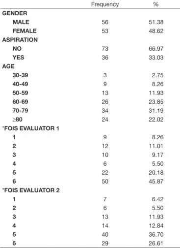
Table 1 presents the descriptive analysis of categorical variables of the sample. The average age was 69 years, being the most frequent age range between 70 and 79 years (31%); 51% of the sample was formed by the male gender.
The prevalence of dysphagia was of 94% and the results found regarding the level of severity, according to DOSS(7) scale
were: 5.5% severe; 3.8% moderately severe; 12.8% moderate;
18.3% mild to moderate; 36.7% mild; 22.7% classiied with
deglutition within functionality limits and 0% with regular deglutition.
It was observed the presence of laringotracheal aspiration in 33% of the patients with dysphagia.
The evaluators classiied the feeding functionality between the levels 1 and 6 and could not ind classiication of regular
deglutition or total via oral diet without restrictions (equivalent to level 7 in both scales).
The association analysis between the scales DOSS(7) and
FOIS(9) of the different evaluators is described on Tables 2 and 3.
It was veriied signiicant association between the scores of the
two scales (p < 0.001), in other words, the higher the score in DOSS(7) scale, higher the score at FOIS(9) scale; according to
both evaluators’ analysis, the higher the dysphagia severity, lower the level of food oral intake.
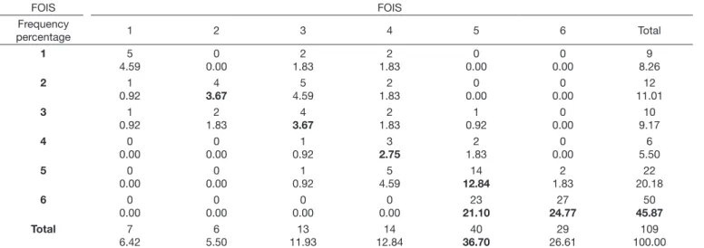
Through the symmetry test, it was veriied signiicant classiication difference for FOIS(9) scale, in which evaluator 1
had more frequency of level 6 and evaluator 2 more frequency of level 5 (S = 25,88; GL = 15; P = 0,039), being the moderate
Table 1. Descriptive analysis of the categorical variables Frequency % GENDER
MALE 56 51.38
FEMALE 53 48.62
ASPIRATION
NO 73 66.97
YES 36 33.03
AGE
30-39 3 2.75
40-49 9 8.26
50-59 13 11.93
60-69 26 23.85
70-79 34 31.19
≥80 24 22.02
*FOIS EVALUATOR 1
1 9 8.26
2 12 11.01
3 10 9.17
4 6 5.50
5 22 20.18
6 50 45.87
*FOIS EVALUATOR 2
1 7 6.42
2 6 5.50
3 13 11.93
4 14 12.84
5 40 36.70
6 29 26.61
matching between the evaluators’ classiication for FOIS(9)
scale (Table 4).
The correlation among the scores of FOIS(9) scale and
between the analyses of the two evaluators are shown on Graph 1.
There was signiicant difference among all the scores of the
scales of both evaluators (p < 0,001; r = 0,839).
DISCUSSION
In the 1990s, the incidence of oropharyngeal dysphagia was studied by several researchers, with the objective of determining the prevalence of this symptom in the neurological etiologies,
viewing at the need to deine suitable therapeutic plans appropriate
for the different dysphagic populations(10).
Among the neurologic diseases, VCA is the one with the
highest incidence of dysphagia, with a proile of incidence
between 30% and 96%, depending on the study method for this variation. The factors considered are injury time, location and range of the cerebral injury, type of VCA and the diagnostic
method of the deglutition disorder(11-13). In the present study, the
incidence of dysphagia found, considering the factor of VCA
presence, was of 94%, according to the classiication of the
DOSS(7) scale, corroborating the incidence studies.
The predominance regarding age above 70 years and occurrence of VCA in individuals of male gender found in this study is pertinent to the one found in literature(12-16).
The speech-language pathologist follow up on the dysphagia patient requires the discussion with the multi professional team regarding the indication of indirect therapies (without food introduction) or direct (with the use of food), related to the releasing moment of the oral intake in a systematic way, the indication of full oral intake diet, with the possibility of weaning of the alternative feeding intake. For this, clinical and instrumental parameters are used in which scales to measure such progression or regression are of extreme importance at the professional clinic practice.
For VSS quantitative interpretation, DOSS(7) scale assumes
the assessment regarding the dysphagia presence and severity,
Table 2. Analysis of the association among scale scores of evaluator 1
*FOIS **DOSS
Frequency 1 2 3 4 5 6 Total
1 5 83.33 0 0.00 2 14.29 2 10.00 0 0.00 0 0.00 9 2 1 16.67 3 75.00 4 28.57 2 10.00 2 5.00 0 0.00 12 3 0 0.00 1 25.00 5 35.71 2 10.00 2 5.00 0 0.00 10 4 0 0.00 0 0.00 2 14.29 2 10.00 2 5.00 0 0.00 6 5 0 0.00 0 0.00 1 7.14 11 55.00 9 22.50 1 4.00 22 6 0 0.00 0 0.00 0 0.00 1 5.00 25 62.50 24 96.00 50
Total 6 4 14 20 40 25 109
*FOIS: Functional Oral Intake Scale; **DOSS: Dysphagia Outcome and Severity Scale Caption: FISHER EXACT TEST: P < 0.001
Table 3. Analysis of the association among scale scores evaluator 2
*FOIS **DOSS
Frequency 1 2 3 4 5 6 Total
1 6 100.00 0 0.00 1 7.14 0 0.00 0 0.00 0 0.00 7 2 0 0.00 3 75.00 3 21.43 0 0.00 0 0.000 0 0.00 6 3 0 0.00 1 25.00 8 57.14 4 20.00 0 0.00 0 0.00 13 4 0 0.00 0 0.00 2 14.29 4 45.00 3 7.50 0 0.00 14 5 0 0.00 0 0.00 0 0.00 7 35.00 33 82.50 0 0.00 40 6 0 0.00 0 0.00 0 0.00 0 0.00 4 10.00 25 100.00 29
Total 6 4 14 20 40 25 109
combined with the observation of the subjective phenomena presented during the exam and with the diet in taken by the individual/patient.
The study that originated DOSS(7) scale evaluated 135 patients
with different clinical deiciencies, from these, 81% neurological, being also included in its sample post VCA patients. It classiied
them regarding the level of dysphagia severity in: 7% severe; 12.6% moderately severe; 15.6% moderate; 22% mild to moderate; 21% mild; 16% with deglutition within the functionality limits; and 5% with regular deglutition(7).
Data related to severe, moderate and mild to moderate dysphagia levels of the present study seem to be close to the study that originated DOSS scale.
The lack or presence of aspiration has been one of the video
luoroscopic signals of higher relevance, being this signal essential
for decision making at the management of oral food intake and for the indication of the most adequate consistency in each case(9-17). The aspiration incidence in VCA neurogenic dysphagia
framework is described in studies between 155 and 50%(18,19).
In the present study it was veriied the aspiration presence in 33% of the cases, conirming data found in other studies for
this population.
FOIS(9) scale complements the evaluation of the deglutition
disorder, suggesting the procedure regarding the feeding way
for the adequate and eficient handling of the post VCA affected
patients, having dysphagia as a consequence. In this study, the higher frequency in this scale was of the levels 5 and 6, with moderate agreement between the evaluators. The study that originated the scale FOIS(9) evaluated 302 post VCA patients
in acute phase and after the period between 1 and 6 months of
the event and classiied the level of functionality in different
moments. At the admission and after a month from VCA level 4 was the most frequent and, after 6 months, level 7(9).
In a study performed in 2012 with patients whose VCA diagnostic occurred between 24h and 48h before, evaluated by means of clinical and objective deglutition assessment through deglutition video endoscopy – DVE, there was predominance of level 7 of FOIS(9,20) scale.
Such divergence found in these studies(9,20) that evaluated
deglutition at the acute phase post VCA can be explained by the presence of different associated comorbidities and also by the use of different evaluation instruments, considering the limitations of DVE exams in relation to DVF, once DVE makes it possible the superior visualization from the oropharynges (what makes it impossible the visualization via oral), while in the DVF it is possible to observe the food from the chewing until the esophageal phase(21).
In the present study levels 5 and 6 were of higher frequency, differently from the ones found in those previous studies, what can be explained by the fact that the post VCA patients addressed to perform the DVF had presented former complaints
of deglutition dificulty, without considering the time of the
cerebrovascular event.
Food functionality veriied in FOIS(9) scale is also an
important parameter to measure the level of oral intake in pre and post speech language therapy at the oropharyngeal
Table 4. Analysis of matching between evaluators for scale FOIS
FOIS FOIS
Frequency
percentage 1 2 3 4 5 6 Total
1 5
4.59
0 0.00
2 1.83
2 1.83
0 0.00
0 0.00
9 8.26
2 1
0.92
4 3.67
5 4.59
2 1.83
0 0.00
0 0.00
12 11.01
3 1
0.92
2 1.83
4 3.67
2 1.83
1 0.92
0 0.00
10 9.17
4 0
0.00
0 0.00
1 0.92
3 2.75
2 1.83
0 0.00
6 5.50
5 0
0.00
0 0.00
1 0.92
5 4.59
14 12.84
2 1.83
22 20.18
6 0
0.00
0 0.00
0 0.00
0 0.00
23 21.10
27 24.77
50 45.87
Total 7
6.42
6 5.50
13 11.93
14 12.84
40 36.70
29 26.61
109 100.00
Caption: BOWKER SYMMETRY TEST: S = 25.88; GL = 15; P = 0.039; KAPA: 0.665; IC 95%: (0.585; 0.745); FOIS: Functional Oral Intake Scale
Caption: FOIS scale - evaluator 1 vs evaluator 2; r = Spearman’s correlation coefficient; P = p-value; FOIS: Functional Oral Intake Scale
dysphagia, indicating the changes occurred at the feeding during the therapeutic process(3,22,23).
This study indicates a relevant inding for the classiication
of the dysphagia level associated to DOSS(7) and FOIS(9) scales,
either by considering the aspiration presence or the safer consistency of food to be indicated. At the analysis performed
it was veriied a difference statistically signiicant between the
evaluators, suggesting that these are reproducible at the clinical practice, and therefore, can be used and associated at large scale.
Besides the classiication scales of dysphagia severity and
ingestion functionality by oral intake being used in a complementary way to the evaluation tools, they can also be aggregated as benchmarks of dysphagia progression or regression(7,9), being
possible to relate their levels, in other words, the worse the individual’s dysphagia level in DOSS(7) scale, the higher food
ingestion restriction within the consistencies by oral intake at FOIS(9) scale he will have.
The matching between these two scales is described in a current study(24) however, the value attribution of this association
was not found in the literature or other published studies. Some limitations of the study must be indicated, in other words, regarding the use of FOIS(9) functionality scale had
been done from the analysis of DVF exam, it is clear that, at
the clinical evaluation, there is important data to deine the
level of functionality, such as: cognition level, collaboration on performing maneuvers and complementary clinical information.
Regarding the moderate agreement between the evaluators there is the divergence regarding the especial preparation or not of the food, preserving the multiplicity of food consistency.
It was also observed the restriction of the database analysis due to the lack of diagnosed information that could contribute to the understanding of cerebrovascular diseases and neurogenic dysphagia.
CONCLUSION
It was veriied that there was signiicant association between
DOSS and FOIS scales in post VCA patients and these can be used together as evaluative benchmarks and in face of speech-language therapy clinical intervention, considering that this indication must be conciliated to the clinical evaluation results.
REFERENCES
1. Cruz MR, Martins S, Brondani R. Doenças cerebrovasculares. . In: Duncan BB, Schmidt MI, Giugliani ERJ, Ducan MS, Giugliani C. Medicina ambulatorial: condutas de atenção primária baseada em evidências. 4. ed. Porto Alegre: ArtMed; 2013. p. 703-18.
2. Brasil. Ministério da Saúde. DATASUS: Departamento de Informática do SUS [Internet]. Informações de saúde. Brasília: Ministério da Saúde; 2010 [citado em 2015 Outubro 8]. Disponível em: http://www2.datasus.gov.br/ DATASUS/index.php?area=02
3. Rofes L, Vilardell N, Clavé P. Post-stroke dysphagia: progress at last.
Neurogastroenterol Motil. 2013;25(4):278-82. PMid:23480388. http:// dx.doi.org/10.1111/nmo.12112.
4. Estrela F, Schneider FL, Aquini MG, Marrone ACH, Seffani MA, Jotz GP. Controle neurológico da deglutição. In: Jotz GP, Angelis EC, Barros APB.
Tratado da deglutição e disfagia: no adulto e na criança. Rio de Janeiro: Revinter; 2009. p. 20-34.
5. Almeida RCA, Haguette RCB, Andrade ISN. Deglutição com e sem comando verbal: achados videofluoroscópicos. Rev Soc Bras Fonoaudiol. 2011;16(3):291-7. http://dx.doi.org/10.1590/S1516-80342011000300009. 6. Shem KL, Castillo K, Wong SL, Chang J, Kao M, Kolakowsky-Hayner SA. Diagnostic accuracy of bedside swallow evaluation versus videofluoroscopy to assess dysphagia in individuals with tetraplegia. PM R. 2012;4(4):283-9. PMid:22541374. http://dx.doi.org/10.1016/j.pmrj.2012.01.002.
7. O’Neil KH, Purdy M, Falk J, Gallo L. The dysphagia outcome and severity scale. Dysphagia. 1999;14(3):139-45. PMid:10341109. http://dx.doi. org/10.1007/PL00009595.
8. Silva RG, Motonaga SM, Cola PC, Gatto AR, Ribeiro PW, Carvalho LR, et al. Estudo multicêntrico sobre escalas para grau de comprometimento em disfagia orofaríngea neurogênica. Rev Soc Bras Fonoaudiol. 2012;17(2):167-70. http://dx.doi.org/10.1590/S1516-80342012000200011.
9. Crary M, Mann GDC, Groher ME. Initial psychometric assessment of a functional oral intake scale for dysphagia in stroke patient. Arch Phys Med Rehabil. 2005;86(8):1516-20. PMid:16084801. http://dx.doi.org/10.1016/j. apmr.2004.11.049.
10. Furkim AM, Behlau MS, Weckx LLM. Avaliação clínica e videofluoroscopica da deglutição em crianças com paralisia cerebral tetraparética espástica.
Arq Neuropsiquiatr. 2003;61(3-A):611-6. PMid:14513167. http://dx.doi. org/10.1590/S0004-282X2003000400016.
11. Paixão CT, Silva LD, Camerini FG. Perfil da disfagia após um acidente vascular cerebral: uma revisão integrativa. Rev Rene Fortaleza. 2010;11(1):181-90. 12. Oliveira ARS, Costa AGS, Morais HCC, Cavalcante TF, Lopes MVO,
Araujo TL. Fatores clínicos preditores do risco para aspiração e aspiração respiratória em pacientes com Acidente Vascular Cerebral. Rev Lat Am Enfermagem. 2015;23(2):216-24. PMid:26039291. http://dx.doi. org/10.1590/0104-1169.0197.2545.
13. Almeida TM, Cola PC, Magnoni D, França JID, Silva RG. Prevalência de disfagia orofaríngea no acidente vascular cerebral após cirurgia cardíaca. Rev. CEFAC. 2015;17(5):1415-9. http://dx.doi.org/10.1590/1982-0216201517520914.
14. Rodrigues ESR, Castro KAB, Rezende AAB, Herrera SDSC, Pereira AM, Takada JAP. Fatores de risco cardiovascular em pacientes com acidente vascular cerebral. Rev Amazônia. 2013;1(2):21-28.
15. Tieges Z, Mead G, Allerhand M, Duncan F, Wijck FV, Fitzsimons C, et al. Sedentary behavior in the first year after stroke: a longitudinal cohort study with objective measures. Arch Phys Med Rehabil. 2015;96(1):15-23. PMid:25220942. http://dx.doi.org/10.1016/j.apmr.2014.08.015. 16. Zhang L, Yan T, You L, Li K. Barriers to activity and participation for
stroke survivors in rural China. Arch Phys Med Rehabil. 2015;96(7):1222-8. PMid:25701640. http://dx.doi.org/10.1016/j.apmr.2015.01.024. 17. Chaves RD, Mangilli LD, Sassi FC, Jayanthi SK, Zilberstein B, Andrade
CRF. Análise videofluoroscópica bidimensional perceptual da fase faríngea da deglutição em indivíduos acima de 50 anos. ABCD Arq Bras Cir Dig. 2013;26(4):274-9. PMid:24510034. http://dx.doi.org/10.1590/S0102-67202013000400005.
18. Chen S, Chie W, Lin Y, Chang Y, Wang T, Lien I. Can the aspiration detected by videofl uoroscopic swallowing studies predict long-term survival in stroke patients with dysphagia? Disabil Rehabil. 2004;26(23):1347-53. PMid:15742979. http://dx.doi.org/10.1080/09638280412331270407. 19. Smithard DG, O’Neill PA, England RE, Park CL, Wyatt R, Martin
DF, et al. The natural history of dysphagia following a stroke. Dysphagia. 1997;12(4):188-93. PMID: 9294937. http://dx.doi.org/10.1007/PL00009535.
20. Nunes MCA, Jurkiewicz AL, Santos RS, Furkim AM, Massi G, Pinto GS, et al. Correlation between brain injury and dysphagia in adult patients with stroke. Int Arch Otorhinolaryngol. 2012;16(3):313-21. PMid:25991951. 21. Stafocher TS, Mourão LF. Estudo do disparo da fase faríngea da deglutição em adultos assintomáticos submetidos à videofluoroscopia e videoendoscopia.
22. Silva RG, Jorge AG, Peres FM, Cola PC, Gatto AR, Spadotto AA. Protocolo
para Controle de Eficácia Terapêutica em Disfagia Orofaríngea Neurogênica
(Procedon). Rev CEFAC. 2010;12(1):75-81. http://dx.doi.org/10.1590/ S1516-18462010000100010.
23. Carnaby-Mann GD, Crary MA. McNeill dysphagia therapy program: a case-control study. Arch Phys Med Rehabil. 2010;91(5):743-9. PMid:20434612. http://dx.doi.org/10.1016/j.apmr.2010.01.013.
24. Dias CJSBS, Lopes IIS, Nogueira DMS. Functional Oral Intake Scale (FOIS): contributo para a validação cultural e linguística para o português
Europeu [dissertação]. Portugal: Escola Superior de Saúde do Alcoitão, Santa Casa da Misericórdia do Alcoitão; 2015. 96 p.
Author contributions