(1) Universidade Federal de Minas Gerais, Belo Horizonte, Brasil;
(2) Departamento de Fonoaudiologia da Universidade Federal de Minas Gerais, Belo Horizonte, Brasil;
(3) Faculdade de Medicina da Universidade Federal de Minas Gerais, Belo Horizonte, Brasil.
Conlict of interest: non-existent
Evolution of swallowing in post-acute stroke:
a descriptive study
Evolução da deglutição no pós-AVC agudo: estudo descritivo
Aline Mansueto Mourão(1)
Erica Oliveira Almeida(1)
Stela Maris Aguiar Lemos(2)
Laélia Cristina Caseiro Vicente(2)
Antonio Lúcio Teixeira(3)
Received on: August 30, 2015 Accepted on: November 24, 2015
Mailing address: Aline Mansueto Mourão
Laboratório Interdisciplinar de Investigação Médica, Sala 281, Faculdade de Medicina da UFMG
Avenida Professor Alfredo Balena, 190 - Santa Eigênia
Belo Horizonte – MG – Brasil CEP: 30130-100
E-mail: alinemmourao@gmail.com altexr@gmail.com
ABSTRACT
Purpose: to analyze the evolution of post-stroke swallowing impairment.
Methods: this is a descriptive exploratory study involving a non-probabilistic sample that evaluated 100 stroke patients admitted at the Hospital Público Regional de Betim. Patients were subjected to a speech pathologist structured evaluation two times: in the irst 48 hours after stroke and at hospital discharge. The Gugging Swallowing Screen, a standardized and validated bed-side tool for evaluating swallowing in stroke patients, was used.
Results: in the initial evaluation, the frequency of dysphagia was 52%, and 28% of patients were classiied as severe dysphagia with a high risk of aspiration. The mean time between the initial evaluation and hospi-tal discharge was 22.1 days. At the hospihospi-tal discharge, only 2.1% of patients still had severe dysphagia. It was observed change in the patient swallowing proile according to the severity of dysphagia.
Conclusion: the frequency of post-stroke dysphagia is high, but there are progressive changes in the swallowing proile of stroke patients during their hospital stay.
Keywords: Stroke; Deglutition Disorders; Clinical Evolution
RESUMO
Objetivo: analisar a evolução da deglutição de pacientes após acidente vascular cerebral.
Métodos: trata-se de estudo exploratório descritivo com amostra não probabilística em que foram acom-panhados 100 pacientes admitidos com o diagnóstico de acidente vascular cerebral no Hospital Público Regional de Betim. Os pacientes foram submetidos à avaliação fonoaudiológica estruturada em dois momentos: nas primeiras 48 horas após acidente vascular cerebral e no momento da alta hospitalar. Utilizou-se a escala Gugging Swallowing Screen que é um instrumento padronizado e validado para ser
utilizado na beira do leito.
Resultados: na avaliação fonoaudiológica inicial, a frequência da disfagia foi de 52%, sendo que 28% dos pacientes foram classiicados como disfagia grave com alto risco de aspiração. A média de tempo entre a avaliação inicial da deglutição e a do momento da alta hospitalar foi de 22,1 dias. Na alta, apenas 2,1% dos pacientes ainda apresentavam disfagia grave. Observou-se mudança do peril de deglutição do paciente de acordo com a gravidade da disfagia e da consistência da dieta oral.
Conclusão: a frequência de disfagia após acidente vascular cerebral é alta, mas há progressiva mudança no peril de deglutição do paciente durante o período de internação.
INTRODUCTION
Dysphagia is clinically diagnosed in 40 to 70%
of patients in the irst three days after stroke, and
the incidence of aspiration of saliva, food and/or
liquid ranges from 20 to 45% in the irst ive days1-3.
Dysphagia is associated with impaired food intake, which can lead to malnutrition during hospital stay and to pulmonary complications, especially pneumonia by aspiration3-7. In addition, it also has potential impact on
emotional aspects of food, as it can lead to withdrawal and isolation of the patients, compromising their quality of life6.
The initial speech evaluation and prophylactic and therapeutic interventions in acute stroke patients are able to reduce the rates of dysphagia-related complications1,7-10.
The swallowing function can be evaluated both
instrumentally and/or clinically. Videoluoroscopy is
a method that enables the objective analysis of the swallowing biomechanics, being considered the gold standard examination in the study of dysphagia. However, it is an expensive procedure not available in most Brazilian hospital services11-16. Thus, in order
to deine speciic procedures during the acute phase
of stroke, tools to investigate swallowing have been developed and validated to identify dysphagia and measure its intensity17-22.
Few studies have systematically evaluated the
evolution of swallowing deicits during the period after
stroke and they showed a great variability of results23-31.
This is possibly due to methodological issues, such as different sample sizes, location, extent and type of stroke, pairing of patients in gender, age and comor-bidities, and use of different and/or not valid protocols.
The purpose of this study is to analyze the evolution of swallowing after stroke.
METHODS
This is a descriptive exploratory study with a non-probabilistic sample. From May to November 2008, all patients of the Regional Public Hospital of Betim
(HPRB), Minas Gerais, with stroke diagnosis conirmed
by neurologists, were invited to participate of this study. It included one hundred patients.
The inclusion criteria were: acute stroke patients, with or without dysphagia and language disorder. The exclusion criteria were: patients in a coma and/or assisted ventilation.
The study was approved in advance by the Board of the HPRB and approved by the Research Ethics Committee of Universidade Federal de Minas Gerais according to the procedure number ETIC 207/08. The individuals of the research or their guardians were duly informed and have authorized the research according to the Free and Cleared Term of Consent.
Clinical and sociodemographic data were collected from medical records and through interviews and/or evaluations. The clinical evaluation of swallowing was carried out by a speech therapist who led the study
in two different times. The irst evaluation took place at the bedside in the irst 48 hours after the stroke. At
the time of hospital discharge, a second evaluation of swallowing was performed in order to observe the evolution of the clinical parameters during the hospital stay.
The Gugging Swallowing Screen - GUSS27 (attached)
was used. It is a standardized and valid tool for stroke patients to be used at the bedside. It is an international scale not validated for Brazilian Portuguese. To date, there is no dysphagia screening protocol in the national literature valid for patients after stroke.
The scale presents two steps, the irst called
“indirect swallowing test or saliva swallow” and the second “direct swallowing test”.
In the indirect swallowing test, the dysphagia criteria are: alertness, voluntary cough and/or throat clearing, saliva swallow, drooling and vocal change.
The direct swallowing test is divided into three sub-steps according to the texture of the food to be evaluated, being semisolid (pudding), liquid and solid in this order. The offered volumes followed the standards suggested in the original article27. For each texture, the
criteria for dysphagia are: swallowing and oral transit time, involuntary cough before, during or after three minutes of the pharyngeal swallowing phase, drooling and voice change.
The dysphagia criteria are scored with variation from 0 to 2 points for each item.
The evaluation steps are sequential, and in each step, the score for the proper swallowing pattern is
equal to ive points. Thus, it is necessary that the
patient swallow the saliva successfully (value equal to 5 points) to proceed with the direct swallowing test
in the irst texture (semisolid). For assessment of the
GUSS scale is 20 points, i.e., patients with adequate swallowing pattern of: saliva, semisolid, liquid and solid texture.
After application of the protocol, and through the obtained score, it is possible to classify the swallowing process in normal/without dysphagia (20), slight dysphagia with low risk of aspiration (15 to 19), moderate dysphagia with risk of aspiration (10 to 14) and severe dysphagia with a high risk of aspiration (0 to 9).
In case of dysphagia (score below 20), the routine rehabilitation of swallowing was performed by speech therapists who led the study, aiming at the release of oral feeding in a safe and effective way. The speech therapy was based on the changes found in the different dysphagia severity ratings, using strategies of indirect and direct therapy: inadequate lip seal,
oral motor incoordination, dificulty in ejection of the
bolus, premature escape of liquid and/or food to the pharyngeal region, incomplete or weakened laryngeal elevation, weakness in the pharyngeal muscles, shortness of breath and changes in the vocal quality.
First, a descriptive analysis of the distribution of frequency for categorical variables and of the measures of central tendency and of dispersion for continuous
variables was made. To compare the result of the irst
swallowing assessment in the hospital admission and at the time of hospital discharge, the chi-square or Fisher’s exact test (when the number of events was less than 5) for categorical variables and the Wilcoxon
test for continuous variables were used. The signii -cance level of 5% was considered. All analyzes were
performed using the Statistical Package for Social Sciences (SPSS version 16.0).
RESULTS
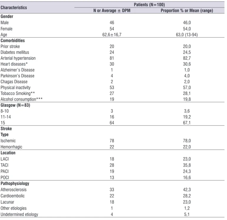
The socio-demographic and clinical characteristics are described in Table 1. There were 78% ischemic strokes and 22% hemorrhagic strokes, mainly involving the territory of the middle cerebral artery (47%). The main pathophysiological mechanism of the ischemic strokes was atherosclerosis (42.3%), followed by cardioembolic (28.3%) and lacunar strokes (18.0%). The most common comorbidity was hypertension (82.7%) and the history of previous stroke was present in 20% of the studied population32.
The average time in days between the initial speech evaluation and the evaluation held at the time of hospital discharge was 22.1 days (minimum of 8 days, maximum of 37 days). During this period there were
ive deaths.
Among the evaluations, there was an increase in the average score of the GUSS, indicating improvement of the swallowing ability. At the same time, a change
in the distribution proile of the severity of dysphagia
can be seen. In the initial evaluation, 28% of the patients presented severe dysphagia with a high risk of aspiration. At the time of hospital discharge, only 2.1% of the patients presented high risk of aspiration, being that 32.6% presented slight dysphagia and low risk of aspiration (Table 2).
In the inal evaluation, the proportion of changes
in swallowing was lower for saliva and semisolid, but
higher for liquids and solids. There was a signiicant
Table 1. Socio-demographic, clinical characteristics and comorbidities of post-stroke patients
Characteristics Patients (N=100)
N or Average ± DPM Proportion % or Mean (range)
Gender
Male Female
46
54 46,054,0
Age 62,6±16,7 63,0 (13-94)
Comorbidities
Prior stroke 20 20,0
Diabetes mellitus 24 24,5
Arterial hypertension 81 82,7
Heart diseases* 30 30,6
Alzheimer’s Disease 1 1,0
Parkinson’s Disease 4 4,0
Chagas Disease 2 2,0
Physical inactivity 53 57,0
Tobacco Smoking** 27 28,1
Alcohol consumption*** 19 19,8
Glasgow (N=83)
8-10 3 3,6
11-14 16 19,2
15 64 67,1
Stroke Type
Ischemic Hemorrhagic
78 22
78,0 22,0
Location
LACI TACI PACI POCI
18 28 19 13
23,0 35,8 24,3 16,6
Pathophysiology
Atherosclerosis Cardioembolic Lacunar Other etiologies Undetermined etiology
33 22 18 1 4
42,3 28,2 23,0 1,2 5,1 N =number of patients; DPM= average standard deviation
*IAM (N=9), Atrial ibrillation (N=8), Valvular disease (N=13) ** Less than 1 packet of cigarettes/day (N=7), 1 packet of cigarettes/day (N= 16) plus 1 packet of cigarettes/day (N= 4) *** Eventual use (N=11), abusive use (N= 8) .In mmHg; ..bpm (beats per minute); ...mpm (movements per minute)
It is noteworthy that 20% of the patients have a history of stroke prior to hospitalization and can
inluence this high number of patients with dysphagia
in the initial swallowing assessment. This is because
previous stroke is deined in the literature as a predictor
of dysphagia after stroke2,13,30.
However, regardless of the presence or not of previous stroke, the evolution of swallowing during hospital stay showed a reduction in the frequency
of dysphagia and change in the swallowing proile, conirming previous works28-31. This indicates the
impor-tance of clearly deining the moment of evaluation of
swallowing, which is not always clear in the studies10-16,
since its proile changes according to the stroke phase.
The GUSS27 is a simple evaluation that allows the
classiication of dysphagia with individualized and
serial evaluations of the three basic textures. The GUSS
DISCUSSION
According to data from national and international literature, the most common stroke in this sample was ischemic, mainly involving atherosclerotic mechanism and anterior circulation32.
In Brazilian studies, a variation between 48 and 91% was observed in the frequency of post-stroke
dysphagia, possibly relecting different diagnostic
protocols and moments of evaluation of swallowing (acute x subacute x chronic phase)10-16.
In this study, a high frequency of post-stroke dysphagia (52%) was observed among patients, being that 28% of them were diagnosed with severe dysphagia with high risk of aspiration. At the time of hospital discharge, there was a decrease in the severity of dysphagia.
Table 2. Severity of dysphagia obtained by total score in the Gugging Swallowing Screen scale in the initial swallowing assessment and
at the time of hospital discharge of post-stroke patients
GUSS Total score
Initial evaluation N=100
Hospital discharge N=95** N or Proportion % or
Average ± DPM Mean (range)
N or Proportion % or Average ± DPM Mean (range)
0 to 9 28 28,0 2 2,1 10 to 14 12 12,0 21 22,1 15 to 19 12 12,0 31 32,7 20 48 48,0 41 43,1 Total 100 100,0 95 100 Average 14,6 ± 6,9 19,5 (0,0-20,0) 17,9 ± 3,4 20,0 (2,0-20,0)
P value* 0,01
N = number of patients; DPM: average standard deviation ** 5 patients died
* Wilcoxon test
Table 3. Score obtained for saliva swallowing and three food textures by the Gugging Swallowing Screen scale in the initial swallowing
assessment and at the time of hospital discharge of post-stroke patients
GUSS Score in each step
Saliva Semisolid Liquid Solid
Initial assessment
Hospital discharge
Initial assessment
Hospital discharge
Initial assessment
Hospital discharge
Initial assessment
Hospital discharge
N=100% N=95% N=86% N=93% N=74% N=92% N=61% N=72%
0 6 6,0 - - - - - - - - - - - - -1 4 4,0 1 1,0 5 5,8 1 1,0 - - - - 3 4,9 20 27,7 2 1 1,0 1 1,0 1 1,1 - - 3 4,0 4 4,3 - - -3 2 2,0 - - 3 3,4 - - 9 12,1 15 16,3 3 4,9 1 1,0 4 1 1,0 - - 3 3,4 - - 1 1,3 1 1,0 7 11,4 10 13,8 5 86 86,0 93 98,0 74 86,0 92 98,0 61 82,4 72 78,0 48 78,6 41 57,0
P value* 0,46 1,00 0,72 0,01
does not classify dysphagia as a change in oral and/ or pharyngeal phase, unlike other tools concerned with data on the speech organs and swallowing biome-chanics9,11-16. This is because the dysphagia study in
post-stroke patients in the acute phase should mainly focus the evaluation of the risk of bronchial aspiration
and subsequent deinition of a safer and more effective
oral feeding1,3-5,9,27. In this study, the GUSS scale allowed
not only the identiication of dysphagia, but also the classiication of its severity and clinical changes of the
acute phase of stroke.
However, as the GUSS scale is a screening protocol as well as other clinical tools, it cannot identify silent aspiration, which is evidenced by objective tests2,17,27,29.
Some researchers support the idea that the screening
performed by the speech therapist is signiicantly more
accurate when compared to other professionals as it
minimizes the error in the identiication of the patient
with or without dysphagia27,29.
Some studies show that dysphagia of neurological origin treated in the acute phase of the disease usually has very positive results9,24. It must be considered that,
in the acute phase of the disease, in addition to rehabili-tation, some other processes are involved such as partial regression of the damage and the transience of symptoms, resulting in their improvement. Apart from that, the early evaluation and the speech rehabilitation are essential because even if the dysphagia is transient, it can be reversed more quickly and with fewer compli-cations, minimizing the risk of aspiration8,19,25.
Regarding the evaluated textures, most patients
(86%) have beneited from oral feeding with semisolid food in the irst evaluation. The intermediate texture
(semisolid) seems to be ideal at this time as the
patient does not need a reined oral motor control
for the cohesion of the liquid in the oral cavity and neuromuscular energy to perform the chewing of solid food10,16,24-27. Some independent studies corroborate
this statement23-27. Studies with videoluoroscopy show
that acute stroke patients present more changes in the swallowing of liquids than in other textures23-27.
This indicates the need to analyze not only the liquid diet as in most of the screening tools17-22 but also other
textures27.
This study highlights the signiicant number of
patients involved, the early evaluation of swallowing and the use of a valid simple tool, able to test different textures. In this context, the speech evaluation with GUSS has allowed the early release of semisolid food and, consequently, the maintenance of post-acute
stroke patients exclusively with oral feeding, without the need for an alternative way of feeding. This gradual approach not only considers the severity of dysphagia, but also emphasizes the quality of life of the patient and reduction of hospital costs2,6,23-25,27.
Therefore, it is suggested that swallowing should be traced in all individuals with stroke by the team of speech therapists through a structured and valid protocol for the studied population. The early
evalu-ation (within 48 hours) allows the identiicevalu-ation of the
signs and symptoms of dysphagia and the individu-alized treatment planning required for intervention.
CONCLUSION
The early speech evaluation using a structured protocol allows the treatment planning required for an intervention to mitigate the severity of post-stroke dysphagia, enabling the safe use of oral pathway and prevention of pulmonary complications.
REFERENCES
1. Perry L, Love CP. Screening for dysphagia and aspiration in acute stroke: a system review. Dysphagia. 2001;16:7-18.
2. Ramsey DJC, Smithard DG, Kalra L. Early assessments of dysphagia and aspiration risk in acute stroke patients. Stroke. 2003;34:1252-7. 3. Ickenstein GW, Riecker A, Höhlig C, Müller R,
Becker U, Reichmann H et al. Pneumonia and in-hospital mortality in the context of neurogenic oropharyngeal dysphagia (NOD) in stroke and a new NOD step-wise concept. J Neurol. 2010; 257:1492–9.
4. Rodrigue N, Cote R, Kirsch C, Germain C, Couturier C, Fraser R. Meeting the nutritional needs of patients with severe dysphagia following a stroke: an interdisciplinary approach. Axone. 2002;23(3):31-7. 5. DeLegge MH. Aspiration pneumonia: incidence,
mortality, and at-risk populations. J Parenter Enteral Nutr. 2002;26(6):19-24.
6. Farri A, Accornero A, Burdese C. Social importance of dysphagia: its impact on dyagnosis and therapy. Acta Otorhinolaryngol. 2007;27(2):83-6.
7. Tohara H, Saitoh E, Mays KA, Kuhlemeier K, Palmer JB. Three tests for predicting aspiration without
videoluorography. Dysphagia. 2003;18(2):126-34.
9. Falsetti P, Acciai C, Palilla R, Bosi M, Carpinteri F. Oropharyngeal dysphagia after stroke: incidence, diagnosis, and clinical predictors in patients admitted to a neurorehabilitation unit. Journal of stroke and cerebrovascular diseases: the
oficial journal of National Stroke Association.
2009;18:329-35.
10. Schelp AO, Cola PC, Gato AR. Incidência de disfagia orofaríngea após acidente vascular encefálico em hospital público de referencia. Arq Neuropsiquiatr. 2004;62:503-6.
11. Doria S, Abreu MAB, Buch R, Assumpção R, Nico MAC, Ekcley CA, et al. Estudo comparativo
da deglutição com nasoibrolaringoscopia e
videodeglutograma em pacientes com acidente vascular cerebral. Rev. Bras. Otorrinolaringol. 2003;69(5):636-42.
12. Xerez DR, Carvalho YSV, Costa MMB. Estudo
clínico e videoluoroscópico da disfagia na fase
subaguda do acidente vascular encefálico. Radiol Bras. 2004;37(1):9-14.
13. Barros AFF, Fábio SRC, Furkim AM. Correlação entre os achados clínicos da deglutição e os
achados da tomograia computadorizada de
crânio em pacientes com acidente vascular cerebral isquêmico na fase aguda da doença. Arq Neuropsiquiatr. 2006;64(4):1009-14.
14. Gatto AR, Rehder MIBC. Comparação entre queixas
de deglutição e achados videoluoroscópicos no
paciente pós acidente vascular encefálico. Rev. CEFAC. 2006;8(3):320-7.
15. Gomes GF, Campos ACL, Pisani JC. Sonda nasoenteral, aspiração traqueal e pneumonia aspirativa em pacientes hospitalizados com doença cérebro-vascular complicada por disfagia orofaríngea. ABCD arq. bras. cir. dig. 2003;16(4):189-92.
16. Padovani AR, Moraes DP, Mangili LD, Andrade CRF. Protocolo Fonoaudiológico de Avaliação do Risco para Disfagia (PARD). Rev Soc Bras Fonoaudiol. 2007;12(3):199-205.
17. Nishiwaki K, Tsuji T, Liu M, Hase K, Tanaka N,
Fujiwara T. Identiication of a simple screening tool
for dysphagia in patients with stroke using factor analysis of multiple dysphagia variables. J Rehabil Med. 2005;37(4):247-51.
18. Wu MC, Chang YC, Wang TG, Lin LC. Evaluating swallowing dysfunction using a 100-ml water swallowing test. Dysphagia. 2004;19(1):43-7.
19. Sudo E, Tanuma S, Sudo E, Takahashi Y, Yoshida A, Kobayashi C et al. The usefulness of
the water swallowing test and videoluorography
in swallowing rehabilitation in patients with cerebrovascular disease. Nihon Ronen Igakkai Zasshi. 2002;39(4):427-32.
20. Marik PE, Kaplan D. Aspiration pneumonia and dysphagia in the elderly. Chest. 2003;124(1):328-36. 21. Lim SHB, Lieu PK, Phua SY, Seshadri R,
Venketasubramanian N, Lee SH et al. Accuracy of bedside clinical methods compared with
iberoptic endoscopic examination of swallowing
in determining the risk of aspiration in acute stroke patients. Dysphagia. 2001;16:1-6.
22. Massey R, Jedlicka D. The Massey Bedside Swallowing Screen. J Neurosci Nurs. 2002;34:257-60.
23. Leder SB, Espinosa JF. Aspiration risk after acute stroke: comparison of clinical examination and
iberoptic endoscopic evaluation of swallowing.
Dysphagia. 2002;17:219.
24. Crary MA, Mann GD, Groher ME. Initial psychometric assessment of a functional oral intake scale for dysphagia in stroke patients. Arch Phys Med Rehabil. 2005;86:1516-20.
25. Doggett DL, Tappe KA, Mitchell MD, Chapell R, Coates V, Turkelson CM. Prevention of pneumonia in elderly stroke patients by systematic diagnosis and treatment of dysphagia: an evidence-based comprehensive analysis of the literature. Dysphagia. 2001;16:279-95.
26. Steele CM, Van Lieshout PH. Inluence of bolus consistency on lingual behaviors in sequential swallowing. Dysphagia. 2004;19(3):192-206.
27. Trapl M, Enderle P, Nowotny M, Teuschl Y, Matz K, Dachenhausen A et al. Dysphagia bedside screening for acute-stroke patients: the Gugging Swallowing Screen. Stroke. 2007;38:2948-52.
28. Smithard DG, O’Neill PA, England RE, Park CL, Wyatt R, Martin DF et al. The natural history of dysphagia following a stroke. Dysphagia. 1997;12:188-93.
29. Nilsson H, Ekberg O, Olsson R, Hindfelt B. Dysphagia in stroke: a prospective study of quantitative aspects of swallowing in dysphagic patients. Dysphagia. 1998;13:32-8.
31. Finestone HM, Woodbury MG, Foley NC, Teasell RW, Greene-Finestone LS. Tracking clinical improvement of swallowing disorders after stroke. J Stroke Cerebrovasc Dis. 2002;11(1):23-7.
32. Almeida EO, Faleiros BE, Martins C, Lemos SMA,
Teixeira AL. Características clínico-demográicas