Effect of electrolytic lesion of the
dorsal raphe nucleus on water
intake and sodium appetite
Departamento de Ciências Fisiológicas, Instituto de Biologia, Universidade Federal Rural do Rio de Janeiro, Seropédica, RJ, Brasil E.L. Olivares,
R.H. Costa-e-Sousa, H.R. Cavalcante-Lima, H.R.C. Lima, P.L. Cedraz-Mercez and L.C. Reis
Abstract
The present study determined the effect of an electrolytic lesion of the dorsal raphe nucleus (DRN) on water intake and sodium appetite. Male Wistar rats weighing 290-320 g with a lesion of the DRN (L-DRN), performed two days before experiments and confirmed by histology at the end of the experiments, presented increased sensitivity to the dehydration induced by fluid deprivation. The cumulative water intake of L-DRN rats reached 23.3 ± 1.9 ml (a 79% increase, N = 9) while sham-lesioned rats (SL-DRN) did not exceed 13.0 ± 1.0 ml (N = 11, P < 0.0001) after 5 h. The L-DRN rats treated with isoprote-renol (300 µg kg-1 ml-1, sc) exhibited an increase in water intake that persisted throughout the experimental period (L-DRN, 15.7 ± 1.47 ml, N = 9 vs SL-DRN, 9.3 ± 1.8 ml, N = 11, P < 0.05). The L-DRN rats also showed an increased spontaneous sodium appetite during the entire period of assessment. The intake of 0.3 M NaCl after 12, 24, 36 and 72 h by the L-DRN rats was always higher than 20.2 ± 4.45 ml (N = 10), while the intake by SL-DRN was always lower than 2.45 ± 0.86 ml (N = 10, P < 0.00001). Sodium- and water-depleted L-DRN rats also exhibited an increased sodium appetite (13.9 ± 2.0 ml, N = 11) compared to SL-DRN (4.6 ± 0.64 ml, N = 11) after 120 min of observation (P < 0.02). The sodium preference of L-DRN rats in both conditions was always higher than that of SL-DRN rats. These results suggest that electrolytic lesion of the DRN overcomes a tonic inhibi-tory component of sodium appetite.
Correspondence L.C. Reis
Departamento de Ciências Fisiológicas, IB, UFRuralRJ Rodovia BR465, km 7 23890-000 Seropédica, RJ Brasil
Fax: +55-21-2682-1763 E-mail: [email protected]
Research supported by CAPES and Departamento de Ciências Fisiológicas, IB, UFRRJ.
Received February 19, 2003 Accepted August 7, 2003
Key words
•Sodium appetite •Water intake •Dorsal raphe nucleus •Serotonergic system •Electrolytic lesion •Rats
Introduction
Serotonergic neurons of the mesencepha-lic dorsal raphe nucleus (DRN), located in its medial portion, project to prosencephalic re-gions involved in hydroelectrolytic and car-diocirculatory homeostasis (1-4).
the short and long term, the changes in vol-ume, pressure and sodium concentration in the extracellular fluid, evokes electrophysi-ological changes in the DRN (7). In this context, it has been shown that the microin-jection of serotonergic agonists of the 5HT2A and 5HT2C receptors into the SFO excites angiotensin II- (ANG II) sensitive neurons (8). In addition, structures of the lamina terminalis, including the SFO and those of the wall of the anteroventral region of the third ventricle (AV3V), the organum vas-culosum laminae terminalis (OVLT), and the median preoptic nucleus (MnPO) con-stitute areas related to hydroelectrolytic and cardiovascular regulation (4,9-11). Relevant observations have shown that stim-ulation of the AV3V region by intracerebro-ventricular (icv) administration of serotonin, serotonin-releasing agents or 5HT2A/2C ago-nists increases the urinary excretion of so-dium (12,13). All of these stimulation condi-tions under fluid deprivation, cholinergic or angiotensinergic stimulation of the AV3V region and central or systemic ß-adrenergic stimulation also caused a de-crease in water intake (14-16). These obser-vations were later confirmed in part using different experimental paradigms in which the 5HT2B/2C agonist, 1-3-chlorophenyl-piperazine (mCPP), was administered by the icv route (17). The authors reported an in-hibition of water intake after dipsogenic challenges induced by fluid deprivation, hypertonic saline overload or hypovole-mia. Furthermore, it has been shown that basal c-Fos expression in serotoninergic neurons of the DRN was decreased after sodium depletion induced by peritoneal dialysis and was increased after spontane-ous and induced sodium intake, suggest-ing that there is a tonic inhibition of so-dium appetite by serotoninergic cells of this nucleus (18).
The observation that the recruitment of ascending serotonergic pathways by flow of information originated from prosencephalic
structures implied hydroelectrolytic and cardiocirculatory regulation (4,5,7). Taken together with the evidence that electrolytic lesions of the DRN reduce the basal or stim-ulated plasma levels of atrial natriuretic pep-tide (ANP), these data suggest that ascend-ing pathways of the raphe integrate signals concerned with the volume of body fluid homeostasis through the control of renal water and electrolyte excretion, as well as of water intake (3).
No systematic studies involving the di-rect manipulation of the DRN and its impli-cation in the expression of sodium appetite and water intake have been reported. There has been only a single report which demon-strated that electrolytic lesion of the DRN caused a chronic increase in water intake when only this fluid was offered (3).
The present study was designed to assess the effect of electrolytic lesion of the DRN on water intake in the experimental para-digms that involve an increase of extracellu-lar fluid tonicity by fluid deprivation (os-motic thirst) and brain signaling induced by the systemic production of ANG II in dl-isoproterenol-treated rats (4,19-21). In addi-tion, spontaneous sodium appetite was in-vestigated in DRN-lesioned rats following a simultaneous offer of water and hypertonic saline for three consecutive days.
Material and Methods
Animals
Electrolytic lesions
In order to perform the electrolytic le-sions, stereotaxic coordinates were obtained according to the parameters defined in the atlas of Paxinos and Watson (22), using the anteroposterior (AP) coordinates = 7.6-7.8 mm, posterior to bregma, lateral = 0 mm and vertical (V) = 6.4-6.6 mm, below the top of the skull.
The lesions were produced by passing an anodal current of 1 mA for 10 s. Sham-operated rats were used as controls. At the end of the experiment the rats were sacri-ficed under deep anesthesia and transcardiac perfusion was performed with 10% formal-dehyde. The location of the lesions was con-firmed by histological examination of serial coronal sections (10 µm) through the brain-stem stained by the Nissl method. A prophy-lactic dose of 30,000 IU penicillin (Fort Dodge Saúde Animal Ltda., Campinas, SP, Brazil) was administered im to operated rats. Data for rats with lesions outside the dorso-ventromedial region of the DRN were cluded from statistical analysis. All the ex-perimental protocols and animal procedures were carried out in accordance with current Brazilian legislation.
Statistical analysis
Data were analyzed statistically by two-way analysis of variance with repeated measures, and the significance of differ-ences between means was determined by the Newman-Keuls test. The difference between DRN-lesioned rats (L-DRN) and sham-lesioned rats (SL-DRN) was calcu-lated in all experiments. The level of sig-nificance was set at 5%. Data are reported as mean ± SEM.
Experimental procedures
Water intake. Water intake was deter-mined in rats submitted to 16 h of fluid
deprivation and in rats treated with dl-iso-proterenol (300 µg kg-1 ml-1, sc; Aldrich) for
5 h at hourly intervals. Under these condi-tions, thirst and water intake are related to an increased tonicity of the extracellular fluid and the systemic production of ANG II, re-spectively (4). In this set of experiments rats were used with an electrolytic lesion of the DRN (L-DRN, N = 9) or sham lesion (SL-DRN, N = 11) produced 2 days before treat-ment with isoproterenol or isotonic saline.
Spontaneous (basal) sodium appetite. In order to assess the sodium appetite, 0.3 M NaCl was offered simultaneously with dis-tilled water to the rats and the intake of fluids was determined at 3, 6, 9, 12 and 24 h (on the first day) and on a daily basis thereafter, up to 3 days post-lesion (N = 10 for SL-DRN and L-DRN rats).
Induced sodium appetite. Additionally, SL-DRN and L-DRN rats (N = 11 for both groups) were treated with a combination of furosemide (10 mg kg-1 ml-1, sc; Aventis
Pharma, Suzano, SP, Brazil) + captopril (5 mg kg-1 ml-1, sc; Aldrich). After 60 min
without fluids or food, water and 0.3 M NaCl were made available in metabolic cages. Fluid
intake was determined over 2 h at 30-min intervals. In this situation, the effect of the ANG-converting enzyme inhibitor captopril increases plasma levels of ANG I available to the circumventricular organs of the lamina terminalis, probably the SFO, in which the local conversion to ANG II occurs, leading to the stimulation of water intake and of sodium appetite (4,23-25). Sodium prefer-ence was calculated according to the follow-ing formula: volume of 0.3 M NaCl intake/ volume of 0.3 M NaCl intake + volume of water intake.
Results
The electrolytic lesions of the DRN suit-able for the investigation of ingestive re-sponses extended from the dorsomedial to the ventromedial portion of the nucleus be-tween the coordinates AP = 7.6-8.4 mm and V = 6.2-6.6 mm (Figure 1).
There was an increased sensitivity of L-DRN rats to the dehydration induced by fluid deprivation with increased water
in-take throughout the period of observation (Figure 2A). After 5 h the cumulative water intake by L-DRN rats reached 23.3 ± 1.9 ml (a 79% increase) while that of SL-DRN rats did not exceed 13.0 ± 1.0 ml (P < 0.0001). Similar results were observed in L-DRN rats treated with isoproterenol, in which the in-crease in water intake persisted throughout the experimental period (Figure 2B). After 5 h, L-DRN rats had ingested 15.7 ± 1.47 ml (a 69% increase, P < 0.05) and SL-DRN rats, 9.3 ± 1.8 ml.
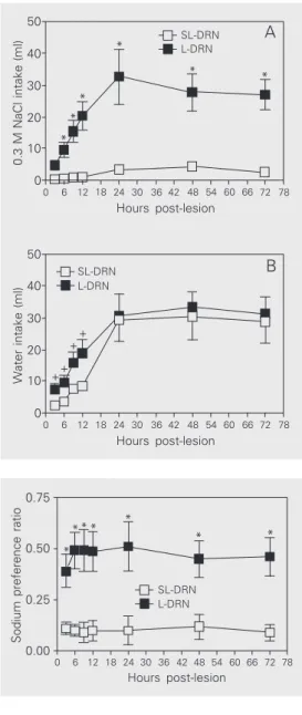
L-DRN rats also exhibited an increased spontaneous sodium appetite throughout the period of assessment, while their water in-take was higher than that by SL-DRN rats only during the first 12 h (Figure 3, panels A and B). The intake of 0.3 M NaCl after 12, 24, 36 and 72 h was 20.2 ± 4.45, 32.6 ± 8.6, 27.7 ± 5.8 and 26.9 ± 4.7 ml in L-DRN rats, and 0.97 ± 0.31, 3.3 ± 1.1, 4.2 ± 1.3 and 2.45 ± 0.86 ml in SL-DRN rats, respectively (P < 0.00001 or less, at all time points). For com-parative purposes, water and 0.3 M NaCl intake was measured cumulatively during the first 24 h. Spontaneous sodium prefer-ence was significantly higher in L-DRN rats than in SL-DRN rats, ranging from 0.39 ± 0.08 3 h lesion to 0.51 ± 0.12 24 h post-lesion, maintaining a level 0.45 up to the 72 h of assessment (P < 0.02 or less, at all time points) (Figure 4).
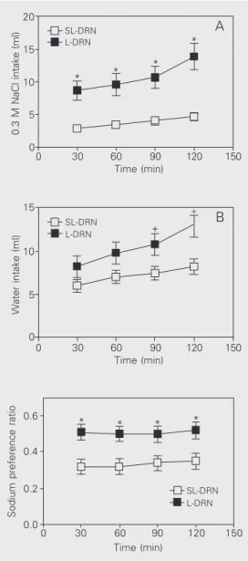
The L-DRN rats submitted to sodium and water depletion, through the combined ad-ministration of furosemide + captopril, also exhibited an increased sodium appetite throughout the experiment compared to SL-DRN (P < 0.02 or less, at all time points) (Figure 5A). After 120 min, the intake of 0.3 M NaCl reached 13.9 ± 2.0 ml, while among SL-DRN rats the intake was 4.6 ± 0.64 ml. The differences in mean water intake be-tween SL-DRN and L-DRN rats were not significant (Figure 5B). Sodium preference ranged from 0.51 ± 0.045 at 30 min to 0.52 ± 0.047 at 120 min among depleted L-DRN rats, while it never exceeded 0.35 ± 0.044 (at
Figure 2. A, Water intake by dor-sal raphe nucleus-lesioned (L-DRN, filled squares) and sham-lesioned (SL-DRN, open squares) rats deprived of water for 16 h. B, Water intake by isoproterenol (ISO)- (L-DRN, filled circles; SL-DRN, open circles) or isotonic saline- (L-DRN, filled triangles; SL-DRN, open triangles) treated rats. Data are reported as means ± SEM. *P < 0.0001 compared to the SL-DRN group (panel A).
+P < 0.05 compared to the
SL-DRN group (panel B) (two-way ANOVA followed by the New-man-Keuls post-test).
Water intake (ml)
+
+ + +
+
0 60 120 180 240 300 360
Time (min)
SL-DRN - ISO L-DRN - ISO
SL-DRN - saline L-DRN - saline
20
10
0 15
5
B
30
* *
* *
*
Water intake (ml)
20
10
0
0 60 120 180 240 300 360
Time (min)
SL-DRN L-DRN
120 min, P < 0.002, or less) among SL-DRN rats (Figure 6).
Discussion
The results of the present study reveal that electrolytic lesions of the DRN pro-duced an increase in sensitivity to osmotic stimulation by dehydration induced by fluid deprivation and ß-adrenergic stimulation with isoproterenol. This observation is consistent with the hypothesis that neurons of the DRN, which are probably serotonergic, exert a modulatory influence on the thirst related to an increase in the tonicity of extracellular fluid or on the thirst signaled by increased plasma levels of ANG II (4,19-21).
Neurons of the lamina terminalis (e.g., SFO, OVLT and MnPO) constitute the neu-roanatomical substrate for the primary inte-gration of thirst induced by osmotic stimula-tion and by ß-adrenergic stimulastimula-tion (4,26, 27). Serotonergic innervation originating in the mesencephalic raphe and receptors for serotonin have been identified through-out the lamina terminalis (1,2,4-6,8).
The effects induced by DRN lesion sug-gest that ascending serotonergic pathways are involved in the modulation of the thirst induced by dehydration and brain angioten-sinergic stimulation. The present observa-tions support previous reports, which dem-onstrated that icv administration of the 5HT2C agonist, MK212, modulates water intake induced by fluid deprivation obtained by central microinjection of ANG II or car-bachol as well as by central or systemic administration of the ß-adrenergic agonist isoproterenol (14-16). More recently, these observations were extended through the use of other dipsogenic challenges in which the icv administration of a 5HT2B/2C agonist, mCPP, inhibited water intake induced by fluid deprivation, acute overload with hy-pertonic saline and hypovolemia (17).
Similarly, homeostatic integration of so-dium appetite depends on structures in the
lamina terminalis (4). The SFO and OVLT constitute convergent sites for signals re-lated to volume depletion (4,18,28,29). As mentioned previously, these structures are innervated by serotonergic neurons, are rich in serotonin receptors and reciprocally trans-mit signals to the raphe (1,2,4-6,8). It is quite likely that these signals reflect adjustments concerning variations in volume, electro-lytic composition of the extracellular fluid and cardiovascular parameters (4,5,7,30).
Lesions of the DRN induced an increase in sodium appetite both under basal condi-tions and in the paradigm of sodium and
Sodium preference ratio
0.75 0.50 0.25 0.00 78 72 66 60 54 48 42 36 30 24 18 12 6 0 Hours post-lesion L-DRN SL-DRN * * * * * * * 20 30 40 50 10 0 78 72 66 60 54 48 42 36 30 24 18 12 6 0
0.3 M NaCl intake (ml)
Hours post-lesion 20 30 40 50 10 0 78 72 66 60 54 48 42 36 30 24 18 12 6 0
Water intake (ml)
Hours post-lesion * * * * * * A B L-DRN SL-DRN L-DRN SL-DRN ++ + +
Figure 3. A, Intake of 0.3 M NaCl by dorsal raphe nucleus-le-sioned (L-DRN, filled squares) and sham-lesioned rats (SL-DRN, open squares) under ba-sal conditions. B, Water intake of L-DRN (filled squares) and SL-DRN (open squares) rats under basal conditions. Data are re-ported as means ± SEM. *P < 0.00001 for 0.3 M NaCl intake and +P < 0.05 for water intake,
compared to the SL-DRN group (two-way ANOVA followed by the Newman-Keuls post-test).
water depletion provoked by combined ad-ministration of furosemide and a low dose of captopril. These data show that under basal conditions without a natriorexigenic chal-lenge, electrolytic lesion of the DRN pro-motes the suppression of a tonic modulatory pathway of sodium appetite. Recent evidence has shown that sodium depletion induced by the combination of furosemide with a low dose of captopril causes an increase in c-Fos expression in the nucleus tractus solitarii (NTS), which is suppressed by renal deaffer-entation (31). Following the same line of reasoning, evoked potentials were recorded in serotonergic neurons of the DRN after the
isoosmotic loss of volume by hemorrhage and, more specifically, induction of c-Fos expression was determined in serotonergic neurons of the DRN and in structures of the lamina terminalis after sodium depletion by peritoneal dialysis (7,18).
These observations permit us to propose that moment-to-moment adjustments of re-nal sodium load and extracellular fluid vol-ume generate signals that are transmitted to the NTS and, from there, to the DRN. Alter-natively, other pertinent observations sup-port the hypothesis that volume depletion (and possibly the serial changes in arterial pressure) activates neurons of the SFO sen-sitive to ANG II, which project to form synapses with serotonergic neurons of the DRN (7,30). As a result of this monitoring of the circulating levels of ANG II, signals would be generated and transmitted by as-cending serotonergic pathways of the DRN with the objective of modulating sodium appetite, with subsequent regulation of ex-tracellular fluid volume. According to this hypothesis, the removal of a tonic modula-tory pathway would imply a greater sensitiv-ity/activity of angiotensinergic mechanisms in the lamina terminalis implicated in the central control of dipsogenesis and of so-dium appetite.
Another hypothesis is based on the widely accepted concept that ANP is involved in the modulation of thirst induced by dehydration or by central angiotensinergic stimulation, and of sodium appetite provoked by sodium depletion (32,33). This same group showed that lesions of the wall of the AV3V region drastically reduce the plasma levels of ANP, mediating the expansion of blood volume (34). Taken together, these observations lead to the conclusion that structures in the lamina terminalis constitute the substrate which in-tegrates signals designed to regulate the re-lease of ANP. In view of the observations cited above, regarding the neural interac-tions between structures of the lamina termi-nalis and mesencephalic raphe, we suggest
Sodium preference ratio
150 Time (min)
0.6
SL-DRN L-DRN
0.4
0.2
0.0
120 90 60 30 0
* * * *
10 15 20
5
0
150 120 90 60 30 0
0.3 M NaCl intake (ml)
Time (min)
* A L-DRN
SL-DRN
* * *
10 15
5
0
150 120 90 60 30 0
Water intake (ml)
Time (min)
+ B
L-DRN SL-DRN
+
Figure 5. Intake of 0.3 M NaCl and water (panels A and B, re-spectively) by dorsal raphe nucleus-lesioned (L-DRN, filled squares) and sham-lesioned rats (SL-DRN, open squares) submit-ted to sodium and water deple-tion induced by treatment with furosemide + captopril. Data are reported as means ± SEM. *P < 0.02 for 0.3 M NaCl intake and
+P < 0.05 for water intake
com-pared to the SL-DRN group (two-way ANOVA followed by the Newman-Keuls post-test).
that electrolytic lesions of the DRN remove an excitatory serotonergic component in-volved in the release of ANP and, thus caus-ing dysfunction of the system responsible for adjustments in volume and the mainte-nance of homeostasis, with a subsequent increase in sodium preference (3,35).
The oxytocin produced by the paraven-tricular nucleus is a candidate for a central mediator of satiety for sodium (4,36-38). Alternatively, this may constitute another modulatory pathway for sodium appetite dependent on serotonergic ascending activa-tion, since serotonergic innervation of the paraventricular nucleus has been well estab-lished (1,2,8,39). In addition, an increase in Fos immunoreactivity was shown in oxyto-cinergic neurons during the process of so-dium satiety in rats previously depleted of sodium by peritoneal dialysis (37).
These conclusions are consistent with the current literature. The existence of
re-dundancy of multiple effector reactions con-cerned with the process of sodium satiety might be explained by the insertion of sero-tonergic circuits in a polymodal system of recruitment/homeostatic activation at differ-ent levels of physiological disturbances of tonicity and extracellular fluid volume. In view of the hypotheses presented, the results obtained are compatible with the assump-tion that electrolytic lesion of the DRN sup-presses a tonic inhibitory component of so-dium appetite.
Acknowledgments
The authors are grateful to Dr. Karla Consort Ribeiro, Laboratório de Cardiologia Celular e Molecular, Instituto de Biofísica Carlos Chagas Filho, Universidade Federal do Rio de Janeiro, for preparing the photo-micrographs, and to Dr. Wellington da Silva Côrtes for help with the English text.
References
1. Azmitia EC & Segal M (1978). An autoradiographic analysis of the differential ascending projections of the dorsal and median raphe nuclei in the rat. Journal of Comparative Neurology, 179: 641-668. 2. Steinbusch HWM (1981). Distribution of
serotonin-immunoreactiv-ity in the central nervous system of the rat cell bodies and termi-nals. Neuroscience, 6: 557-618.
3. Reis LC, Ramalho MJ, Favaretto AL, Gutkowska J, McCann SM & Antunes-Rodrigues J (1994). Participation of the ascending seroto-nergic system in the stimulation of atrial natriuretic peptide release. Proceedings of the National Academy of Sciences, USA, 91: 12022-12026.
4. Fitzsimons JT (1998). Angiotensin, thirst, and sodium appetite. Physiological Reviews, 78: 583-686.
5. Lind RW (1986). Bi-directional, chemically specified neural connec-tions between the subfornical organ and the midbrain raphe sys-tem. Brain Research, 384: 250-261.
6. Bosler O & Descarries L (1988). Monoamine innervation of the organum vasculosum laminae terminalis (OVLT): a high resolution radioautographic study in the rat. Journal of Comparative Neurol-ogy, 272: 545-561.
7. Tanaka J, Okumura T, Sakamati K & Miyakubo H (2001). Activation of serotonergic pathways from the midbrain raphe system to the subfornical organ by hemorrhage in the rat. Experimental Neurol-ogy, 169: 156-162.
8. Scrogin KE, Johnson AK & Schmid HA (1998). Multiple receptor subtypes mediate the effects of serotonin on rat subfornical organ neurons. American Journal of Physiology, 275: R2035-R2042.
9. Bealer SL (1988). Acute hypertensive and natriuretic responses following preoptic hypothalamic lesions. American Journal of the Medical Sciences, 295: 346-351.
10. De Luca Jr LA & Menani JV (1996). Preoptic-periventricular tissue (AV3V): central cholinergic-induced hydromineral and cardiovascu-lar responses, and salt intake. Revista Brasileira de Biologia, 56 (Suppl 1): 233-238.
11. De Luca Jr LA, Sugawara AM & Menani JV (2000). Brain versus peripheral angiotensin II receptors in hypovolemia: behavioural and cardiovascular implications. Clinical and Experimental Pharmacolo-gy and PhysioloPharmacolo-gy, 27: 437-442.
12. Stein JM, Lind RW & Johnson AK (1987). Central serotonergic influences on renal electrolyte and water excretion. Neuropharma-cology, 26: 1685-1692.
13. Reis LC, Ramalho MJ & Antunes-Rodrigues J (1991). Effect of central administration of serotoninergic agonists on electrolyte ex-cretion control. Brazilian Journal of Medical and Biological Research, 24: 633-641.
14. Reis LC, Ramalho MJ & Antunes-Rodrigues J (1990). Central sero-tonergic modulation of drinking behavior induced by water depriva-tion. Effect of serotonergic agonist (MK-212) administered intrace-rebroventricularly. Brazilian Journal of Medical and Biological Re-search, 23: 1335-1338.
Biologi-cal Research, 23: 1339-1342.
16. Reis LC, Ramalho MJ & Antunes-Rodrigues J (1992). Brain seroto-ninergic stimulation reduces the water intake induced by systemic and central beta-adrenergic administration. Brazilian Journal of Medi-cal and BiologiMedi-cal Research, 25: 529-536.
17. Castro L, Maldonado I, Campos I, Varjão B, Angelo AL, Athanazio RA, Barbetta MC, Ramos AC, Fregoneze JB & De Castro e Silva E (2002). Central administration of mCPP, a serotonin 5-HT(2B/2C) agonist, decreases water intake in rats. Pharmacology, Biochemis-try and Behavior, 72: 891-898.
18. Franchini LF, Johnson AK & Vivas L (2002). Sodium appetite and Fos activation in serotonergic neurons. American Journal of Physiol-ogy, 282: R235-R243.
19. Phillips MI, Hoffman WE & Bealer SL (1982). Dehydration and fluid balance: central effects of angiotensin. Federation Proceedings, 41: 2520-2527.
20. Lind RW, Thunhorst RL & Johnson AK (1984). The subfornical organ and the integration of multiple factors in thirst. Physiology and Behavior, 32: 69-74.
21. Ramsay DJ & Thrasher TN (1986). Hyperosmotic and hypovolemic thirst. In: de Caro G, Epstein AN & Massi M (Editors), The Physiolo-gy of Thirst and Sodium Appetite. Plenum Press, New York, 83-96. 22. Paxinos G & Watson C (1986). The Rat Brain in Stereotaxic
Coordi-nates. 2nd edn. Academic Press, New York.
23. Fitts DA & Masson DB (1989). Forebrain sites of action for drinking and salt appetite to angiotensin or captopril. Behavioral Neurosci-ence, 103: 865-872.
24. Menani JV, Thunhorst RL & Johnson AK (1996). Lateral parabrachial nucleus and serotonergic mechanisms in the control of salt appetite in rats. American Journal of Physiology, 270: R162-R168.
25. Rauch M & Schmid HA (1999). Functional evidence for subfornical organ intrinsic conversion of angiotensin I to angiotensin II. Ameri-can Journal of Physiology, 276: R1630-R1638.
26. Thrasher TN & Keil LC (1987). Regulation of drinking and vaso-pressin secretion: role of organum vasculosum laminae terminalis. American Journal of Physiology, 253: R108-R120.
27. Fitts DA (1994). Angiotensin II receptors in SFO but not in OVLT mediate isoproterenol-induced thirst. American Journal of Physiolo-gy, 267: R7-R15.
28. Vivas L, Pastuskovas CV & Tonelli L (1995). Sodium depletion
induces Fos immunoreactivity in circumventricular organs of the lamina terminalis. Brain Research, 679: 34-41.
29. Pastuskovas C & Vivas L (1997). Effect of intravenous captopril on c-fos expression induced by sodium depletion in neurons of the lamina terminalis. Brain Research Bulletin, 44: 233-236.
30. Tanaka J, Ushigome A, Hori K & Nomura M (1998). Response of raphe nucleus projecting subfornical organ neurons to angiotensin II in rats. Brain Research Bulletin, 45: 315-318.
31. Fitch GK & Weiss ML (2000). Activation of renal afferent pathways following furosemide treatment. II. Effect of angiotensin blockade. Brain Research, 861: 377-389.
32. Antunes-Rodrigues J, McCann SM & Samsom WK (1985). Atrial natriuretic factor inhibits dehydration- and angiotensin II-induced water intake in the conscious, unrestrained rat. Proceedings of the National Academy of Sciences,USA, 82: 8720-8723.
33. Antunes-Rodrigues J, McCann SM, Rogers LC & Samsom WK (1986). Central administration of atrial natriuretic factor inhibits sa-line preference in the rat. Endocrinology, 118: 1726-1728. 34. Antunes-Rodrigues J, Ramalho MJ, Reis LC, Menani JV, Turrin MQ,
Gutkowska J & McCann SM (1991). Lesions of the hypothalamus and pituitary inhibit volume-expansion-induced release of atrial na-triuretic peptide. Proceedings of the National Academy of Sciences, USA, 88: 2956-2960.
35. Reis LC (1993). Participação do sistema serotoninérgico central na regulação do equilíbrio hidroeletrolítico. Doctoral thesis, Faculty of Medicine of Ribeirão Preto, São Paulo University, Ribeirão Preto, SP, Brazil.
36. Johnson AK & Thunhorst RL (1997). The neuroendocrinology of thirst and salt appetite: visceral sensory signals and mechanisms of central integration. Frontiers in Neuroendocrinology, 18: 292-353. 37. Franchini LF & Vivas L (1999). Distribution of Fos immunoreactivity
in rat brain after sodium consumption induced by peritoneal dialy-sis. American Journal of Physiology, 276: R1180-R1187.
38. Amico JA, Morris M & Vollmer RR (2001). Mice deficient in oxytocin manifest increased saline consumption following overnight fluid deprivation. American Journal of Physiology, 281: R1368-R1373. 39. Van de Kar LD, Rittenhouse PA, Li Q, Levy AD & Brownfield MS