ISSN 1414-431X
www.bjournal.com.br
www.bjournal.com.br
Volume 45 (12) 1102-1340 December 2012
Braz J Med Biol Res, December 2012, Volume 45(12) 1112-1118
10.1590/S0100-879X2012007500130
doi:
Overexpression of Fc receptor-like 1 associated with B-cell
activation during hepatitis B virus infection
Ke Wang, Hao Pei, Biao Huang, Run-Lin Yang, Hang-Yuan Wu, Xue Zhu and Lan Zhu
Institutional Sponsors
The Brazilian Journal of Medical and Biological Research is partially financed by
Faculdade de Medicina de Ribeirão Preto Campus
Ribeirão Preto
Explore High - Performance MS Orbitrap Technology In Proteomics & Metabolomics
analiticaweb.com.br S C I E N T I F I C
BIOMEDICAL SCIENCES
AND
Overexpression of Fc receptor-like 1
associated with B-cell activation during
hepatitis B virus infection
Ke Wang
1, Hao Pei
2, Biao Huang
1, Run-Lin Yang
1, Hang-Yuan Wu
2,
Xue Zhu
1and Lan Zhu
11Key Laboratory of Nuclear Medicine, Ministry of Health, Jiangsu Key Laboratory of Molecular Nuclear Medicine,
Jiangsu Institute of Nuclear Medicine, Wuxi, Jiangsu Province, China
2Wuxi Hospital of Infectious Disease, Wuxi, Jiangsu Province, China
Abstract
The role of B cells in the pathogenesis of hepatitis B virus (HBV) infection has not been explored in depth.In the present study, the activation status of B cells from peripheral blood of healthy controls (N = 20) and patients with acute hepatitis B (AHB, N = 15) or chronic hepatitis B (CHB, N = 30) was evaluated by measuring the expression levels of B-cell activation markers CD69
and CD86, using quantitative real-time PCR and flow cytometry. Moreover, the potential mechanism underlying B-cell activation during HBV infection was further investigated by analyzing the expression profile of FCRL1, an intrinsic activation molecule
of B cells. An elevation in the levels of B-cell activation markers including CD69 and CD86 was observed in the AHB patients (44.31 ± 9.27, 27.64 ± 9.26%) compared to CHB patients (30.35 ± 11.27, 18.41 ± 6.56%, P < 0.05), which was still higher than healthy controls (12.23 ± 7.84, 8.22 ± 3.43%, P < 0.05). Furthermore, the expression of FCRL1 was found to be similar to B-cell activation markers,which was highest in AHB patients (70.15 ± 17.11%), lowest in healthy donors (36.32 ± 9.98%, P < 0.05) and half-way between these levels in patients with CHB (55.17 ± 12.03%, P < 0.05). The results were positively associated with aberrant B-cell activation. These data suggest that B cells can play a role in HBV infection, and therefore more effort should be devoted to exploring their functions.
Key words: Hepatitis B virus; B-cell activation; Fc receptor-like 1; Gene expression
Introduction
Correspondence: Biao Huang, Key Laboratory of Nuclear Medicine, Jiangsu Institute of Nuclear Medicine, No. 20 Qianrong Street, Wuxi 214063, Jiangsu Province, China. Fax: +86-510-8552-0070. E-mail: [email protected]
Received March 6, 2012. Accepted August 3, 2012. Available online August 17, 2012. Published December 17, 2012.
Hepatitis B virus (HBV) is the most common cause of liver disease worldwide (1). It is estimated that two billion people have been infected with HBV and more than 350 million suffer chronic liver infections. About 5% of adult pa-tients with acute HBV (AHB) infection may develop chronic hepatitis B (CHB) infection (2,3). HBV infection can cause necroinflammation that with time can progress to hepatic fibrosis, cirrhosis and even hepatocellular carcinoma (4-6). Extensive investigations have focused on the mechanisms
of HBV-specific T-cell dysfunction, whereas B-cell dys
-function during HBV infection has been less studied (7,8). Previous studies have found that HBV antigen binds to B
cells in vitro, resulting in B-cell activation through a
T-cell-dependent or T-cell-inT-cell-dependent pathway. However, studies on the activation status of B cells in HBV-infected patients are not available (9-11).
The understanding of the B-cell activation mechanisms has been advanced by identification of Fc receptor-like 1 (FCRL1), also known as FcRH1 (12,13). FCRL1 is a member of the Fc receptor homologue family encoded by a gene on chromosome 1q21-23 and preferentially expressed in B cells (14-16). This molecule has two immunorecep-tor tyrosine-based activation motifs (ITAM-like motifs), suggesting that it may play a key role in B-cell activation. Compelling evidence indicates that FCRL1 is an intrinsic activation molecule that has the potential to amplify B-cell antigen receptor-induced activation. Abnormal expression of FCRL1 has been reported in diseases such as chronic lymphocytic leukemia, hairy cell leukemia and B-cell non-Hodgkin lymphoma, but its particular role in the progression of the HBV infection remains unknown (17,18).
FCRL1 and B cells during HBV infection 1113
was studied in human peripheral blood lymphocytes from AHB- or CHB-infected individuals by analyzing the expres-sion profiles of B-cell activation markers including CD69 and CD86. The expression level of FCRL1, an intrinsic activation molecule of B cells, and its correlation with the activation status of B cells during HBV infection were also investigated. An attempt was made to elucidate the role of B lymphocytes in HBV-induced disease progression.
Patients and Methods
Patients
The study was approved by the Ethics Committee of Wuxi Hospital of Infectious Disease and Jiangsu Institute of Nuclear Medicine. All donors gave written informed consent to donate blood. Sixty-five subjects were recruited from Wuxi Hospital of Infectious Disease between 2009 and 2010, 20 were healthy donors, 15 were AHB patients and 30 were CHB patients. Patients with autoimmune hepatitis or hepatitis C virus (HCV) co-infection were excluded from the study by screening with HCV and antinuclear antibodies. None of the patients had been treated with antiviral agents during a 12-month period before enrollment in the study.
Cell isolation
Peripheral blood mononuclear cells (PBMC) were sepa-rated by Ficoll density centrifugation (Cedarlane Labs, USA) as mentioned before (12). B lymphocytes were further puri-fied from PBMC by negative selection with magnetic bead-conjugated antibody (StemCell, Canada). The homogeneity of cells was confirmed by flow cytometry (>95%).
Quantitative real-time polymerase chain reaction (RT-PCR)
Total RNA was extracted from purified B lymphocytes with Trizol and first-strand cDNA was obtained with the
PrimeScript™RT reagents Kit (Takara, Japan). RT-PCR was
performed using SYBR green PCR Master Mix (Takara) with the ABI PRISM 7700 detection system. Primers for this study were designed using the Primer Express Software version 2.0 (Table 1). To assure specific amplification,
the melting curves of each reaction were monitored and each sample was processed in duplicate. Human glyceraldehyde-3-phos-phate dehydrogenase was included as the housekeeping gene. Relative quantification of the target gene expression was analyzed using the Q-Gene software.
Flow cytometry
All blood samples were processed on the day of collection. PerCP-conjugated CD19, FITC-conjugated CD69 or anti-CD86 and FITC-conjugated anti-FCRL1 [BD Biosciences (USA) and R&D Systems (USA)]
were used to evaluate the activation status of B cells and FCRL1-positive B cells, respectively. After staining, cells were resuspended in fluorescence-activated cell sorting solution, fixed in 1% paraformaldehyde and analyzed with the FACS Calibur system (BD Pharmingen, USA).
Statistical analysis
Data are reported as means ± SD. The unpaired Student
t-test and the chi-square test were used for data analysis. A
P value of <0.05 was considered to be statistically signifi
-cant. The analyses were performed using the SPSS 15.0 software (USA).
Results
Clinical characteristics of the groups studied
Three groups of subjects were included in the present
study (Table 2). The first consisted of 20 health controls
with no evidence of any liver diseases and with negative
tests for HBV markers. The second consisted of 15 patients
diagnosed with AHB and with no history of chronic liver disease. The third consisted of 30 patients who had been HBsAg-positive for more than 6 months and whose clinical
appearance showed active CHB. Patient age and gender
data and biochemical tests for serum bilirubin, albumin, aspartate aminotransferase and alanine transaminase were also obtained.
Activation status of B cells in HBV-induced diseases
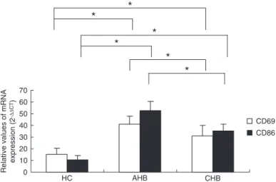
To explore the activation status of B cells from healthy controls and HBV patients, the expression profiles of B-cell surface markers, including early lymphocyte activation marker CD69 and co-stimulatory molecule CD86, were analyzed by real-time PCR and flow cytometry. As shown in Figure 1, B cells from HBV-infected patients had rela-tively higher mRNA expression levels of CD69 and CD86 compared to those from healthy donors (P < 0.05) and AHB-infected individuals exhibited a much higher level than the CHB group (P < 0.05). The frequency of CD69 and CD86 protein expression from 65 samples was examined by flow
Table 1. Primers used to measure the relative gene expression by RT-qPCR.
Genes Primers Sequences (5’-3’) Amplicon size (bp)
CD69 CD69-F TGGTGATGAAGACCACATTCA 129
CD69-R AGAACAGCTCTTTGCATCCG
CD86 CD86-F AGAGGAGCAGCACCAGAGAG 125
CD86-R ATGAGTGGGGTCATTTCCAG
FCRL1 FCRL1-F CCTACCTCAACTCACCTAC 127
FCRL1-R TCTGCTGCTACTGATTCC
GAPDH GAPDH-F AACAGCCTCAAGATCATCAGC 199
GAPDH-R GGATGATGTTCTGGAGAGCC
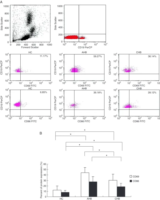
cytometry. Representative plots of CD69 and CD86 expression in CD19 B cells from PBMC of 1 healthy donor, 1 AHB patient and 1 CHB patient are shown in Figure 2A. Compared to healthy controls (12.23 ± 7.84, 8.22 ± 3.43%), a higher frequency of expres-sion was observed in AHB patients (44.31 ± 9.27, 27.64 ± 9.26%) and CHB patients (30.35 ± 11.27,
18.41 ± 6.56%) and there was also a significant dif
-ference between the two patient groups (P < 0.05; Figure 2B). In addition, the protein expression level of these molecules was significantly correlated with their corresponding mRNA expression level (data not shown).
FCRL1 is up-regulated in B cells from HBV-infected patients
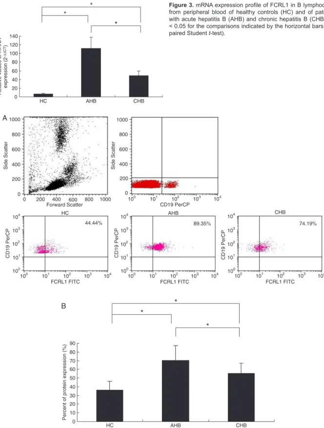
The expression profiles of FCRL1 in B cells from healthy controls and HBV-infected patients were also analyzed using real-time PCR and flow cytometry. The results revealed that, compared to healthy controls, on average, FCRL1 mRNA expres-sion was 18-fold higher in AHB and 8-fold higher in CHB (P < 0.05; Figure 3). In addition, the variance was significant different among the patients of the two groups (P < 0.05). The protein expression level of FCRL1 in B cells from the three groups was also evaluated. As shown in Figure 4, the level of FCRL1 expression was highest in AHB patients (70.15 ± 17.11%), lowest in healthy donors (36.32 ± 9.98%) and half-way between these levels in patients with CHB (55.17 ± 12.03%; P < 0.05). Overall, the protein expression pattern was similar to that obtained by real-time PCR.
Correlation between FCRL1 expression and activation status of B cells
The PBMC employed in these experiments were simul-taneously used to examine the expression levels of B-cell activation markers and FCRL1. Up-regulation of FCRL1 was then associated with the up-regulation of B-cell
activa-tion markers including CD69 (R2 = 0.708; P < 0.05) as well
as CD86 (R2 = 0.740, P < 0.05) by correlation analysis.
These findings indicated that the protein expression level of FCRL1 in the total CD19 B cells is positively correlated with the activation status of B cells in HBV-induced progres-sion (Figure 5).
Discussion
The mechanism of HBV infection is not clear and previ-ous studies have focused on virus-specific T-cell immune responses (7,8). B cells, important antigen-presenting cells, also may play an important role in HBV infection (19,20). Some data suggest that abnormal B-cell activation may be involved in the process of immune responses to HBV
antigen (21). To determine the activation status of peripheral blood B cells from HBV patients, the expression profiles of activation markers including CD69 and CD86 were exam-ined in circulating B cells of patients, being considered to be nearly absent from circulating B cells of healthy controls. The expression profile of FCRL1, a key molecule in the activation pathway of B cells, was also assessed.
In the present study, we collected 65 samples from 20 healthy donors, 15 patients with AHB infection and 30 pa-tients with active CHB infection. The percentage of CD19 B cells did not differ significantly between healthy controls and the two patient groups (data not shown). Higher expression levels of the early lymphocyte activation marker CD69 and the co-stimulatory molecule CD86 in B cells isolated from patients with HBV infection suggested that an increased B-cell activation status may play an important role during
HBV infection. Moreover, we observed an increaseinthe
fraction of positive cells after the acute phase of infection, compared to the active chronic phase of infection. This
Figure 1. mRNA expression profiles of B-cell activation markers includ -ing CD69 and CD86 in B lymphocytes from peripheral blood of healthy controls (HC) and of patients with acute hepatitis B (AHB) and chronic hepatitis B (CHB). *P < 0.05 for the comparisons indicated by the horizon-tal bars (unpaired Student t-test).
Table 2. Clinical characteristics of the subjects enrolled in the study.
Parameters HC (N = 20) AHB (N = 15) CHB (N = 30)
Age (years) 34.2 ± 8.3 30.5 ± 4.3 42.5 ± 8.3
Gender (M/F) 14/6 10/5 19/11
Serum bilirubin (mg/mL) 0.4 ± 0.4 10.5 ± 9.2 1.4 ± 6.1
Albumin (g/dL) 4.5 ± 1.9 5.4 ± 3.8 4.3 ± 2.8
AST (IU/L) 19.4 ± 4.2 496.6 ± 145.3 46.0 ± 57.1
ALT (IU/L) 16.3 ± 3.8 1199.8 ± 628.2 98.8 ± 62.3
FCRL1 and B cells during HBV infection 1115
Figure 2. Protein expression profiles of B-cell activation markers including CD69 and CD86 in B lymphocytes from peripheral blood
Figure 3. mRNA expression profile of FCRL1 in B lymphocytes
from peripheral blood of healthy controls (HC) and of patients with acute hepatitis B (AHB) and chronic hepatitis B (CHB). *P < 0.05 for the comparisons indicated by the horizontal bars (un-paired Student t-test).
Figure 4. Protein expression profile of FCRL1 in B lymphocytes from peripheral blood of healthy controls (HC) and of patients with
FCRL1 and B cells during HBV infection 1117
dynamic modulation of the antiviral immune response via activation of B lymphocytes might support the healing process at the site of acute infection. Previous studies have also reported that the majority of acute virus-infected patients exhibited activated B cells, but few data have been reported about the difference in B-cell activation status between patients with acute and chronic infection (22). Our results indicated that HBV-infected subjects who developed acute hepatitis showed a much stronger B-cell response to the virus than did the chronic cases.
The mechanisms underlying B-cell activation dur-ing HBV infection have been further investigated in our study. Previous studies have reported that FCRL1, an intrinsic activation molecule in B cells, was found in all the circulating B cells and showed a much higher expression than activation markers including CD69 and CD86 (12,23). Our results indicated that FCRL1 was highest in AHB patients, lowest in healthy donors and half-way between these two extremes in patients with CHB. The mean values were significantly different among the three groups. In addition, up-regulation of FCRL1 was positively correlated with the corre-sponding up-regulation of activation markers. The biological meaning of this overexpression deserves careful interpretation and additional investigation. It may represent a pathogenic phenomenon of activated B cells that overexpress FCRL1 or an intrinsic factor that stimulates aberrant B-cell activation.
A relationship between abnormal B-cell activation and FCRL1 overexpression is evident, but how this regulatory pathway acts in the development of acute and chronic HBV infection is unknown. Further study is needed to investigate the cellular and molecular basis of this phenomenon. The understanding of this mechanism might be helpful for the
treatment of this disease.
Acknowledgments
The authors are very grateful to the Natural Science Foundation of Jiangsu Province (#BK2011168).
References
1. Te HS, Jensen DM. Epidemiology of hepatitis B and C vi-ruses: a global overview. Clin Liver Dis 2010; 14: 1-21, vii. 2. Lledo JL, Fernandez C, Gutierrez ML, Ocana S.
Manage-ment of occult hepatitis B virus infection: an update for the clinician. World J Gastroenterol 2011; 17: 1563-1568. 3. Vierling JM. The immunology of hepatitis B. Clin Liver Dis
2007; 11: 727-viii.
4. Chisari FV, Isogawa M, Wieland SF. Pathogenesis of hepa-titis B virus infection. Pathol Biol 2010; 58: 258-266. 5. Ni YH. Natural history of hepatitis B virus infection: pediatric
perspective. J Gastroenterol 2011; 46: 1-8.
6. Nguyen VT, Law MG, Dore GJ. Hepatitis B-related hepa-tocellular carcinoma: epidemiological characteristics and disease burden. J Viral Hepat 2009; 16: 453-463.
7. Bertoletti A, Maini MK, Ferrari C. The host-pathogen inter-action during HBV infection: immunological controversies.
Antivir Ther 2010; 15 (Suppl 3): 15-24.
8. Jung MC, Pape GR. Immunology of hepatitis B infection.
Lancet Infect Dis 2002; 2: 43-50.
9. Milich DR, Chen M, Schodel F, Peterson DL, Jones JE, Hughes JL. Role of B cells in antigen presentation of the hepatitis B core. Proc Natl Acad Sci U S A 1997; 94: 14648-14653.
10. Lazdina U, Alheim M, Nystrom J, Hultgren C, Borisova G, Sominskaya I, et al. Priming of cytotoxic T cell responses to exogenous hepatitis B virus core antigen is B cell depen-dent. J Gen Virol 2003; 84: 139-146.
11. Lazdina U, Cao T, Steinbergs J, Alheim M, Pumpens P, Peterson DL, et al. Molecular basis for the interaction of the hepatitis B virus core antigen with the surface immunoglobu-lin receptor on naive B cells. J Virol 2001; 75: 6367-6374. 12. Leu CM, Davis RS, Gartland LA, Fine WD, Cooper MD.
FcRH1: an activation coreceptor on human B cells. Blood
2005; 105: 1121-1126.
13. Du X, Nagata S, Ise T, Stetler-Stevenson M, Pastan I. FCRL1 on chronic lymphocytic leukemia, hairy cell
mia, and B-cell non-Hodgkin lymphoma as a target of im-munotoxins. Blood 2008; 111: 338-343.
14. Mechetina LV, Najakshin AM, Volkova OY, Guselnikov SV, Faizulin RZ, Alabyev BY, et al. FCRL, a novel member of the leukocyte Fc receptor family possesses unique structural features. Eur J Immunol 2002; 32: 87-96.
15. Ehrhardt GR, Leu CM, Zhang S, Aksu G, Jackson T, Haga C, et al. Fc receptor-like proteins (FCRL): immunomodulators of B cell function. Adv Exp Med Biol 2007; 596: 155-162. 16. Davis RS. Fc receptor-like molecules. Annu Rev Immunol
2007; 25: 525-560.
17. Kazemi T, Asgarian-Omran H, Memarian A, Shabani M,
Sharifian RA, Vossough P, et al. Low representation of Fc
receptor-like 1-5 molecules in leukemic cells from Iranian patients with acute lymphoblastic leukemia. Cancer Immunol Immunother 2009; 58: 989-996.
18. Kazemi T, Asgarian-Omran H, Hojjat-Farsangi M, Shabani
M, Memarian A, Sharifian RA, et al. Fc receptor-like 1-5 mol -ecules are similarly expressed in progressive and indolent clinical subtypes of B-cell chronic lymphocytic leukemia. Int
J Cancer 2008; 123: 2113-2119.
19. Farci P, Diaz G, Chen Z, Govindarajan S, Tice A, Agulto L, et al. B cell gene signature with massive intrahepatic produc-tion of antibodies to hepatitis B core antigen in hepatitis B virus-associated acute liver failure. Proc Natl Acad Sci U S A 2010; 107: 8766-8771.
20. Wu C, Liu Y, Zhao Q, Chen G, Chen J, Yan X, et al. Soluble CD40 ligand-activated human peripheral B cells as surro-gated antigen presenting cells: A preliminary approach for anti-HBV immunotherapy. Virol J 2010; 7: 370.
21. Oliviero B, Cerino A, Varchetta S, Paudice E, Pai S, Ludovisi S, et al. Enhanced B-cell differentiation and reduced prolif-erative capacity in chronic hepatitis C and chronic hepatitis B virus infections. J Hepatol 2011; 55: 53-60.
22. Bachmann MF, Kopf M. The role of B cells in acute and chronic infections. Curr Opin Immunol 1999; 11: 332-339. 23. Llinas L, Lazaro A, de Salort J, Matesanz-Isabel J, Sintes J,
Engel P. Expression profiles of novel cell surface molecules on B-cell subsets and plasma cells as analyzed by flow