O R I G I N A L A R T I C L E
Antimicrobial effect of
Dinoponera quadriceps
(Hymenoptera: Formicidae) venom against
Staphylococcus
aureus
strains
D.B. Lima1, A.F.C. Torres1, C.P. Mello1, R.R.P.P.B. de Menezes2, T.L. Sampaio2, J.A. Canuto1,
J.J.A. da Silva3, V.N. Freire4, Y.P. Quinet5, A. Havt2, H.S.A. Monteiro2, N.A.P. Nogueira1and A.M.C. Martins1
1 Department of Clinical and Toxicological Analysis, Faculty of Pharmacy, Federal University of Ceara, Fortaleza, Ceara, Brazil 2 Department of Physiology and Pharmacology, Faculty of Medicine, Federal University of Ceara, Fortaleza, Ceara, Brazil 3 Federal Rural University of the Semi-Arid, Natal, Rio Grande do Norte, Brazil
4 Department of Physics, Science Center, Federal University of Ceara, Fortaleza, Ceara, Brazil 5 Institute of Biomedical Sciences, State University of Ceara, Fortaleza, Ceara, Brazil
Keywords
action mechanism, antimicrobial activity,
Dinoponera quadriceps,Staphylococcus aureus, venoms.
Correspondence
Alice Maria Costa Martins, Department of Clinical and Toxicological Analysis, Faculty of Pharmacy, Federal University of Ceara, 1210 Cap Francisco Pedro Street, Rodolfo Teofilo, Fortaleza, Ceara 60431-372, Brazil. E-mail: +martinsalice@gmail.com
2013/2565: received 23 December 2013, revised 17 April 2014 and accepted 17 May 2014
doi:10.1111/jam.12548
Abstract
Aims: Dinoponera quadriceps venom (DqV) was examined to evaluate the antibacterial activity and its bactericidal action mechanism against Staphylococcus aureus.
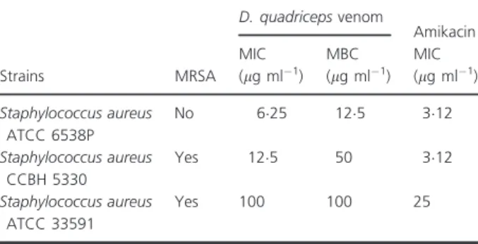
Methods and Results: DqV was tested against a standard strain of methicillin-sensitive Staphylococcus aureus (MSSA), Staph. aureus ATCC 6538P and two standard strains of methicillin-resistant Staphylococcus aureus (MRSA), Staph. aureus ATCC 33591 and Staph. aureus CCBH 5330. The minimum inhibitory concentration (MIC) and minimum bactericidal concentration (MBC), the rate of kill and pH sensitivity of the DqV were determined by microdilution tests. Bactericidal and inhibitory concentrations of DqV were tested to check its action on Staph. aureus membrane permeability and cell morphology. The MIC and MBC of DqV were 625 and 125lg ml 1 for Staph. aureus ATCC 6538P, 125 and 50lg ml 1 for Staph. aureus CCBH
5330 and 100 and 100lg ml 1 for Staph. aureus ATCC 33591, respectively.
Complete bacterial growth inhibition was observed after 4 h of incubation with the MBC of DqV. A lowest MIC was observed in alkaline pH. Alteration in membrane permeability was observed through the increase in crystal violet uptake, genetic material release and morphology in atomic force microscopy.
Conclusions: The results suggest antibacterial activity of DqV against Staph. aureusand that the venom acts in the cell membrane.
Significance and Impact of the Study: Alteration in membrane permeability may be associated with the antimicrobial activity of hymenopteran venoms.
Introduction
The indiscriminate use of antibiotics has caused a selec-tive pressure of micro-organisms, resulting in frequent emergence of resistant bacteria. Microbial resistance is a global concern in the treatment of infectious diseases, resulting in conventional therapy failure, prolonged ill-ness and higher risk of death (Heinemann et al. 2000; Pittet 2005; Bandyopadhyayet al.2013).
approach of biological researches is the discovery of new therapeutic options for treatment of microbial infections caused by multiresistant bacteria (Kanetiet al.2013).
Venom from the Hymenoptera order (ants, wasps and bees) displays a wide range of functions and biological roles. They are complex mixtures of different substances that include enzymes, peptides, biogenic amines, neuro-toxins, amino acids and lipids with a range of pharmaco-logical functions (Palma 2006; Chen and Lariviere 2010), which have great biotechnological potential.
It is generally suspected that the venom of social hymenopterans, such as ants, has properties that allow them to regulate microbial infections in their nests. Venoms produced by ants of the subfamilies Paraponeri-nae, EctatommiParaponeri-nae, PseudomyrmeciParaponeri-nae, MyrmeciiParaponeri-nae, Myrmicinae and Ponerinae are rich in peptides with myotoxic, neurotoxic, cytotoxic or antimicrobial proper-ties (Akre and Reed 2002; Palma 2006; Moreau 2013). Pachycondyla goeldii, Myrmecia pilosula, Crematogaster pygmaeaand Dinoponera australis ant venoms have anti-microbial activity against Gram-negative and Gram-posi-tive bacterial strains (Orivel et al. 2001; Zelezetsky et al. 2005; Johnsonet al.2010; Quinetet al.2012).
Dinoponera quadriceps (Ponerinae) is a primitive ant found in north-eastern Brazil (Paiva and Brand~ao 1995). Dinoponera quadriceps venom (DqV) biological action is poorly known. However, a few studies showed antinoci-ceptive (Souza et al. 2012) and antiseizure potentials in DqV (Lopeset al. 2013). In this context, the aim of this work was to study the antimicrobial activity of DqV against differentStaphylococcus aureusstrains and suggest the antimicrobial action mechanism.
Materials and methods
Dinoponera quadricepsvenom
Ant nests were collected (authorization from the SISBIO— licence nr. 28794-1) in ‘Serra de Maranguape’, a small mountain range in the state of Ceara, north-eastern Brazil. The animals were maintained at the Entomology Labora-tory at the State University of Ceara, in standard condi-tions. To collect venom, ants were seized in the thorax region and its sting was introduced in a tube to induce venom secretion. DqV was then transferred to a tube con-taining 10 mmol l 1 ammonium acetate buffer (pH 68), lyophilized and maintained at 20°C until further use (Souzaet al.2012). For experimental assays, aliquots were diluted (at final concentrations of 200, 100, 50, 25, 125, 625, 312, 156 and 078lg ml 1) with sterile
phosphate-buffered saline (PBS;137 mmol l 1 NaCl, 27 mmol l 1 KCl, 10 mmol l 1 Na2HPO4, 2 mmol l 1 KH2PO4; VE-TEC, S~ao Paulo, Brazil; pH 74).
Microbial strains
Methicillin-sensitive Staphylococcus aureus (MSSA) ATCC 6538P, Staphylococcus aureus CCBH 5330 (MRSA) and Staphylococcus aureus ATCC 33591 (MRSA) strains were donated by the Laboratory for Reference Materials of Oswaldo Cruz Foundation, FIOCRUZ. Bacteria cultures were maintained on Nutrient agar (HiMedia Laboratories, Mumbai, India) slope at 4°C and subcultured every 4 weeks. For experimental use, they were subcultured in Brain Heart Infusion (BHI, HiMedia Laboratories, Mumbai, India) broth at 35°C until reaching exponential growth.
Determination of minimum inhibitory concentration (MIC) and minimum bactericidal concentration (MBC)
Minimum inhibitory concentration was determined using the broth microdilution method as described in the CLSI (2009). Briefly, microbial strains were added to BHI broth and incubated at 35°C until they reached a visible turbidity equivalent to 05 on the McFarland scale (approximately 108CFU ml 1). Cultures were diluted to 106CFU ml 1 in M€uller–Hinton broth (HiMedia Laboratories, Mumbai, India). An inoculum of 100ll of microbial culture was
added to 20ll of each concentration of DqV in each wells
containing 80ll of medium and incubated with DqV for
24 h at 37°C in 96-well plates (Techno Plastic Products AG, Transadingen, Switzerland). Sterile PBS and amikacin were used as negative and positive controls, respectively. The MIC was the lowest venom concentration required to inhibit visible turbidity. Experimental groups that showed no observed visible turbidity were subcultured on the surface of a Plate Count Agar (HiMedia Laboratories, Mumbai, India) for colony counting. MBC was considered as the lowest concentration that could inhibit 99% of bacterial growth (Pearsonet al.1980).
Rate of kill assessment
The rate of kill of Staph. aureus treated with DqV was evaluated as described by Goncßalves et al. (2012). Over-night broth cultures were adjusted to a concentration of 106CFU ml 1. Aliquots of experimental and untreated controls incubated at 37°C were removed at intervals of 0, 2, 4, 6, 8 and 24 h after the addition of 125 lg ml 1
of DqV. Serial dilutions were plated onto plate count agar to count viable cells. Results were expressed as log of colony-forming units (CFU) per ml (log CFU ml 1).
Crystal violet assay
Staphylococcus aureus suspensions were prepared in BHI broth. Cells were harvested at 4500g for 5 min at 4°C,
washed twice and resuspended in PBS at pH 74. DqV (625, 125 and 25 lg ml 1), antibiotics (amikacin,
312lg ml 1; Sigma-Aldrich, St. Louis, MO), positive
control (ethylenediaminetetraacetic acid—EDTA 025 mol l 1; Isofar Industria e Comercio, Rio de Janeiro, Brazil) and negative control (sterile PBS) were added to bacterial suspensions and incubated for 30 min at 37°C. Groups were centrifuged at 9300g for 5 min and
resus-pended in PBS containing crystal violet (10 lg ml 1;
Cro-mato Produtos Quımicos, S~ao Paulo, Brazil). After 10 min at 37°C, groups were again centrifuged at 13 400g for
15 min, the optical density at 590 nm of the supernatant was measured using a microplate reader (Model synergy HT, Biotek, Winooski, VT), and the percentage of crystal violet uptake in the samples was calculated.
Genetic material-release assay
Release of intracellular DNA and RNA contents in experi-mental groups was evaluated by increase in optical density at 260 nm, after incubation of DqV with Staph. aureus. Cells were harvested by centrifugation at 400g for
15 min, the supernatant was discarded, and the pellet was washed twice and then resuspended in PBS (pH 74). Sam-ples were adjusted to 20 on the McFarland scale and incu-bated with DqV (625, 125 and 25lg ml 1), amikacin
(312lg ml 1) and negative control (sterile PBS) for
60 min at 37°C. Groups were centrifuged at 13 400g for
15 min, and the absorbance at 260 nm of supernatants was determined (Deviet al.2010).
Atomic force microscopy
Changes in bacterial morphology induced by DqV were examined using atomic force microscopy (AFM; Braga and Ricci 1998). Staphylococcus aureuscultures were pre-pared in BHI broth and treated with DqV (625 and 312lg ml 1) for 4 h. Groups were harvested at 4500g
for 5 min at 4°C and washed twice in PBS (pH 74). An aliquot of the suspension was placed on a circular cover-slip and air-dried for 15 min. The samples were analysed in a Multimode Atomic Force Microscope Nanoscope III-a (Digital Instruments, Santa Barbara, CA). Scans were performed in air, and amplitude images were acquired by intermittent contact mode using crystalline silicon cantilevers (Veeco-probes) with a spring constant of approximately 40 N m 1, resonance frequency of 24238 kHz and tip radius of 15 nm. The amplitude images were used to better evidence cell borders and their shape. Cultures of untreated Staph. aureus were used as controls.
pH sensitivity assay
The effect of pH on the antibacterial activity of DqV againstStaph. aureus was assessed by pH sensitivity assay (Goncßalves et al. 2012). The MIC was determined on Muller-Hinton broth that was previously prepared and calibrated to different pH ranges (from 55 to 90).
Statistical analysis
The data were expressed as mean SEM and analysed usingANOVA with Tukey’s post-test. The statistical signifi-cance was considered with aP-value of 005.
Results
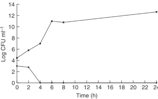
All tested strains were susceptible toD. quadricepsvenom. The values of MIC and MBC obtained in this study are shown in Table 1. The experiments to investigate DqV action mechanism against this bacterium were performed withStaph. aureusATCC 6538P strain, the most sensitive among all tested strains. Treatment with DqV (125lg ml 1- MBC) was able to kill all the bacteria after
4 h of incubation, as shown in Fig. 1. Additionally, the uptake of crystal violet by Staph. aureus was 47% in the absence of DqV, but increased to 60–68% after treatment with MIC, MBC and 29MBC of DqV, comparable with the chelating agent (disodium EDTA, 025 mol l 1), in which case the uptake increased to 60–65% (Fig. 2). After treatment with MIC, MBC and 29MBC of DqV, the optical density at absorbance of 260 nm increased in all concentrations. These results suggest that DqV damages the cytoplasmic membrane and causes release of genetic material (Fig. 3). A pH-dependent inhibitory effect of DqV over Staph. aureus was observed. The lowest MIC was found in alkaline pH (75–90), as shown in Table 2.
Table 1 Minimum inhibitory concentration and minimum bactericidal concentration ofDinoponera quadricepsvenom against Staphylococ-cus aureusstrains
Strains MRSA
D. quadricepsvenom
Amikacin MIC
(lg ml 1)
MBC (lg ml 1)
MIC (lg ml 1)
Staphylococcus aureus
ATCC 6538P
No 625 125 312
Staphylococcus aureus
CCBH 5330
Yes 125 50 312
Staphylococcus aureus
ATCC 33591
Yes 100 100 25
According to atomic force microscopy, the morphology of untreated Staph. aureus was regular and smooth with an intact cell membrane, disclosing its characteristic shape, resembling a ‘brunch of grapes’. Fig. 4B,C shows the loss of membrane integrity and cell-to-cell adherence after 4 h of treatment with MIC and MIC/2 of DqV (Fig. 4).
Discussion
Different components found in animal venoms have been studied and identified as important biological active com-pounds (Pimenta and Lima 2005). These substances have great biotechnological interest, as they are useful to iden-tify new therapeutic targets to treat infections, as well molecular models to development of new drugs, with improvement in efficacy and toxicity.
Several fractions with antimicrobial properties have been isolated from Hymenoptera venoms. Peptides from the venom ofAgelaia pallipes pallipes (Protonectin),Vespa crab-ro(Crabolin),Anoplius samariensis(Anoplin),Apis mellifera (Melittin), Melecta albifrons (Melectin), Polybia paulista (polybia-MP1 and polybia-CP),Vespa tropica (Mastoparan and Vespid chemotactic peptides), Tetramorium bicarina-tum (Bicarinalin), Panurgus calcaratus (Panurgines) have shown antimicrobial activity (Krishnakumari and Nagaraj 1997; Konno et al. 2001; Bucki et al. 2004; Mendes et al. 2004; Cerovsky et al. 2008; Riffletet al. 2012; Wang et al. 2012, 2013b; Cujov a et al. 2013; Yang et al. 2013). Alka-loids from the venom of theSolenopsis invictaant and anti-microbial peptides (pilosulins) found in the venom of the Australian jumper antMyrmecia pilosula exhibit antibacte-rial activity against Gram-negative and Gram-positive bac-teria (Jouvenazet al.1972; Inagakiet al.2004).
Fifteen different antimicrobial peptides (AMPs), called ponericins, were purified from Pachycondyla goeldii venom, and their amino acid sequences were character-ized. According to their first most frequent aminotermi-nal amino acid, these AMPs can be classified into three families: ponericin G (seven peptides), W (six peptides) and L (two peptides). Ponericins G1 and G3 have a broad antibacterial and antifungal spectrum. With exception of W6, ponericins W are active against Gram-positive and Gram-negative bacteria and yeast and have expressive haemolytic and insecticidal actions. Ponericin L2 are active only against bacteria, but not against fungi (Orivel et al. 2001). Recently, six peptides (dinoponeratoxins) were isolated from the venom of Di-noponera australis. Two of them (Da-3105 and Da-3177) showed 929% sequence similarity with ponericin G2 (Johnsonet al.2010).
0 2 4 6 8 10 12 14 16 18 20 22 24
0 2 4 6 8 10 12 14
Time (h)
Log CFU ml
–1
Figure 1 Effect ofDinoponera quadricepsvenom onStaphylococcus aureus ATCC 6538P viability. (125lg ml 1: minimum bactericidal
concentration of Dinoponera quadriceps venom; control: untreated cells). Control; 125lg ml 1.
Control Amikacin EDTA 6·25 12·5 25
0 20 40 60 80
DqV (µg ml–1)
* * *
% of uptake
Figure 2 Crystal violet uptake ofDinoponera quadriceps venom-trea-tedStaphylococcus aureus ATCC 6538P. (DqV: Dinoponera quadri-cepsvenom; control: cells treated with PBS; EDTA: cells treated with disodium EDTA 025 mol l 1; amikacin: cells treated with amikacin
312lg ml 1). The meanSEM for three replicates is shown,
*P<0.05.
25 12·5 6·25 Control Amikacin 0·00
0·05 0·10 0·15
* * *
DqV (µg ml–1)
OD
260 nm
Figure 3Genetic material release in the supernatants of Staphylococ-cus aureusATCC 6538P treated with 625, 125 and 25lg ml 1of
Cologna et al. (2013) demonstrated the presence of compounds similar to antimicrobial peptides found in venoms of other animals, such as temporin-like, dem-aseptins-like and ponericins-like in D. quadriceps venom. It was also observed a wide variation in the composition of venoms produced by ants from different locations, suggesting that environmental characteristics have great influence on the composition of these venoms. An important fact is that ponericin is one of few common compounds between these ants. It was also observed anti-microbial potential over bacterial and fungi strains in synthetic peptides similar to ponericins obtained after proteomic analysis ofD. quadricepsvenom.
In this study, we demonstrated the antimicrobial effect of DqV against three different Staph. aureusstrains, sug-gesting the presence of antimicrobial peptides in this venom.
Recently, our group produced a D. quadriceps venom gland cDNA library to investigate the toxin repertoire in the venom used in this work. It was observed that dinopon-eratoxins are major components, but also peptides pilosu-lin-like were found, and these peptides reported primarily for its antimicrobial effect (Torreset al. 2014). However, these antimicrobial peptides have been subjected to further analysis and molecular modelling in order to reduce its toxic effects, such as the pilosulins (Zelezetskyet al.2005).
The greater susceptibility of the bacteria at higher pHs is a valuable characteristic, as it represents good evidence of a possible effectiveness in inhibiting bacterial growth in physiologic pH. Furthermore, this venom was able to completely kill the bacteria after a short incubation time (4 h), suggesting a quick action mechanism, such as membrane leakage. To determine the effect of DqV on bacterial membrane, crystal violet uptake and intracellular
328·3 mV 74·3 mV 509·7 mV
–366·4 mV –67·6 mV
–305·2 mV
1·0 µm
1·0 µm
Height Height 1·0 µm Height 1·0 µm
971·6 nm
–344·5 nm –51·7 nm
211·3 nm 427·6 nm
–91·5 nm
Amplitude Amplitude 1·0 µm Amplitude 1·0 µm
(A) (B) (C)
(a) (b) (c)
Figure 4 Atomic force microscopy images ofStaphylococcus aureusATCC 6538P without exposure toDinoponera quadriceps venom(A and a), after exposure to 625lg ml 1ofD. quadriceps venom(B and b) and after exposure to 312lg ml 1ofD. quadriceps venom(C and c).
Ampli-tude images show the morphology of cells in A, B and C. The scale bar in volts corresponds to changes in the oscillation ampliAmpli-tude of the free cantilever. The three-dimensional aspect of the groups is shown in a, b and c.
Table 2 Influence of pH on minimum inhibitory concentration ofDinoponera quadricepsvenom againstStaphylococcus aureusATCC 6538P
pH 50 55 60 65 70 75 80 85 90
DqV MIC (lg ml 1) 125 125 125 125 125 625 625 625 625
content release assays were performed. The crystal violet method verifies the increase in membrane permeability through its uptake after incubation with DqV. Crystal violet has low penetrability in normal cells, but in the presence of membrane lesions, it accumulates in micro-organisms and other cells. Releasing of intracellular con-tent was determined by increase in absorbance at 260 nm in culture supernatant after 30 min. DqV caused an apparent alteration in the structure of Staph. aureus membrane, as about 60% of crystal violet uptake was observed, as well significant release of DNA and RNA contents. These effects suggest that DqV cause rupture or formation of pores in Staph. aureus membrane. Amika-cin, an antimicrobial which does not act on membrane molecules, did not show any effect in these experiments. Usually, over 50% of amino acids found in antimicrobial peptides are hydrophobic, which implicates in their inter-action with cell membranes in different micro-organisms (Hancock and Diamond 2000; Teixeiraet al.2012).
Images obtained by AFM confirmed that DqV caused important changes in Staph. aureus membrane structure, as well loss of cell-to-cell adherence. Other Hymenoptera-isolated antimicrobial peptides such as melittin (Leeet al. 2013), polybia-MPI (Wang et al. 2013b) polybia-CP (Wang et al. 2012) and protonectin (Wang et al. 2013a) also show important effects on microbial membranes.
In conclusion, D. quadriceps venom showed potential antibacterial activity against Staphy. aureus strains. The venom was able to alter membrane permeability at bacte-riostatic and bactericidal concentrations. DqV is therefore good candidates for future researches.
Acknowledgements
We thank National Council of Technological and Scien-tific Development (CNPq) and Coordination for Enhancement of Higher Education Personnel (CAPES) for their financial support.
Conflict of interest
No conflict of interest declared.
References
Akre, R.D. and Reed, H.C. (2002) Ants, Bees, and Wasps (Hymenoptera), Chapter 19. InMedical Veterinary Entomologyed. Mullen, G. and Durden, L. pp 383–408. Pullman, WA: Elsevier Science.
Bandyopadhyay, S., Lee, M., Sivaraman, J. and Chatterjee, C. (2013) Model membrane interaction and DNA-binding of antimicrobial peptide Lasioglossin II derived from bee venom.Biochem Biophys Res Commun430, 1–6.
Braga, P.C. and Ricci, D. (1998) Atomic force microscopy: application to investigation ofEscherichia colimorphology before and after exposure to cefodizime.Antimicrob Agents Chemother42, 18–22.
Bucki, R., Pastore, J.J., Randhawa, P., Vegners, R., Weiner, D.J. and Janmey, P.A. (2004) Antibacterial activities of rhodamine B-conjugated gelsolin-derived peptides compared to those of the antimicrobial peptides cathelicidin LL37, magainin II, and melittin.Antimicrob Agents Chemother48, 1526–1533.
Cerovsky, V., Hovorka, O., Cvacka, J., Voburka, Z.,
Bednarova, L., Borovickova, L., Slaninova, J. and Fucık, V. (2008) Melectin: a novel antimicrobial peptide from the venom of the cleptoparasitic beeMelecta albifrons. ChemBioChem9, 2815–2821.
Chen, J. and Lariviere, W.R. (2010) The nociceptive and anti-nociceptive effects of bee venom injection and therapy: a double-edged sword.Prog Neurobiol92, 151
–183.
CLSI (2009)Performance Standards for Antimicrobial
Susceptibility Testing; 18th Informational Supplement. CLSI M07-A8. Wayne, PA: Clinical and Laboratory Standards Institute, pp. 15–18.
Cologna, C.T., Cardoso, J.D., Jourdan, E., Degueldre, M., Upert, G., Gilles, N., Uetanabaro, A.P., Costa Neto, E.M. et al.(2013) Peptidomic comparison and characterization of the major components of the venom of the giant ant Dinoponera quadriceps collected in four different areas of Brazil.J Proteomics94C, 413
–422.
Cujova, S., Slaninova, J., Monincova, L., Fucık, V., Bednarova, L.,Stokrova, J., Hovorka, O., Voburka, Z.et al.(2013) Panurgines, novel antimicrobial peptides from the venom of communal beePanurgus calcaratus(Hymenoptera: Andrenidae).Amino Acids45, 143–157.
Devi, K.P., Nisha, S.A., Sakthivel, R. and Pandain, S.K. (2010) Eugenol (an essential oil of clove) acts as antibacterial agent againstSalmonella typhiby disrupting the cellular membrane.J Ethnopharmacol130, 107–115.
Goncßalves, T.B., Braga, M.A., de Oliveira, F.F., Santiago, G.M., Carvalho, C.B., Brito e Cabral, P., de Melo Santiago, T., Sousa, J.S.et al.(2012) Effect of subinihibitory and inhibitory concentrations ofPlectranthus amboinicus (Lour.) Spreng essential oil onKlebsiella pneumoniae. Phytomedicine19, 962–968.
Hancock, R.E.W. and Diamond, G. (2000) The role of cationic antimicrobial peptides in innate host defences.Trends Microbiol8, 402–410.
Heinemann, J.A., Ankenbauer, R.G. and Amabile-Cuevas, C.F. (2000) Do antibiotics maintain antibiotic resistance?Drug Discov Today5, 195–204.
Inagaki, H., Akagi, M., Imai, H.T., Taylor, R.W. and Kubo, T. (2004) Molecular cloning and biological
characterization of novel antimicrobial peptides, pilosulin 3 and pilosulin 4, from a species of the Australian ant genus Myrmecia. Arch Biochem Biophys 428, 170
Johnson, S.R., Copello, J.A., Evans, M.S. and Suarez, A.V. (2010) A biochemical characterization of the major peptides from the venom of the giant neotropical hunting antDinoponera australis.Toxicon55, 702–710.
Jouvenaz, D.P., Blum, M.S. and MacConnell, J.G. (1972) Antibacterial activity of venom alkaloids from the imported fire ant, Solenopsis invicta Buren.Antimicrob Agents Chemother2, 291
–293.
Kaneti, G., Sarig, H., Marjieh, I., Fadia, Z. and Mor, A. (2013) Simultaneous breakdown of multiple antibiotic resistance mechanisms inS. aureus.FASEB J12, 4834–4843.
Klein, E., Smith, D.L. and Laxminarayan, R. (2009)
Community-associated methicillin resistantStaphylococcus aureusin outpatients, United States, 1999–2006.Emerg
Infect Dis15, 1925–1930.
Konno, K., Hisada, M., Fontana, R., Lorenzi, C.C., Naoki, H., Itagaki, Y., Miwa, A., Kawai, N.et al.(2001) Anoplin, a novel antimicrobial peptide from the venom of the solitary waspAnoplius samariensis.Biochim Biophys Acta 1550, 70–80.
Krishnakumari, V. and Nagaraj, R. (1997) Antimicrobial and hemolytic activities of crabolin, a 13-residue peptide from the venom of the European hornet,Vespa crabro,and its analogs.J Pept Res50, 88
–93.
Lee, M.T., Sun, T.L., Hung, W.C. and Huang, H.W. (2013) Process of inducing pores in membranes by melittin.Proc Natl Acad Sci U S A110, 14243–14248.
Lopes, K.S., Rios, E.R., Lima, C.N., Linhares, M.I., Torres, A.F.C., Havt, A., Quinet, Y.P., Fonteles, M.M.et al.(2013) The effects of the Brazilian antDinoponera quadriceps venom on chemically induced seizure models.Neurochem Int63, 141–145.
Mendes, M.A., de Souza, B.M., Marques, M.R. and Palma, M.S. (2004) Structural and biological characterization of two novel peptides from the venom of the neotropical social waspAgelaia ‘pallipes pallipes.Toxicon44, 67–74.
Moreau, S.J.M. (2013) ‘It stings a bit but it cleans well’: venoms of Hymenoptera and their antimicrobial potential. J Insect Physiol59, 186
–204.
Orivel, J., Redeker, V., Le Caer, J.P., Krier, F., Revol-Junelles, A.M., Longeon, A., Chaffotte, A., Dejean, A.et al.(2001) Ponericins, new antibacterial and insecticidal peptides from the venom of the antPachycondyla goeldii.J Biol Chem276, 17823
–17829.
Paiva, R.V.S. and Brand~ao, C.R.F. (1995) Nests, worker population, and reproductive status of workers, in the giant queenless ponerine ant Dinoponera Roger (Hymenoptera: Formicidae).Ethol Ecol Evol7, 297–312.
Palma, M.S. (2006) Insect venom peptides, Chapter 56. In: Handbook of Biologically Active Peptidesed. Kastin, A. pp. 389–396. Los Angeles, CA: Elsevier.
Pearson, R.D., Steigbigel, R.T., Davis, H.T. and Chapman, S.W. (1980) Method of reliable determination of minimal lethal antibiotic concentrations.Antimicrob Agents Chemother18, 699
–708.
Pimenta, A.M.C. and Lima, M.E. (2005) Small peptides, big world: biotechnological potential in neglected bioactive peptides from arthropod venoms.J Pept Sci11, 670–676.
Pittet, D. (2005) Infection control and quality health care in the new millennium.Am J Infect Control33, 258–267.
Plata, K., Rosato, A.E. and Wegrzyn, G. (2009)Staphylococcus aureusas an infectious agent: overview of biochemistry and molecular genetics of its pathogenicity.Acta Biochim Pol56, 597–612.
Quinet, Y., Vieira, R.H.S.F., Sousa, M.R., Evangelista-Barreto, N.S., Carvalho, F.C.T., Guedes, M.I.F., Alves, C.R., de Biseau, J.C.et al.(2012) Antibacterial properties of contact defensive secretions in neotropicalCrematogaster ants.J Venom Anim Toxins Incl Trop Dis18, 441–445.
Rifflet, A., Gavalda, S., Tene, N., Orivel, J., Leprince, J., Guilhaudis, L., Genin, E., Vetillard, A.et al.(2012) Identification and characterization of a novel antimicrobial peptide from the venom of the ant Tetramorium bicarinatum.Peptides38, 363–370.
Souza, P.L., Quinet, Y., Ponte, E.L., Vale, J.F., Torres, A.F.C., Pereira, M.G. and Assreuy, A.M.S. (2012) Venom’s antinociceptive property in the primitive antDinoponera quadriceps.J Ethnopharmacol144, 213
–216.
Teixeira, V., Feio, M.J. and Bastos, M. (2012) Role of lipids in the interaction of antimicrobial peptides with membranes. Prog Lipid Res51, 149–177.
Torres, A.F., Huang, C., Chong, C.M., Leung, S.W., Prieto-da-Silva, A.R., Havt, A., Quinet, Y.P., Martins, A.M.et al. (2014) Transcriptome analysis in venom gland of the predatory giant ant Dinoponera quadriceps: insights into the polypeptide toxin arsenal of hymenopterans.PLoS ONE9, e87556.
Wang, K., Yan, J., Chen, R., Dang, W., Zhang, B., Zhang, W., Song, J. and Wang, R. (2012) Membrane-active action mode of polybia-CP, a novel antimicrobial peptide isolated from the venom ofPolybia paulista.Antimicrob Agents Chemother56, 3318–3323.
Wang, K., Dang, W., Yan, J., Chen, R., Liu, X., Yan, W., Zhang, B., Xie, J.et al.(2013a) Membrane perturbation action mode and structure-activity relationships of protonectin, a novel antimicrobial peptide from the venom of neotropical social waspAgelaia pallipes pallipes. Antimicrob Agents Chemother57, 4632–4639.
Wang, K., Yan, J., Dang, W., Liu, X., Chen, R., Zhang, J., Zhang, B., Zhang, W.et al.(2013b) Membrane active antimicrobial activity and molecular dynamics study of a novel cationic antimicrobial peptide polybia-MPI, from the venom of Polybia paulista.Peptides39, 80–88.
Yang, X., Wang, Y., Lee, W.H. and Zhang, Y.
(2013) Antimicrobial peptides from the venom gland of the social waspVespa tropica.Toxicon74, 151
–157.
Zelezetsky, I., Pag, U., Antcheva, N., Sahl, H.G. and Tossi, A. (2005) Identification and optimization of an antimicrobial peptide from the ant venom toxin pilosulin.Arch Biochem Biophys434, 358