INTERNATIONAL ARCHIVESOF MEDICINE SECTION: ANATOMIC PATHOLOGY
ISSN: 1755-7682 International
Medical Society http://imedicalsociety.org
1
2017
Vol. 10 No. 190 doi: 10.3823/2460
© Under License of Creative Commons Attribution 3.0 License This article is available at: www.intarchmed.com and www.medbrary.com
Abstract
Introduction: Congenital diaphragmatic hernia is a poor forma-tion of the diaphragm characterized by the presence of an intestinal malrotation, It is related to abnormal position of the intestine in the thorax.
Objective and Result: This case report a stillborn at 32 weeks, that was observe anatomical abnormalities associated with congenital dia-phragmatic hernia, which occurred in the left antero-posterior region, such as intestinal malrotation, hepatomegaly and nephromegaly, with the presence of a hernial ring that occupied 80% of the left side of the diaphragm, besides a hypertrophied heart, deviated to the right, bilateral pulmonary hypotrophy.
Conclusion This case illustrates a rare case of diaphragmatic hernia with intestinal changes of clinical and surgical importance.
Congenital Diaphragmatic Hernia Associated with
Uncommon Abnormalities
CASE REPORT
Helson Freitas da Silveira1, Howard Lopes Ribeiro Junior2, Jalles Dantas De Lucena1, Osvaldo Pereira da Costa Sobrinho2, Luane Macedo de Sousa1, Kalina Kelma Oliviera de Sousa2, João Erivan Façanha Barreto2, Roberta Silva Pessoa2, Ariel Gustavo Scafuri1,2, Delane Viana Gondim1,2, Gilberto Santos Cerqueira1,3
1 Post-Graduate Program in
Morphofunctional Sciences, Federal University of Ceara, Fortaleza, Ceara, Brazil.
2 Department of Morphology, Federal University of Ceará, Fortaleza, Ceara, Brazil.
3 Department of Medicine, Federal University of Piauí, Parnaíba, Piaui, Brazil.
Contact information:
Gilberto Santos Cerqueira.
Address: Av. Delmiro de Farias, s/n. Campus Porangabussu- Fortaleza/Ceará, Brazil. CEP: 64204-035.
Tel: +55 (85) 98648-9899.
[email protected]Keywords
Anatomy; Hernia; Medicine; Congenital Hernia.
INTERNATIONAL ARCHIVESOF MEDICINE SECTION: ANATOMIC PATHOLOGY
ISSN: 1755-7682
2017
Vol. 10 No. 190 doi: 10.3823/2460
This article is available at: www.intarchmed.com and www.medbrary.com
2
the umbilical cord hernia and definitive fixation. [2] Over 80% of reported cases are diagnosed as pos-tero-lateral left-side CDH being reported that 40-50% of patients are affected with other congenital malformations. [3]
We report a stillbirth that presented unusual ab-normalities (i.e. cardiac hypertrophy, several pulmo-nary hypoplasia, hepatomegaly, nephromagaly and cranial splenic torsion) associated to anteroposterior left-side CDH with IM.
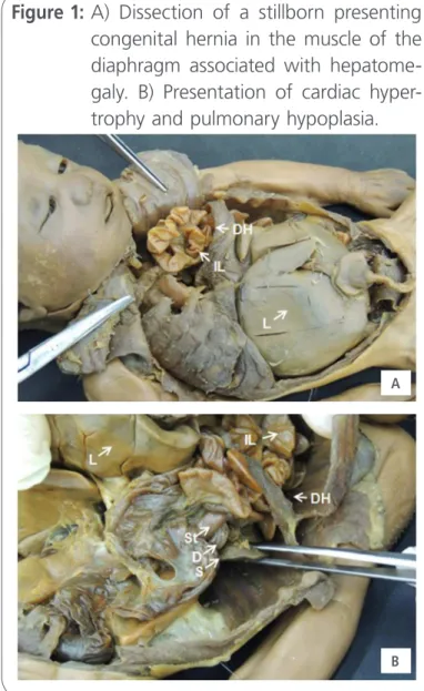
A 32-weeks-old boy stillborn, at autopsy, was diagnosed with a clinical condition of large ante-roposterior left-side CDH associated with IM and the presence of five unusual abnormalities. First, after dissection of the thoracic cage, was possible to visualize the intestinal loops displaced from the abdomen to the thorax as from the congenital her-nia in the diaphragm muscle (Figure 1A). The right diaphragmatic cupula is normal. In the mediastinum is possible to visualize the heart slightly deflected to the right, later covered by the pericardium, presen-ting cardiac hypertrophy when compared to fetal age (Figure 1B).
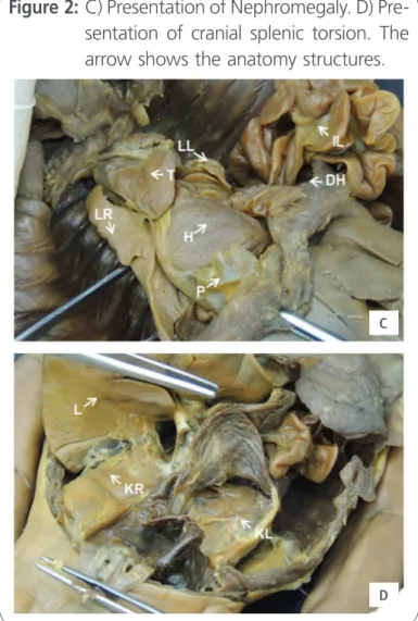
Additionally, laterally to mediastinum, it is possi-ble to visualize the pulmonary hypoplasia, charac-terized more sternly in the left lung, and being still possible to identify the oblique fissure and the pul-monary lingula (Figure 1B). After dissection of the abdomen, was possible to identify a hepatomegaly and nephromegaly severe condition and having the liver and kidneys almost totally occupying the ab-dominal cavity (Figure 1A, 1B and 2C, respectively). Additionally, we verified the presence of a cranial splenic torsion toward the CDH (Figure 2D). It was also possible to identify the ileocecal junction in the upper left quadrant of the abdominal cavity (Figure 2D). The large intestine was located in the left me-dial region of the abdominal cavity being possible to identify the descending and sigmoid colon in anato-mical position. It is possible to identify the umbilical vein moving toward the umbilicus, was continued as the umbilical cord (Figure 2D).
To the best of our knowledge, this is the first case report which identifies the presence of cardiac hypertrophy, several pulmonary hypoplasia, hepa-tomegaly, nephromagaly and cranial splenic torsion associated to anteroposterior left-side CDH with IM in a stillborn. In this report we note that the con-genital diaphragmatic hernia can be seen that the intrathoracic displacement of the intestinal loops im-pairs cardiac development and lung often resulting in neonatal death by respiratory insufficiency due to pulmonary hypoplasia and hypertension. Additional comorbidities, such as those cited in this study, may have further aggravated the gestational develop-ment of the fetus. It is important to develop-mention that
Figure 1: A) Dissection of a stillborn presenting congenital hernia in the muscle of the diaphragm associated with hepatome-galy. B) Presentation of cardiac hyper-trophy and pulmonary hypoplasia.
A
INTERNATIONAL ARCHIVESOF MEDICINE SECTION: ANATOMIC PATHOLOGY
ISSN: 1755-7682
2017
Vol. 10 No. 190 doi: 10.3823/2460
© Under License of Creative Commons Attribution 3.0 License
3
recent advances in fetal surgery have developed new surgical methods for increasing the survival of the newborn especially in relation to pulmonary hypoplasia. [4]
This case illustrates a rare case of diaphragmatic hernia with intestinal changes of clinical and sur-gical importance. Thus, this report demonstrates the importance of knowledge of diaphragmatic congenital malformations for the surgical planning of perinatal urgency, describing parameters to aid in the repair of acute diaphragmatic lesions using laparoscopy or conventional surgeries, as well as an anatomic-topographic description.
Abbreviations
L: Liver; DH: Diaphragmatic Hernia; IL: Intestinal Loop; S: Spleen; St: Stomach; D: Duodenum; H: Heart; P: Pericardium; LL: Lung Left; LR: Lung Right; T: Thyme; DC - Descending Colon; SC: Sigmoid Co-lon
Declarations of Interests
The authors declare that they have no competing interests.
Financial support No.
References
1 Salmanian B, Shamshirsaz AA, Cass DL, Javadian P, Ruano R, Ayres NA, Mehollin-Ray A, Belfort MA. Fetal cardiac tamponade in a case of right-side congenital diaphragmatic hernia. Obstet Gynecol. 2014 Feb; 123(2 Pt 2 Suppl 2):447-50. http://journals. lww.com/greenjournal/pages/articleviewer.aspx?year=2014&iss ue=02002&article=00012&type=abstract
2 Baoquan Q, Diez-Pardo JA, Tovar JA. Intestinal rotation in experimental congenital diaphragmatic hernia. J Pediatr Surg. 1995 Oct; 30(10):1457-62.
3 Bohn, D. Congenital Diafragmatic Hernia. Am J Respir Crit Care Med 2002 (166): 911-915. http://www.atsjournals.org/doi/ abs/10.1164/rccm.200204-304CC
4 Yao W, Elangovan H, Nicolaides K. Design of a flexible fetoscopy manipulation system for congenital diaphragmatic hernia. Med Eng Phys. 2014 Jan;36(1):32-8. Epub 2013 Sep 24. https:// fetalmedicine.org/var/pdf/publications/760.pdf
Figure 2: C) Presentation of Nephromegaly. D) Pre-sentation of cranial splenic torsion. The arrow shows the anatomy structures.
C
D
International Archives of Medicine is an open access journal publishing articles encompassing all aspects of medical scien-ce and clinical practiscien-ce. IAM is considered a megajournal with independent sections on all areas of medicine. IAM is a really international journal with authors and board members from all around the world. The journal is widely indexed and classified Q2 in category Medicine.