Morphological prognostic factors in nosocomial pneumonia:
an autopsy study*
Determinantes morfológicos de prognóstico em
pneumonia nosocomial: um estudo em autópsias
Luiz Mário Baptista Martinelli, Paulo José Fortes Villas Boas, Thais Thomaz Queluz, Hugo Hyung Bok Yoo
Abstract
Objective: To determine the prevalence of nosocomial pneumonia in autopsies at a public university hospital; to identify the risk factors for nosocomial pneumonia and the potential prognostic factors associated with fatal nosocomial pneumonia and with fatal aspiration pneumonia; and to determine whether anatomopathological findings correlate with nosocomial pneumonia or aspiration pneumonia. Methods: A retrospective study involving 199 autopsied patients, older than 1 year of age, who had been admitted to the São Paulo State University Botucatu School of Medicine Hospital das Clínicas and died of nosocomial pneumonia (underlying or contributing cause), between 1999 and 2006. Demographic, clinical and anatomopathological variables were tested regarding their association with the outcomes (fatal nosocomial pneumonia and fatal aspiration pneumonia). The significant variables were analyzed using multivariate analysis. Results: The mean age was 59 ± 19 years. The prevalence of nosocomial pneumonia in autopsies was 29%, and the disease was the cause of death in 22.6% of the autopsied patients. Fatal nosocomial pneumonia correlated with the following anatomopathological findings: tobacco-associated structural lesions (OR = 3.23; 95% CI: 1.26-2.95; p = 0.02) and bilateral pneumonia (OR = 3.23; 95% CI: 1.26-8.30; p = 0.01). None of the variables were found to be significantly associated with fatal aspiration pneumonia. Conclusions: In our sample, there was a high prevalence of nosocomial pneumonia, which was responsible for almost 25% of all of the deaths. Smoking-related structural lesions and bilateral pneumonia all favored mortality. These findings corroborate the results of various clinical studies on nosocomial pneumonia.
Keywords: Autopsy; Risk factors; Prognosis; Pneumonia, aspiration; Pneumonia/mortality.
Resumo
Objetivo: Determinar a prevalência de pneumonia nosocomial nas autópsias em um hospital público universi-tário; identificar os fatores de risco relacionados à pneumonia nosocomial e os potenciais fatores prognósticos relacionados à ocorrência de pneumonia nosocomial fatal; e correlacionar os achados anatomopatológicos com a ocorrência de pneumonia nosocomial e/ou pneumonia aspirativa. Métodos: Estudo retrospectivo de 199 pacientes autopsiados, maiores de 1 ano de idade, internados no Hospital das Clínicas da Faculdade de Medicina de Botucatu da Universidade Estadual Paulista entre 1999 e 2006, cuja causa de morte (causa básica ou associada) foi pneumonia nosocomial. Testou-se a associação dos dados demográficos, clínicos e anatomopatológicos com os desfechos pneumonia nosocomial fatal e pneumonia aspirativa fatal. As variáveis significativas entraram na análise multivariada. Resultados: A idade média foi de 59 ± 19 anos. A prevalência de pneumonia nosocomial em autópsias foi 29%, e essa foi a causa mortis de 22,6% dos pacientes autopsiados. A pneumonia nosocomial fatal correlacionou-se com os achados anatomopatológicos de alterações estruturais tabágicas (OR = 3,23; IC95%: 1,26-2,95; p = 0,02) e acometimento pulmonar bilateral (OR = 3,23; IC95%: 1,26-8,30; p = 0,01). Não houve asso-ciações significativas entre as variáveis e pneumonia aspirativa fatal. Conclusões: Em nossa amostra, a pneumonia nosocomial teve prevalência elevada e foi responsável por quase 25% das mortes. A mortalidade é favorecida por alterações estruturais tabágicas e pneumonia bilateral. Esses achados corroboram os resultados de diversos estudos clínicos sobre pneumonia nosocomial.
Descritores: Autopsia; Fatores de risco; Prognóstico; Pneumonia aspirativa; Pneumonia/mortalidade.
* Study carried out at the Botucatu School of Medicine, Universidade Estadual Paulista – UNESP, São Paulo State University – Botucatu, Brazil.
Correspondence to: Hugo Hyung Bok Yoo. Disciplina de Pneumologia do Departamento de Clínica Médica, Faculdade de Medicina de Botucatu - UNESP, CEP 18618-970, Botucatu, SP, Brasil.
Tel 55 14 3882-2969. E-mail: hugo@fmb.unesp.br
Financial support: This study received financial support in the form of a Young Investigator Grant from the Fundação de Amparo à Pesquisa do Estado de São Paulo (FAPESP, Foundation for the Support of Research in the State of São Paulo; grant no. 07/51267-6).
Paulista (UNESP, São Paulo State University) Botucatu School of Medicine Hospital das Clínicas and died of NP (underlying or contrib-uting cause), between January of 1999 and December of 2006.
The autopsy studies were performed jointly by professors and residents of the Department of Pathology, following a well-established, traditional routine, consisting of sequential and systematic procedures, by means of which the topography and alterations of the organs are analyzed in locus, followed by dissection and detailed analysis of the organs.
The study was based on the autopsy reports, in which pneumonia was macroscopically or microscopically documented. The patient charts were reviewed, and those indicating a diag-nosis of NP were selected. From the charts and the autopsy reports, the following data were extracted:
• Demographic data: gender, age and ethnicity
• Clinical data: hospital ward of origin and associated diseases
• Anatomopathological data: location of the pneumonia and diagnosis of the remaining alterations observed
The cases of NP were subdivided into fatal, when NP was the principal cause of death (i.e., when no other cause of death was found), and nonfatal, when the immediate cause of death was another disease. The cases in which there was evidence of aspiration (presence of vegetal cells or muscle fibers within the pulmonary alveoli with neutrophilic inflammatory infiltrate) were designated aspiration NP (ANP) and were also subdivided into fatal and nonfatal, according to the criteria cited above.
Due to the great quantity of clinical data collected, the most common data were selected, and some data were grouped by the systems affected, becoming the following clinical variables:
• Diabetes mellitus
• Diseases of the circulatory system (systemic arterial hypertension, congestive heart failure, acute myocardial infarction and stroke)
• Respiratory diseases (COPD and interstitial lung disease)
• Alcoholism • Renal failure
Introduction
Nosocomial pneumonia (NP) is defined as the development of pneumonia, which was not incubating at the time of hospital admission, at least 48 h after the patient has been admitted to hospital. It is currently the most common noso-comial infection, accounting for 13-18% of all such infections in Brazil, as well as for the highest morbidity and mortality rates, the latter being as high as 60%.(1,2) In ICUs, the mortality rate among patients with NP is approximately 50%, whereas that among patients without pulmo-nary infection is drastically lower (3.5%).(3)
According to the National Nosocomial Infection Surveillance System of the United States, the principal risk factors for the development of NP are as follows: endotracheal intubation or mechanical ventilation; a drop in the level of consciousness; COPD; age > 70 years; and aspi-ration of microorganisms from the oropharynx, the principal entry point for bacteria to reach the lower respiratory tract.(4,5)
In contrast, the contributing causes of aspiration, the most relevant consequence of which is aspiration pneumonia (AP), followed by the supine position, abnormalities in swal-lowing mechanisms leading to dysphagia, a drop in the level of consciousness, enteral nutri-tion, instrumentation of the respiratory tract and instrumentation of the gastrointestinal tract.(4-8) Therefore, it is believed that aspiration is the leading cause of ICU-acquired NP.(5,6)
Although pneumonia is the principal infec-tious disease found in autopsies,(9) there have been no autopsy studies determining whether anatomopathological findings correlate with NP. Aspiration, which is histologically documented by the presence of vegetal cells or muscle fibers within the pulmonary alveoli with neutrophilic inflammatory infiltrate, deserves special atten-tion due to its key role in causing NP. The objectives of the present study were as follows: 1) to determine the prevalence of NP in autop-sies at a public teaching hospital; 2) to identify the risk factors for NP and the potential prog-nostic factors associated with fatal NP; and 3) to determine whether anatomopathological find-ings correlate with NP or AP.
Methods
(n = 30). Each of these groups was subsequently subdivided into fatal and nonfatal cases.
The study design was approved by the Research Ethics Committee of the UNESP Botucatu School of Medicine.
The data obtained were initially described in terms of discrete or continuous quantitative variables and transcribed into an instrument developed to store them.
The mean ages of the groups were compared by means of the Student’s t-test. To perform a preliminary exploratory analysis, the vari-ables were transformed into binary varivari-ables. Subsequently, the association of the variables with the outcomes fatal nANP and fatal ANP was tested by the chi-square test (OR), applied to each variable separately. Only the variables that had a significant effect on the outcomes (p < 0.05) were maintained. The multivariate analysis with logistic regression was performed by introducing each variable in the model in decreasing order, one by one. In the final model, only the variables that were statistically related to the outcomes were maintained. The set of steps described above allowed the analysis of confounding and interacting variables.
To calculate the comorbidities/patient ratio, all of the comorbidities of the patients were summed and divided by the number of patients in the group.
Results
During the period under study, there were 6,016 deaths, 765 of which (12.7%) were submitted to autopsy. The prevalence of NP in autopsies was 29%.
The frequencies of the demographic and clinical variables studied are shown in Table 1. Among the 199 cases studied, the mean age was 59 ± 19 years; the mean length of hospital stay was 15 ± 22 days; the mean length of use of a nasogastric feeding tube was 7 ± 13 days; the mean length of stay on mechanical ventilation was 6 ± 12 days.
Table 2 shows the frequencies of the prin-cipal autopsy findings. Fatal nANP was found in 26 (13.1%) of the cases, and fatal ANP was found in 19 (9.5%) of the cases.
The mean numbers of comorbidities per patient, according to the hospital ward of origin, were as follows: 3.3 comorbidities/patient in the clinical ward; 3.5 comorbidities/patient • Neoplasia
• Smoking • Tracheostomy
• Use of a nasogastric tube
• Use of antacids or proton pump inhibitors • Mechanical ventilation
Similarly, the anatomopathological findings that had a frequency ≥ 5 in each of the systems studied were selected and grouped as follows:
• Lesions caused by orotracheal intubation: acute erosive tracheitis; erosive laryngitis; tracheal edema; ulcerations of the trachea; ulcerations of the larynx; contact ulcers of the larynx; ulcers of the trachea; and ulcers of the larynx, all of which were consi-dered part of this group when the patient reported orotracheal intubation
• Signs suggestive of pulmonary throm -boembolism: pulmonary embolism or pulmonary artery embolism; pulmonary microembolism; and pulmonary infarction • Signs suggestive of diffuse alveolar
damage: diffuse alveolar damage, ARDS; and interstitial damage
• Smoking-related structural lesions: emphy -sema; chronic bronchitis; bronchiectasis; and anthracosis (when the patient had a history of smoking)
• Bronchial disease: presence of a secretion plug; increased local secretion; architec-tural distortion; and bronchiectasis when the patient had no history of smoking • Signs of previous tuberculosis: scars, cavi
-tations and pulmonary fibrosis in patients with a history of tuberculosis
• Signs of inflammation of the digestive tract: esophagitis; gastritis; duodenitis; peptic ulcer; ileitis; and colitis
• Changes in the central nervous system: cortical atrophy; stroke (ischemic or hemorrhagic); diffuse cerebral softening; cortical depression; and tumors
Table 4 presents the results for the variables after multivariate analysis to test the degree of association with fatal nANP.
Discussion
The present study was carried out at the Botucatu School of Medicine Hospital das Clínicas, the largest public institution of the Unified Health Care System in the central-west region of the state of São Paulo. It is a tertiary teaching hospital and a referral hospital that treats a large number of patients with severe diseases, patients with chronic diseases and in the surgical ward; and 3.3 comorbidities/
patient in the ICU. There were few differences between the mean quantity of comorbidities/ patient when the hospital ward of origin was related to groups nANP and ANP. The following values were observed, respectively: clinical ward, 3.3 and 3.1 comorbidities/patient; surgical ward, 3.5 and 3.5 comorbidities/patient; and ICU, 3.2 and 3.5 comorbidities/patient. Patients with nANP who were over the age of 60 presented, on average, 11.3 comorbidities/ patient; patients with nANP who were under the age of 60 presented, on average, 3.5 comorbidi-ties/patient; patients with ANP who were over the age of 60 presented, on average, 3.9 comor-bidities/patient; and patients with ANP who were under the age of 60 presented, on average, 2.5 comorbidities/patient.
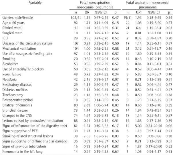
Table 3 shows the principal risk factors and pathological findings in the autopsies that were associated with the outcomes fatal nANP and fatal ANP. The statistical analysis using univariate regression showed that the variables “smoking-related structural lesions” and “bilat-eral pneumonia” correlated with fatal nANP, whereas the variable “hospitalization in clinical ward” correlated with fatal ANP. The variable “ethnicity” was not significantly associated with any of the outcomes (p = 0.96 and p = 0.24 for nANP and ANP, respectively).
Table 1 - Frequencies of the demographic and clinical variables studied.
Variable n %
Gender, M/F 127/72 64.0/36.0
Ethnicity, W/Af/MU/As 159/14/24/2 80.0/7.0/12.0/1.0
Age > 60 years 114 57.0
Clinical ward/surgical ward/ICU 134/20/36 67.0/10.0/18.0
Mechanical ventilation 125 63.0
Diseases of the circulatory system 124 62.0 Use of a nasogastric tube 116 58.0
Smoking 83 41.0
Alcoholism 58 29.0
Renal failure 56 28.0
Use of antacids/H2 blockers 54 27.0
Neoplasia 49 24.5
Respiratory diseases 33 16.5
Diabetes mellitus 33 16.5
Tracheostomy 29 14.5
Postoperative period 27 13.5
W: White; Af: African; MU: Mulatto or undetermined; As: Asian; and H2 blockers: proton pump inhibitors.
Table 2 - Frequencies of the principal alterations found in the autopsies of patients with nosocomial pneumonia.
Alteration n % Bilateral pneumonia 94 47.0 Bronchial disease 92 46.0 Changes in the central nervous system 91 45.7 Lesions caused by orotracheal intubation 84 42.0 Signs of inflammation of the digestive tract 73 36.5 Smoking-related structural lesions 44 22.0 Signs suggestive of pulmonary
thromboembolism
the need for a better understanding of the risk factors for NP and of their mechanisms, as well as the adoption of strong measures to prevent NP. Data from two other studies on the preva-lence of pneumonia in autopsies were similar to those of the present study, ranging from 21.8% to 29.5%.(9,10) In the elderly, infectious pneu-monitis was found in 40.6% of the autopsied cases,(11) and, in a study on ventilator-associated pneumonia,(12) it was reported that NP (identified in autopsies) accounted for 60% of the deaths due to nosocomial infections.
The present study had the objective of filling a gap in the literature, since there have been few autopsy studies of pneumonia, and none of them specifically on the topic of NP. We observed that patients with clinical conditions related to the
various fields of medicine.
The objective of the present study was to contribute to a more accurate identification and better understanding of the risk factors for and prognostic factors of NP (a disease of high inci-dence, morbidity and mortality) by studying a series of 199 autopsies in which the cause of death (underlying or contributing cause) was listed as NP.
Our results show that the prevalence of NP in autopsies was 29%, and that nANP and ANP were, respectively, the principal causes of death in 13.1% and 9.5% of the 199 cases in the study sample. This is a high mortality rate, as previ-ously reported in the literature, which justifies
Table 3 - Analysis of the prognostic factors and autopsy findings related to fatal nonaspiration nosocomial pneumonia and fatal aspiration nosocomial pneumonia.*
Variable Fatal nonaspiration nosocomial pneumonia
Fatal aspiration nosocomial pneumonia n OR 95% CI p n OR 95% CI p Gender, male/female 108/61 1.12 0.47-2.66 0.47 19/11 1.93 0.38-9.69 0.34 Age > 60 years 92 1.71 0.71-4.09 0.15 22 1.05 0.19-5.60 0.63 Clinical ward 113 1.41 0.55-3.59 0.31 21 6.4 1.15-35.4 0.03 Surgical ward 18 1.11 0.29-4.15 0.54 2 0.81 0.61-1.08 0.12 ICU 29 0.85 0.27-2.70 0.52 7 0.32 0.58-1.87 0.20 Diseases of the circulatory system 107 0.91 0.38-2.16 0.50 17 1.14 0.25-5.11 0.57 Mechanical ventilation 104 1.00 0.42-2.36 0.58 21 3.12 0.61-15.7 0.16 Use of a nasogastric feeding tube 97 1.01 0.43-2.36 0.57 19 1.80 0.39-8.34 0.35 Smoking 70 0.86 0.36-2.03 0.45 13 0.48 0.10-2.19 0.28 Alcoholism 53 0.96 0.39-2.39 0.57 5 0.84 0.11-6.03 0.61 Use of antacids/H2 blockers 50 0.85 0.33-2.18 0.47 4 0.52 0.06-4.41 0.47 Renal failure 48 0.72 0.27-1.92 0.34 8 5.83 0.61-55.7 0.10 Neoplasia 42 2.16 0.89-5.24 0.07 7 0.71 0.12-3.99 0.51 Respiratory diseases 29 1.18 0.40-3.44 0.47 4 0.52 0.06-4.41 0.47 Diabetes mellitus 29 1.18 0.40-3.44 0.47 4 0.52 0.64-4.41 0.47 Tracheostomy 23 1.18 0.36-3.82 0.48 6 0.50 0.08-3.06 0.38 Postoperative period 18 0.66 0.14-3.06 0.45 9 1.23 0.23-6.35 0.57 Bilateral pneumonia 80 2.39 1.00-5.74 0.03 14 0.60 0.13-2.70 0.39 Bronchial disease 78 1.44 0.62-3.31 0.26 14 1.94 0.42-8.91 0.31 Changes in the CNS 74 1.64 0.69-3.73 0.18 17 1.14 0.25-5.11 0.57 Lesions caused by orotracheal intubation 68 0.91 0.38-2.16 0.51 16 1.65 0.37-7.36 0.39 Signs of inflammation of the digestive tract 61 1.64 0.70-3.82 0.17 12 5.00 0.84-29.56 0.06 Signs suggestive of PTE 39 1.27 0.49-3.31 0.38 3 1.18 0.97-1.44 0.23 Smoking-related structural lesions 38 2.56 1.05-6.26 0.03 6 0.50 0.08-3.06 0.38 Signs suggestive of diffuse alveolar damage 35 0.89 0.31-2.57 0.53 7 0.71 0.12-3.99 0.51 Signs of previous tuberculosis 15 0.89 0.84-0.94 0.07 4 1.87 0.17-20.60 0.53 Pneumonia in the left lung 14 0.91 0.19-4.32 0.63 1 1.05 0.94-1.17 0.63
might have influenced our findings is the possi-bility that cases of chronic bronchitis/emphysema and other chronic respiratory diseases had not been clinically diagnosed and were only identi-fied during the autopsy. Therefore, only the cases that presented smoking-related structural lesions at autopsy were at increased risk for developing fatal NP, a finding that corroborates the results obtained by another group of authors,(20) who investigated the association between pulmonary structural diseases and the increase in morbidity and mortality in nosocomial bronchopulmonary infections.
The extent of pulmonary involvement influences the survival of patients with pneu-monia, since bilateral and/or multilobar pneumonia increases the risk for mortality in these patients,(10,13,14) a fact that was corrobo-rated by our finding that bilateral NP was associated with death. One group of authors,(17) who studied the risk factors for and prognosis of NP, reported that bilateral pneumonia seen on chest X-rays was as an independent risk factor for unfavorable prognosis. In cases of commu-nity-acquired pneumonia, multilobar infiltrate seems to be related to early failure of the treat-ment for pneumonia, characterizing more severe cases of the disease.(21)
In our study, contrary to what was expected, we found no association between ANP and the location of the pneumonia, probably due to the small number of ANP cases. However, hospitali-zation in a clinical ward, which was associated with fatal ANP in the univariate analysis, was considered a study bias because of the large number of patients who were originally hospi-talized in such wards.
The results of the present study show no association between the outcomes fatal nANP or fatal ANP and other well-known risk factors for NP, such as cardiovascular disease; respira-tory disease; previous antibiotic therapy; use of antacids or proton pump inhibitors; use of a nasogastric tube, tracheal cannula or trache-ostomy; mechanical ventilation; neurological disease; severity of the underlying disease; and previous surgery, especially thoracic or upper abdominal surgery.(22) However, the frequency of these conditions, especially mechanical ventila-tion, diseases of the circulatory system and the use of a nasogastric feeding tube, was signifi-smoking-related structural lesions and bilateral
pneumonia favored the development of fatal nANP, whereas no relevant association between these variables and fatal ANP was observed. We attribute this fact to the small number of ANP cases (only 30) in our sample.
The association between age and mortality, that is, advanced age as a predictor of high mortality, has been well-established for pneu-monia in general.(12-15) Elderly people present alterations in the defense mechanisms of the respiratory system, with a decrease in mucociliary clearance and in other mechanical barriers, as well as the aging of the immune system and the presence of comorbidities, which facilitate infec-tion with the various microorganisms that cause the disease.(16) There is evidence that being over 65 years of age—presenting or not presenting with the comorbidities that are characteristic of this age bracket(16,17)—is a risk factor for a worse prognosis in NP. In the present study, the mean number of comorbidities/patient in the cases of patients over 60 years of age was 11.3, much higher than the 3.5 observed for patients under 60 years of age, suggesting that the number of comorbidities influences the mortality rate.
Smoking-related structural lesions impair the local defenses of the lung and increase the chances of colonization by Pseudomonas aeruginosa and other gram-negative bacilli,(18,19) thereby increasing the risk of pneumonia with an unfavorable prognosis for individuals with smoking-related sequelae.(16,17) In the present study, clinical data related to smoking or respi-ratory diseases were not found to be associated with the fatal outcomes studied. However, we must underscore that not every smoker develops structural lesions in the respiratory system, since this process also depends on genetic influences of the individual,(18) and that smoking-related structural lesions are not limited to COPD and interstitial lung disease. Another strong bias that Table 4 - Results of the adjustment of the logistic regression model for the variables associated with fatal nonaspiration nosocomial pneumonia.*
Variable n OR 95% CI p Bilateral pneumonia 38 2.95 1.17-2.95 0.02 Smoking-related
structural lesions
80 3.23 1.26-8.30 0.01
References
1. Balthazar AB, Von Nowakonski A, De Capitani EM, Bottini PV, Terzi RG, Araújo S. Diagnostic investigation of ventilator-associated pneumonia using bronchoalveolar lavage: comparative study with a postmortem lung biopsy. Braz J Med Biol Res. 2001;34(8):993-1001. 2. Medeiros EA. Tratamento de pneumonia em
pacientes hospitalizados: resultado de um estudo clínico multicêntrico utilizando uma cefalosporina de quarta geração (cefepima). Rev Assoc Med Bras. 1999;45(1):2-8.
3. Tarantino AB, Silva RF, Salluh J. Pneumonias. In: Tarantino AB, editor. Doenças Pulmonares. Rio de Janeiro: Guanabara Koogan; 2002. p. 179-250. 4. Marik PE, Careau P. The role of anaerobes in patients
with ventilator-associated pneumonia and aspiration pneumonia: a prospective study. Chest. 1999;115(1):178-83.
5. Lucchesi FR, Taketani G, Elias Jr J, Trad CS. O papel da radiologia na Unidade de Terapia Intensiva. Medicina (Ribeirão Preto). 1998;31(4):517-31.
6. Schelp AO, Cola PC, Gatto AR, Silva RG, Carvalho LR. Incidence of oropharyngeal dysphagia associated with stroke in a regional hospital in São Paulo State - Brazil [Article in Portuguese]. Arq Neuropsiquiatr. 2004;62(2B):503-6.
7. American Thoracic Society; Infectious Diseases Society of America. Guidelines for the management of adults with hospital-acquired, ventilator-associated, and healthcare-associated pneumonia. Am J Respir Crit Care Med. 2005;171(4):388-416.
8. Gleeson K, Eggli DF, Maxwell SL. Quantitative aspiration during sleep in normal subjects. Chest. 1997;111(5):1266-72.
9. Bonds LA, Gaido L, Woods JE, Cohn DL, Wilson ML. Infectious diseases detected at autopsy at an urban public hospital, 1996-2001. Am J Clin Pathol. 2003;119(6):866-72.
10. Soeiro Ade M, Parra ER, Canzian M, Farhat C, Capelozzi VL. Pulmonary histopathological alterations in patients with acute respiratory failure: an autopsy study. J Bras Pneumol. 2008;34(2):67-73.
11. de Oliveira FA, dos Reis MA, Castro EC, da Cunha SF, Teixeira Vde P. Infectious diseases as causes of death in autopsied elderly [Article in Portuguese]. Rev Soc Bras Med Trop. 2004;37(1):33-6.
12. da Silva JM Jr, Rezende E, Guimarães T, dos Campos EV, Magno LA, Consorti L, et al. Epidemiological and microbiological analysis of ventilator-associated pneumonia patients in a public teaching hospital. Braz J Infect Dis. 2007;11(5):482-8.
13. Inoue Y, Koizumi A, Wada Y, Iso H, Watanabe Y, Date C, et al. Risk and protective factors related to mortality from pneumonia among middleaged and elderly community residents: the JACC Study. J Epidemiol. 2007;17(6):194-202.
14. Yoshimoto A, Nakamura H, Fujimura M, Nakao S. Severe community-acquired pneumonia in an intensive care unit: risk factors for mortality. Intern Med. 2005;44(7):710-6.
15. Trotter CL, Stuart JM, George R, Miller E. Increasing hospital admissions for pneumonia, England. Emerg Infect Dis. 2008;14(5):727-33.
cant, which underscores the importance of these factors for the development of NP.
Despite the recognized efficacy of autopsy as a tool for medical education and as a method for the evaluation of diagnostic accuracy in general,(23) the reduction in autopsy rates is a phenomenon observed worldwide,(9-11,23-25) possibly due to the interaction of a number of factors, such as the development of sensitive diagnostic methods, which reduce the need for autopsy to investigate the cause of death; the aging of the population, which makes death be interpreted as a natural part of the process; a lack of interest on the part of the team of professionals, who question the validity of the procedure; the fear of assigning blame to the physician in charge; and the idea that autopsy findings can set a precedent for filing a medical malpractice lawsuit.(9,11,25) Consequently, autopsy studies have become scarce in the literature, and the potential of autopsies for diagnosis, medical education and evaluation of the quality of diag-nosis has been minimized.(9)
Although our hospital had a relatively low autopsy rate in the past decade (12.7%), there was no seasonal influence and we were able to collect the demographic, clinical and anatomopathological variables of a series of 199 patients whose cause of death (underlying or contributing) was NP. However, our study had some potential limitations. The first is the possi-bility that the cases submitted to autopsy were selected due to the uncertainty of the clinical diagnosis, to the severity of the disease or even to the interest of the medical team, although we believe that these factors were diluted in the sample of 199 cases and that the sample had a normal distribution. The second potential limitation is the lack of systematization of the vast amount of information in patient charts. We tried to reduce this limitation by having the same researchers collect all of the data.
with early failure in hospitalized patients with community-acquired pneumonia. Arch Intern Med. 2004;164(5):502-8.
22. Sociedade Brasileira de Pneumologia e Tisiologia. Diretrizes brasileiras para tratamento das pneumonias adquiridas no hospital e das associadas à ventilação mecânica – 2007. J Bras Pneumol, 2007;33(Suppl 1):1-30.
23. Reid WA, Harkin PJ, Jack AS. Continual audit of clinical diagnostic accuracy by computer: a study of 592 autopsy cases. J Pathol. 1987;153(2):99-107.
24. Yoo HH, De Paiva SA, Silveira LV, Queluz TT. Logistic regression analysis of potential prognostic factors for pulmonary thromboembolism. Chest. 2003;123(3):813-21.
25. Roosen J, Frans E, Wilmer A, Knockaert DC, Bobbaers H. Comparison of premortem clinical diagnoses in critically ill patients and subsequent autopsy findings. Mayo Clin Proc. 2000;75(6):562-7.
16. Gomes L. Fatores de risco e medidas profiláticas nas pneumonias adquiridas na comunidade. J Pneumol. 2001;27(2):97-114.
17. Celis R, Torres A, Gatell JM, Almela M, Rodríguez-Roisin R, Agustí-Vidal A. Nosocomial pneumonia. A multivariate analysis of risk and prognosis. Chest. 1988;93(2):318-24.
18. Sociedade Brasileira de Pneumologia e Tisiologia. Diretrizes para cessação do tabagismo. J Bras Pneumol. 2004;30(2):1-76.
19. Rello J, Rodriguez A, Torres A, Roig J, Sole-Violan J, Garnacho-Montero J, et al. Implications of COPD in patients admitted to the intensive care unit by community-acquired pneumonia. Eur Respir J. 2006;27(6):1210-6.
20. Nseir S, Di Pompeo C, Soubrier S, Cavestri B, Jozefowicz E, Saulnier F, et al. Impact of ventilator-associated pneumonia on outcome in patients with COPD. Chest. 2005;128(3):1650-6.
21. Rosón B, Carratalà J, Fernández-Sabé N, Tubau F, Manresa F, Gudiol F. Causes and factors associated
About the authors
Luiz Mário Baptista Martinelli
Medical Student. Botucatu School of Medicine, Universidade Estadual Paulista – UNESP, São Paulo State University – Botucatu, Brazil.
Paulo José Fortes Villas Boas
Assistant Professor. Department of Geriatrics, Botucatu School of Medicine, Universidade Estadual Paulista – UNESP, São Paulo State University – Botucatu, Brazil.
Thais Thomaz Queluz
Full Professor. Department of Pulmonology, Botucatu School of Medicine, Universidade Estadual Paulista – UNESP, São Paulo State University – Botucatu, Brazil.
Hugo Hyung Bok Yoo