Streptococcus pneumoniae
-associated pneumonia
complicated by purulent pericarditis: case series*
Pneumonia por Streptococcus pneumoniae complicada por pericardite purulenta: uma série de casos
Catia Cillóniz1, Ernesto Rangel2, Cornelius Barlascini3,
Ines Maria Grazia Piroddi4, Antoni Torres1, Antonello Nicolini4
Abstract
Objective: In the antibiotic era, purulent pericarditis is a rare entity. However, there are still reports of cases of the disease, which is associated with high mortality, and most such cases are attributed to delayed diagnosis. Approximately 40-50% of all cases of purulent pericarditis are caused by Gram-positive bacteria, Streptococcus pneumoniae in particular. Methods: We report four cases of pneumococcal pneumonia complicated by pericarditis, with different clinical features and levels of severity. Results: In three of the four cases, the main complication was cardiac tamponade. Microbiological screening (urinary antigen testing and pleural fluid culture) confirmed the diagnosis of severe pneumococcal pneumonia complicated by purulent pericarditis. Conclusions: In cases of pneumococcal pneumonia complicated by pericarditis, early diagnosis is of paramount importance to avoid severe hemodynamic compromise. The complications of acute pericarditis appear early in the clinical course of the infection. The most serious complications are cardiac tamponade and its consequences. Antibiotic therapy combined with pericardiocentesis drastically reduces the mortality associated with purulent pericarditis.
Keywords: Pneumonia, pneumococcal/complications; Pneumonia, pneumococcal/mortality; Pericarditis/therapy; Cardiac tamponade; Streptococcus pneumoniae/pathogenicity.
*Study carried out in the Servicio de Neumología, Hospital Clínic de Barcelona, Ciber de Enfermedades Respiratorias – CIBERES – Instituto de Investigación Biomédica Agustí Pi i Sunyer, Universidad de Barcelona, España; at the Facultad de Medicina, Universidad Autónoma de Nayarit, Tepic, México; and at the Ospedale Generale di Sestri Levante, Sestri Levante, Italia.
1. Servicio de Neumología, Hospital Clínic de Barcelona, Ciber de Enfermedades Respiratorias – CIBERES – Instituto de Investigación Biomédica Agustí Pi i Sunyer, Universidad de Barcelona, España.
2. Facultad de Medicina, Universidad Autónoma de Nayarit, Tepic, México.
3. Servizio di Igiene e Sanità Pubblica, Ospedale Generale di Sestri Levante, Sestri Levante, Italia. 4. Servizio di Pneumologia, Ospedale Generale di Sestri Levante, Sestri Levante, Italia.
Correspondence to: Antonello Nicolini. Servizio di Pneumologia, Ospedale Generale di Sestri Levante, via Terzi, 43, 16039, Sestri Levante, Italia.
Tel.: 39 0185 329145. Fax: 39 0185 329121. E-mail: [email protected] Financial support: None.
Submitted: 14 January 2015. Accepted, after review: 20 May 2015.
Introduction
Community-acquired pneumonia is associated with high morbidity and mortality worldwide. Streptococcus pneumoniae is the main causative agent of such pneumonia,(1,2) being responsible for
20-40% of cases. Albeit rare, purulent pericarditis can arise as a complication of pneumococcal pneumonia, significantly increasing morbidity and mortality, especially in patients with predisposing factors such as pre-existing pericardial effusion, immunosuppression, poorly-controlled diabetes mellitus, alcoholism, and chronic autoimmune diseases (e.g., rheumatoid arthritis), as well as in those with a history of cardiac surgery or chest trauma. (3-6) Purulent pericarditis is an acute syndrome
with a rapid course and a mortality rate approaching 100%. Even when the syndrome is diagnosed and
treated promptly, the mortality rate is nearly 40%; death is most often from cardiac tamponade, with or without septic shock and constrictive pericarditis. (6)
Here, we present a case series of pneumococcal pneumonia complicated by pericarditis, together with a brief review of the literature.
Neither the case reports nor the accompanying images contain any information that could be used in order to identify the patients. Surviving patients gave written informed consent.
Case series
Case 1
On the sixth day, thoracic ultrasound showed volume loss of the right hemithorax with minimal pleural effusion. On the seventh day, an exploratory thoracotomy was performed and the findings were as follows: massive hepatization of the right lung; right pleural effusion and fibrosis; fibropurulent pericarditis with diffuse areas of pericardial thickening; and cardiac tamponade (500 cm3 of purulent fluid). In addition, the vena
cava, pulmonary veins, pulmonary arteries, aorta, and pericardium were engulfed by a fibrin layer; the pulmonary arteries were stiff and dilated. The patient died during the procedure. Death was the consequence of complicated sepsis caused by S. pneumoniae, serotype 1.
Case 2
A 53-year-old male with severe pneumonia was transferred from another hospital. He had dry cough, and chest pain. In the last 48 h,
the symptoms had worsened, with tachycardia (140 bpm) and increasing dyspnea. The physical examination revealed hypotension (blood pressure, 90/60 mmHg) and tachypnea (26 breaths/min). Auscultation of the thorax revealed signs of right-sided pleural effusion. Laboratory data revealed marked elevation of inflammatory and infectious parameters: white blood cell count, 29.10 × 10⁹
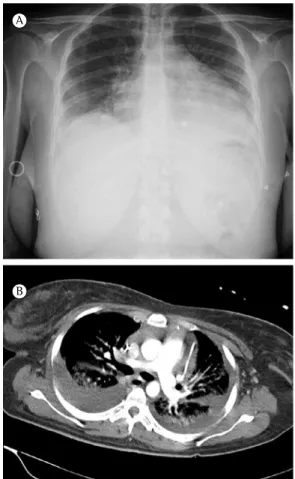
cells/L (94.3% neutrophils, 2.9% lymphocytes, and 2.6% basophils); serum C-reactive protein, 57.3 mg/dL; serum sodium, 120 mEq/L; and serum potassium, 4.7 mEq/L. Arterial blood gas analysis demonstrated severe hypoxemia (PaO2, 39.4 mmHg; PaCO2, 25.2 mmHg; and pH, 7.47). A chest X-ray revealed opacities in the middle and lower fields of the right lobe, accompanied by enlargement of the cardiac silhouette (Figure 1). Empirical antibiotic treatment with ceftriaxone and levofloxacin was started. An electrocardiogram showed sinus rhythm with diffuse ST segment elevation. Thoracentesis showed exudative pleural effusion (pH, 7.17; glucose, 45 mg/dL; protein, 38 g/dL; and lactate dehydrogenase [LDH], 1,425 IU/L). Staining of the pleural fluid revealed gram-positive diplococci. A chest tube was inserted in order to drain the empyema. The patient reported some relief after the drainage; however, because of worsening of respiratory failure and the need to maintain a high FiO2 (80%), he was admitted
to the ICU. Microbiological studies, including urinary antigen testing, sputum culture, and culture of the pleural fluid, were positive for S. pneumoniae that was penicillin- and erythromycin-susceptible at minimum inhibitory concentrations < 0.25 µg/ml and < 0.03 µg/mL, respectively).
Echocardiography showed severe pericardial effusion (without signs of compression or hemodynamic impairment) and mild pulmonary hypertension with tricuspid regurgitation, although the other valves were functioning normally. A chest X-ray performed on the second day of hospitalization showed increased heart size and new pulmonary infiltrate. On the fourth day, tracheal intubation was required because the patient showed worsening of his clinical condition and deterioration of blood gas analysis. The patient was hemodynamically unstable, which called for aggressive measures. A second pericardiocentesis was performed and yielded 480 ml of seropurulent fluid. The condition of the patient improved after an infusion of norepinephrine (0.8 µg/kg per min). Unfortunately, he could not be stabilized.
A
B
a four-day history of flu with cough and fever (39.4°C), having recently complained of dyspnea and progressive edema of the lower extremities. Physical examination revealed hypotension (blood pressure, 80/50 mmHg) and tachycardia (140 bpm). Auscultation of the thorax showed decreased breath sounds in the right lung and crackles in the left lung base. At admission, a chest X-ray revealed right pleural effusion and a CT scan showed right pleural effusion with collapse of the right lower and middle lobes associated with extensive effusion. Laboratory tests at admission revealed a white blood cell count of 30.2 × 10⁹ cells/L (92% neutrophils and
3.6% lymphocytes), a platelet count of 956 × 109/L, a serum C-reactive protein level of 15.84
mg/dL, a serum creatinine concentration of 1.32 mg/dL, a serum sodium level of 135 mEq/L, and a serum potassium level of 5.6 mEq/L.
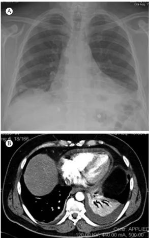
A chest X-ray performed on the second day of hospitalization showed progression of the disease process: an opacity involving nearly all of the right lung and further enlargement of cardiopericardial silhouette (Figure 2A). Echocardiography showed severe pericardial effusion affecting the entire cardiac silhouette. Pericardiocentesis yielded 640 mL of exudative fluid, the culture of which was negative. Pericardial and thoracic drainage was performed; purulent fluid was obtained in a sterile manner from the chest drain. Examination of the pleural fluid showed a pH of 6.95, a glucose level of 5 mg/dL, proteins at 47.4 g/L, and an LDH content of 5,347 U/L. A second chest X-ray showed consolidation in the right lung, minimal pleural fluid, and a normal cardiac silhouette (Figure 2B). A urinary pneumococcal antigen test was negative. Pleural fluid culture was positive for S. pneumoniae that was susceptible to erythromycin, levofloxacin, and penicillin. The isolate was S. pneumoniae serotype 1.
The patient received empirical antibiotic treatment with the piperacillin-tazobactam combination (4.5 g/8 h) and levofloxacin (500 mg twice a day for 5 days). The latter was continued because the pneumococcus was susceptible to it. The patient was discharged on the twelfth day, after withdrawal of the pericardial and pleural drains.
Case 3
A 26-year-old female from Bahrain, with no relevant medical history, was brought to the
A
B
Figure 2 - Chest X-ray showing an opacity involving nearly the entire right lung, together with enlargement of the cardiac silhouette, before and after pericardial and thoracic drainage (A and B, respectively). Note consolidation in the right lower lobe with mild pleural effusion and normal cardiac silhouette after drainage.
emergency room. At presentation, the patient had cough with purulent expectoration, fever, and chest pain. She presented with tachypnea (34 breaths/min), hypotension (blood pressure, 85/55 mmHg), and tachycardia (144 bpm). Physical examination revealed bilateral crackles in the lower lung fields. Laboratory data were remarkable for elevation of inflammatory and infectious parameters: white blood cell count, 11 × 109 cells/L (68.2% neutrophils and 22.3%
lymphocytes); platelet count, 573 × 109/L; serum
C-reactive protein, 6 mg/dL; serum sodium, 142 mEq/L; serum potassium, 3.7 mEq/L; and serum creatinine, 0.83 mg/dL.
fluid. A CT scan of the chest showed pneumonia in the left lower lobe, bilateral pleural effusion, and moderate pericardial effusion (Figure 4B). Thoracentesis was performed and yielded 1,050 mL of exudative fluid. Laboratory tests yielded the following data: white blood cell count, 11.78 × 10⁹ cells/L (84.3% neutrophils, 4.3%
lymphocytes, and 9.1% monocytes); platelet count, 512 × 109/L; serum C-reactive protein,
31.27 mg/dL; serum creatinine, 0.94 mg/dL; serum sodium, 133 mEq/L; and serum potassium, 3.72 mEq/L. Examination of the pleural fluid showed a pH of 7.16, a glucose level of 4.5 mg/ dL, proteins at 49.1 g/L, and an LDH content of 1,385 U/L. A urinary pneumococcal antigen test was positive. Pleural fluid culture was positive for S. pneumoniae. The patient was treated for four weeks with amoxicillin-clavulanate (2.2 g/8 h, i.v.) plus levofloxacin (500 mg twice a day), together with a nonsteroidal anti-inflammatory drug (ibuprofen, 800 mg/day), after which there A CT scan showed bilateral pleural effusion,
abundant pericardial effusion, and bilateral alveolar opacities (Figure 3B). The patient was admitted to the ICU. Supportive treatment was started, as was empirical antibiotic therapy with ceftriaxone (2 g/24 h) plus levofloxacin (500 mg twice per day), as well as a nonsteroidal anti-inflammatory drug (ibuprofen). It was necessary to drain the pericardial effusion multiple times. Various tests were performed: pleural fluid culture; blood culture; serological tests for atypical bacteria; and RT-PCR for respiratory viruses. In addition, we screened for tuberculosis (with a PPD test and enzyme-linked immunospot assay). A urinary pneumococcal antigen test was positive. Cytological studies of the pericardial effusion were negative for malignancy.
On the third day after admission, the clinical condition of the patient worsened and treatment was therefore started with methylprednisolone (1 mg/kg per day), which initially produced a promising response (decrease in the serum C-reactive protein level, a decrease in heart rate, and improvement of general status). On the fifth day, the condition of the patient deteriorated, with increasing pericardial effusion. The patient showed worsening respiratory distress, with an increase in the FiO2 requirement (from 0.24 to 0.60). Drainage of the pleural effusion resulted in no improvement. At that time, she required intubation and mechanical ventilation. On the seventh day, she showed impaired ventilation, caused by decreased lung compliance and difficulties in maintaining oxygenation. Consequently, extracorporeal membrane oxygenation was started. However, because there was no subsequent improvement in the respiratory parameters, the extracorporeal membrane oxygenation was stopped and the patient died.
Case 4
A 57-year-old male presented with fever (38.9°C), chest pain, cough, and progressive dyspnea. The patient exhibited tachypnea (34 breaths/min) and tachycardia (134 bpm). Auscultation revealed decreased breath sounds in both lung bases, with crackles on the left. A chest X-ray revealed bilateral pleural opacities and enlargement of the cardiac silhouette (Figure 4A). Echocardiography showed moderate pericardial effusion affecting the entire cardiac silhouette. Pericardiocentesis yielded 250 mL of exudative
A
B
aggressive, long-term therapy. (11,12) Pericarditis
must be considered a possible complication in pneumococcal infections.(13) Cardiac tamponade
is the most dangerous and common complication of pneumococcal pericarditis; it can be fatal, especially when its diagnosis is delayed.(14-18)
When there are signs of infection and a chest X-ray shows an enlarged cardiac silhouette, echocardiography is mandatory. Aspiration of the purulent fluid by pericardiocentesis facilitates the diagnosis.(15) Recurrent pericardial effusion
or constrictive pericarditis call for pericardial drainage or pericardiectomy.(14-18) In patients with
pericarditis, urinary pneumococcal antigen tests can be successfully used in order to identify the causative agent.(19,20) Recent studies have employed
molecular analysis of pericardial fluid for that purpose.(19) The management of pneumococcal
pericarditis involves prolonged (at least 4 weeks of) antimicrobial therapy,(14) in conjunction with
pericardiocentesis.(15) When antibiotic therapy
is used in combination with pericardiocentesis, mortality can be reduced to 20% or less. The complications of acute pericarditis typically appear early in its clinical course.(16)
Each case in our series showed different clinical features and levels of severity. In three of the four cases, the main complication was cardiac tamponade. The low specificity of the symptoms makes early clinical diagnosis difficult. Too often, the diagnosis is made after the disease has already resulted in severe hemodynamic compromise.(12,13,16)
Using pericardiocentesis to remove excess fluid from the pericardial sac and administering the appropriate antibiotic therapy are of paramount importance in reducing the mortality associated with purulent pericarditis.(11,15,21,22)
In conclusion, our case series demonstrates that, despite advances in diagnostic and treatment modalities, purulent pericarditis continues to be a serious complication of infection with S. pneumoniae.(23) Therefore, a diagnosis of S.
pneumoniae-associated purulent pericarditis should be considered and investigated early in patients presenting with clinical deterioration and showing unsatisfactory improvement during conventional therapy.
References
1. File TM Jr. Streptococcus pneumoniae and community-acquired pneumonia: a cause for concern. Am J Med. 2004;117 Suppl 3A:39S-50S. http://dx.doi.org/10.1016/j. amjmed.2004.07.007
was nearly complete resolution of the alterations seen on the chest X-ray and CT scan.
Discussion
Infectious etiologies are the cause of only 5% of all cases of pericardial effusion.(6) In most
cases, S. pneumoniae infection spreads from an intrathoracic site, whereas the dissemination of Staphylococcus aureus is most often hematogenous. (6-8) Pneumococcal pericarditis
is a rare syndrome. To our knowledge, only 20 cases were reported between 1980 and 2010, the prevalence of the syndrome being similar across all age groups. (8-10) Prior to the advent of
antibiotic therapy, pneumococcal infection was the most common cause of purulent pericarditis. The fact that this is no longer the case might reflect the efficacy of penicillin therapy.(11)
Mortality remains high in cases of pneumococcal pericarditis, even when patients receive
A
B
13. Majid AA, Omar A. Diagnosis and management of purulent pericarditis. Experience with pericardiectomy. J Thorac Cardiovasc Surg. 1991;102(3):413-7.
14. Inkster T, Khanna N, Diggle M, Sonecki P. Diagnosis of pneumococcal pericarditis using antigen testing and polymerase chain reaction. Scan J Infect Dis. 2010;42(10):791-3. http://dx.doi.org/10.3109/00365 548.2010.486002
15. Cakir O, Gurkan F, Balci AE, Eren N, Dikici B. Purulent pericarditis in childhood: ten years of experience. J Pediatr Surg. 2002;37(10):1404-8. http://dx.doi.org/10.1053/ jpsu.2002.35401
16. Sagristà-Sauleda J, Barrabés JA, Permanyer-Miralda G, Soler-Soler J. Purulent pericarditis: review of a 20-year experience in a general hospital. J Am Coll Cardiol. 1993;22(6):1661-5. http://dx.doi. org/10.1016/0735-1097(93)90592-O
17. Geri G, Dupeux S, Pouchot J. Pneumococcal purulent pericarditis. Rev Med Interne. 2008;29(7):568-72. http:// dx.doi.org/10.1016/j.revmed.2007.11.012
18. Thébaud B, Sidi D, Kachaner J. Purulent pericarditis in children: a 15 year-experience [Article in French]. Arch Pediatr. 1996;3(11):1084-90. http://dx.doi.org/10.1016/ S0929-693X(96)89513-3
19. Nakagawa C, Kasahara K, Yonekawa S, Ogawa T, Kutsuna S, Maeda K, et al. Purulent pericarditis due to Streptococcus pneumoniae diagnosed by pneumococcal urinary antigen assay and 16S rDNA sequence of the pericardial fluid. Inter Med. 2010;49(15):1653-6. http:// dx.doi.org/10.2169/internalmedicine.49.3245 20. Le Monnier A, Carbonnelle E, Zahar JR, Le Bourgeois M,
Abachin E, Quesne G, et al. Microbiological diagnosis of empyema in children: comparative evaluations by culture, polymerase chain reaction, and pneumococcal antigen detection in pleural fluids. Clin Infect Dis. 2006;42(8):1135-40. http://dx.doi.org/10.1086/502680 21. Cheatham JE Jr, Grantham RN, Peyton MD, Thompson
WM, Luckstead EF, Razook JD, et al. Hemophilus influenzae purulent pericarditis in children: diagnosis and therapeutic considerations. J Thorac Cardiovasc Surg. 1980;79(6):933-6.
22. Simony CF, Malham M, Kanstrup J, Wojtek P, Lynggaard F, Andersen S. Lifesaving pericardiocentesis due to purulent pericarditis with growth of Gram-negative rods in an immune-competent Inuit male. Int J Emerg Med. 2014;7:21. http://dx.doi.org/10.1186/s12245-014-0021-8 23. Parikh SV, Memon N, Echols M, Shah J, McGuire DK, Keeley
EC. Purulent pericarditis: report of 2 cases and review of the literature. Medicine (Baltimore). 2009;88(1):52-65. http://dx.doi.org/10.1097/MD.0b013e318194432b 2. Mandell LA. Spectrum of microbial etiology of
community-acquired pneumonia in hospitalized patients: Implications for selection of the population for enrolment in clinical trials. Clin Infect Dis. 2008;47 Suppl 3:S189-92. http:// dx.doi.org/10.1086/591403
3. Tan TQ, Mason EO Jr, Wald ER, Barson WJ, Schutze GE, Bradley JS, et al. Clinical characteristics of children with complicated pneumonia caused by Streptococcus pneumoniae. Pediatrics. 2002;110(1 Pt 1):1-6. http:// dx.doi.org/10.1542/peds.110.1.1
4. Hästbacka J, Kolho E, Pettilä V. Purulent pneumococcal pericarditis: a rarity in the antibiotic era. J Crit Care. 2002;17(4):251-4. http://dx.doi.org/10.1053/ jcrc.2002.36758
5. Kan B, Ries J, Normark BH, Chang FY, Feldman C, Ko WC, et al. Endocarditis and pericarditis complicating pneumococcal bacteraemia, with special reference to the adhesive abilities of pneumococci: results from a prospective study. Clin Microbiol Infect. 2006;12(4):338-44. http://dx.doi.org/10.1111/j.1469-0691.2006.01363.x 6. Ferreira dos Santos L, Moreira D, Ribeiro P, Rodrigues
B, Correia E, Nunes L, et al. Purulent pericarditis: a rare diagnosis. Rev Port Cardiol. 2013;32(9):721-7. http:// dx.doi.org/10.1016/j.repce.2013.10.010
7. Koster N, Narmi A, Anand K. Bacterial pericarditis. Am J Med. 2009;122(5):e1-2. http://dx.doi.org/10.1016/j. amjmed.2008.11.012
8. Feinstein Y, Falup-Pecurariu O, Mitrica M, Berezin EN, Sini R, Krimko H, et al. Acute pericarditis caused by Streptococcus pneumoniae in young infants and children: three case reports and a literature review. Int J Infect Dis. 2010;14(2):e175-8. http://dx.doi.org/10.1016/j. ijid.2009.03.033
9. Go C, Asnis DS, Saltzman H. Pneumococcal pericarditis since 1980. Clin Infect Dis. 1998;27(5):1338-40. http:// dx.doi.org/10.1086/517730
10. Vindas-Cordero JP, Sands M, Sanchez W. Austrian’s triad complicated by suppurative pericarditis and cardiac tamponade: a case report and review of the literature. Int J Infect Dis. 2009;13(1):e23-5. http://dx.doi.org/10.1016/j. ijid.2008.04.005
11. Tatli E, Buyuklu M, Altun A. An unusual complication of pneumococcal pneumonia: acute tamponade due to purulent pericarditis. Int J Cardiol. 2007;119(1):e1-3. http://dx.doi.org/10.1016/j.ijcard.2007.02.042 12. Rubin RH, Moellering RC Jr. Clinical microbiologic,