373
AbstractObjectives: High prevalence rates of celiac disease in patients with Down syndrome have been reported in several countries. However, in Brazil there are no data regarding this association. In this study we report the prevalence of celiac disease in Down syndrome children and adolescents from southern Brazil.
Methods: Seventy-one patients (32 female and 39 male, 2-18 years) from Curitiba, Brazil, were studied. Eighty young people (42 male and 38 female, 2-19 years) were used as controls. All subjects were screened for the IgA-antiendomysium antibody (EmA) and IgA anti-tecidual transglutaminase (anti-tTG). EmA was measured by an immunofluorescence assay using umbilical cord as the substrate and anti-tTG by ELISA with tecidual transglutaminase as the antigen. The total IgA serum level was determined by turbidimetry.
Results: Five DS patients (7%) were positive for EmA-IgA, with titers from 1/5 to 1/80 and 14 (17.5%) for anti-tTG (21-340 units). All EmA positive patients also presented anti-tTG antibodies simultaneously. Clinical and histological findings of the intestinal mucosa confirmed celiac disease diagnoses in four patients. The other EmA positive patient was asymptomatic and was not submitted to duodenal biopsy. Patients only positive for anti-tTG presented borderline values (< 25 units) and were asymptomatic. None of the controls were positive for EmA or anti-tTG. No Down syndrome patients or controls presented IgA deficiency.
Conclusions: These data indicate a high prevalence (5.6%) of confirmed celiac disease in Down syndrome patients from southern Brazil.
J Pediatr (Rio J). 2005;81(5):373-6: Antiendomisyum antibodies, anti-transglutaminase antibodies, celiac disease, Down syndrome.
Resumo
Objetivos: Alta prevalência de doença celíaca em pacientes com síndrome de Down tem sido descrita em vários países. No entanto, no Brasil ainda não há relatos mostrando essa associação. O presente estudo teve como objetivo avaliar a prevalência de doença celíaca em crianças e adolescentes com síndrome de Down no sul do Brasil.
Métodos: Setenta e um pacientes (32 do sexo feminino e 39 masculino, 2-18 anos) provenientes de Curitiba, Brasil, foram estuda-dos. Oitenta indivíduos (42 do sexo masculino e 38 feminino, 2-19 anos) foram utilizados como controles do estudo. Todas as amostras foram investigadas para anticorpo anti-endomísio (EmA) e anti-trans-glutaminase tecidual (anti-tTG). O EmA foi pesquisado através de imunofluorescência indireta usando cordão umbilical como substrato e o anti-tTG através da técnica de ELISA, utilizando transglutaminase tecidual como antígeno. As dosagens de IgA foram realizadas por turbidimetria.
Resultados: Cinco pacientes com síndrome de Down (7%) foram positivos para EmA-IgA, com títulos entre 1/5 to 1/80 e catorze (17,5%) para anti-tTG (21-340 units). Todos os pacientes positivos para EmA apresentaram simultaneamente positividade para o anti-tTG. Os achados clínicos e histológicos na mucosa intestinal confimaram doença celíaca em quatro pacientes. O outro paciente EmA positivo não foi submetido a biópsia duodenal. Os pacientes positivos apenas para anti-tTG apresentaram valores limítrofes (< 25 unidades) e eram assintomáticos. Nenhum indivíduo do grupo controle foi positivo para EmA ou anti-tTG. Nenhuma amostra do estudo foi deficiente para IgA. Conclusões: Os dados do presente estudo mostram alta prevalên-cia (5,6%) de doença celíaca confirmada em crianças e adolescentes com síndrome de Down da região sul do Brasil.
J Pediatr (Rio J). 2005;81(5):373-6:Anticorpos anti-endomísio, anticorpos anti-transglutaminase, doença celíaca, síndrome de Down.
Celiac disease in children and adolescents
with Down syndrome
Doença celíaca em crianças e adolescentes com síndrome de Down
Renato M. Nisihara,1 Lorete M. S. Kotze,2 Shirley R. R. Utiyama,3 Nanci P. Oliveira,4 Patrícia T. Fiedler,4 Iara T. Messias-Reason3
0021-7557/05/81-05/373
Jornal de Pediatria
Copyright © 2005 by Sociedade Brasileira de Pediatria
O
RIGINALA
RTICLE1. MSc.Laboratory of Immunopathology, Hospital das Clínicas, Universida-de FeUniversida-deral do Paraná (UFPR), Curitiba, PR.
2. MD. Service of Gastroenterology and Digestive Endoscopy, Hospital Universitário Cajuru, Pontifícia Universidade Católica do Paraná (PUC-PR), Curitiba, PR.
3. PhD. Laboratory of Immunopathology, Hospital das Clínicas, UFPR, Curitiba, PR.
4. MD. Down Syndrome Clinic, Hospital das Clínicas, UFPR, Curitiba, PR.
Manuscript received Sep 21 2004, accepted for publication Apr 13 2005.
Suggested citation: Nisihara RM, Kotze LM, Utiyama SR, Oliveira NP, Fiedler PT, Messias-Reason IT. Celiac disease in children and adoles-cents with Down syndrome. J. Pediatr (Rio J). 2005;81:373-6.
Introduction
Downs syndrome (DS) is the most frequent chromosomal anomaly observed in the general population, affecting 1 in 800 newborns throughout the world and is the main genetic factor for moderate mental retardation.1
Different studies have shown that patients with DS
present several immune dysfunctions,2,3 which lead to
374 Jornal de Pediatria - Vol. 81, Nº5, 2005 CD in children and adolescents with Down syndrome Nisihara RM et alii
authors,4-6 with the most significant association with celiac disease (CD)7-11 and autoimmune thyroiditis.12,13
CD affects individuals of all ages and is characterized by permanent gluten intolerance. In its classic form, CD appears with symptoms and signs of intestinal malabsorption. However, the disease may occur in a silent or latent form.14
According to Marsh,15 the duodenal mucosa can be normal
or present changes ranging from mild alterations to severe atrophy in mucosal architecture. The treatment with a gluten-free diet results in clinical and mucosal recovery, with recurrence of the disease after the return of gluten to the diet.14 The classic and symptomatic form of CD is the presence of diarrhea, vomiting and weight loss, which appears in a minority of cases. On the other hand, most of the patients present unspecific gastroenterologic symptoms such as dyspepsia, abdominal pain, flatulence and alteration of the intestinal rhythm. These characteristics often cause delay in CD diagnosis and the incorrect management of patients.14
In 1975, Bentley reported for the first time the association
between CD and DS.16 In the following years, several
reports showed increased frequency of CD in individuals with DS in different populations, with a prevalence ranging
from 3.2 to 10.3%.7-11 In this study we investigate the
prevalence of CD in children with DS from southern Brazil.
Materials and methods
The institutional ethics committee approved the present investigation.
Patients
Seventy-one consecutive DS patients (32 female and 39 male) with mean age 6.12 years (range 2-18 years) from
the DS clinic of the Hospital de Clínicas Universidade
Federal do Paraná (UFPR) (Curitiba, Brazil) were studied. DS patients older than 18 years were excluded from the study. After formal consenting of DS childrens first-degree relatives, 3 ml of blood were collected from each subject. Samples were centrifuged and the serum separated, aliquoted and immediately stored at -80 °C, until used. Only the EmA-IgA/anti-tTG positive patients were clinically evaluated and submitted to upper gastrointestinal endoscopy. Crypt hyperplasia and villous atrophy were classified as partial (PVA) or total (TVA), according to Marsh.15
Controls
Blood samples of 80 healthy children of the same geographic area of the patients, matched with sex and age with the DS group (38 female and 42 male, mean age 8.02 years), range 2-19 years, were used as controls.
EmA-IgA
All subjects were screened for EmA-IgA according to
Volta et al.,17 using immunofluorescence indirect assay,
with human umbilical cord as substrate and FITC anti-IgA human as conjugate (INOVA, USA).
Anti-tTG antibodies
Anti-tissue transglutaminase IgA antibodies (tTG) were determined by enzyme-linked immunosorbent assay (ELISA) with guinea pig liver tTG as antigen, using a commercial kit (INOVA Diagnostics Inc., San Diego, CA, USA), according to
Dieterich et al.18 Results were considered positive when
higher than 20 units.
Serum IgA
The total serum IgA levels was determined by turbidimetry (Behring, Germany).
Statistical analysis
Data were analyzed with Statistica (Microsoft, USA) software, using the Fisher exact test.
Results
Serological screening for CD based on EmA and tTG antibodies was performed in 71 patients with DS and in 80 healthy children.
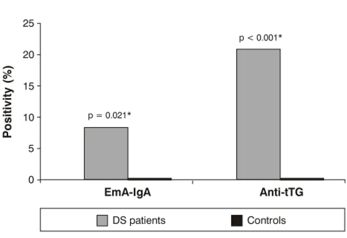
These results for DS patients and controls are shown in Figure 1. A highly significant positivity of EmA-IgA (p = 0.021) and anti-tTG (p < 0.001) was observed in the DS patients when compared to the controls. Five DS patients (7%; 5/71), four male and one female, were positive for EmA-IgA, with titers varying from 1/5 to 1/80. Fourteen patients (17.5%; 14/71), seven male and seven female, were positive for anti-tTG antibodies with values from 21 to 340 U/ml. Five of these patients were concomitantly positive for EmA-IgA and the other nine were only positive for anti-tTG antibodies, presenting borderline values (Table 1). None of the 80 control individuals was positive for EmA-IgA or anti-tTG.
* Fisher exact test
Figure 1 - Anti-endomysial (IgA-EmA) and anti-transglutaminase (tTG) antibodies in Down syndrome patients and controls
P
o
s
it
iv
it
y
(
%
)
EmA-IgA
p = 0.021*
DS patients Controls
p < 0.001*
Anti-tTG
25
20
15
10
5
Jornal de Pediatria - Vol. 81, Nº5, 2005 375
Patient Sex Age EmA-IgA Anti tTG* Symptoms Hystologic
(U/ml) aspects
C.M. M 6 POS 1/5 72 Symptom-free ND
A.M.E. M 8 POS 1/5 30 Dypepsia, growth failure, abdominal pain Partial atrophy
R.D. F 18 POS 1/80 198 Dyarrheia, abdominal pain Total atrophy
R.K M 6 POS 1/80 176 Dyarrheia, growth failure Total atrophy
R.S.A. M 10 POS 1/80 340 Symptom-free Total atrophy
S.R.O. F 4 Negative 22 Symptom-free ND
L.P. F 12 Negative 21 Symptom-free ND
R.I. F 3 Negative 24 Symptom-free ND
A.O.S. F 15 Negative 23 Symptom-free ND
E.H.S. F 3 Negative 24 Symptom-free ND
L.I.H. M 3 Negative 24 Symptom-free ND
D.M.S. M 7 Negative 23 Symptom-free ND
S.R.O. F 5 Negative 31 Symptom-free ND
J.B. M 4 Negative 39 Symptom-free ND
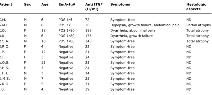
Table 1 - Antiendomisyum and anti-transglutaminase antibodies, symptoms and histologic aspects in DS patients
ND = Not done. * Reference values: < 20 U/ml = Negative; 20 to 30 U/ml = weakly positive, > 30 U/ml = positive.
Among the five patients concomitantly positive for EmA-IgA and anti-tTG antibodies, three showed clinical symptoms such as diarrhea, abdominal pain, anemia and growth failure (Table 1). Four of them underwent an upper digestive endoscopy followed by duodenal mucosa biopsy. Histological aspects of duodenal mucosa compatible with CD were observed in all patients, three of them presented TVA and one PVA. Only one asymptomatic patient (EmA 1/5, anti tTG = 72 UI) did not undergo digestive endoscopy and duodenal biopsy. Thus, the prevalence of confirmed CD among the investigated DS patients was 5.6% (4/71).
Neither the DS patients nor the controls presented IgA deficiency.
Discussion
This is the first report to show the prevalence of CD in children with DS in Brazil. Our findings confirmed the increased prevalence of CD among patients with DS reported by several studies in Europe, North America and Argentina.7-11 The prevalence of CD in the general population in these countries ranges from 1:200 to 1:2,000, and among DS patients it presents a 20-200 fold increase. In Brazil, the prevalence for CD has been estimated to be 0.14% (1:687) in the central-western region19 and 0.1% (1:1,000) in the south region.20
Although not all EmA positive patients in our study underwent an intestinal biopsy, the confirmed CD prevalence in DS patients was still very high (5.6%; 1:18), in comparison with the general population of the same geographical area (1:1,000). If we consider CD prevalence diagnosed by positive EmA, this prevalence would be 7%. The high
association of CD with DS has not yet been clarified, however both, CD and DS patients present more frequent dysfunction in their immune system and are more predisposed to autoimmune diseases, such as thyroid diseases, diabetes mellitus type 1, lupus and arthritis.4,5,6,12 Besides that, this association may be related to shared common genetic markers. It has been shown that the DS patients with CD present the characteristic CD associated with high-risk
human leukocyte antigens (HLA) DR3 and DQ2 alleles.21 In
addition, gene disorders caused by chromosomal imbalance may be related to the enhanced expression of CD in SD patients.
Different studies have shown that the most sensitive and specific serologic test for the diagnosis of CD is the assessment of EmA antibodies. During this investigation, five patients were positive for EmA-IgA (7%) and four of them underwent duodenal biopsy. All patients who underwent intestinal biopsy showed clinical manifestations and histological alterations of intestinal mucosa compatible with CD. The other patient both EmA-IgA and anti tTG positive was asymptomatic and did not undergo biopsy. It is possible that this patient had a latent form of CD, which needs to be followed-up. Thus EmA screening showed high sensitivity and specificity for the diagnosis of CD in the investigated patients. Our results corroborate the widely accepted concept that EmA-IgA is a specific screening test for CD.
376 Jornal de Pediatria - Vol. 81, Nº5, 2005
guinea pig liver extract as antigen in the ELISA kit. Recently, the use of human tissue transglutaminase as purified antigen has been shown to enhance the specificity of this assay.23
In the first 2 years of life, most of the CD patients present classic symptoms and when associated with DS this diagnosis is almost always delayed. Certain characteristics of DS children, such as a distended abdomen and the tendency to consider growth failure as natural consequences of the DS, can be responsible for the difficulty in diagnosing CD in these patients. Thus, several authors consider that the CD is underestimated as the cause of diarrhea, malnutrition or development deficit in patients with DS.9,24
The importance of the CD investigation in DS patients has been showed in recent studies, which recommended serologic tests every 2 years, since negative young individuals
may be positive some years later.9
The data reported in the present study emphasize the value of investigating CD in DS patients, since the correct and early diagnosis will offer a better quality of life to the patient and his family.
Correspondence: Renato Mitsunori Nisihara Laboratório de Imunopatologia
Hospital de Clínicas, Setor de Ciências da Saúde Rua Padre Camargo, 280
CEP 80060-240 Curitiba, PR E-mail: [email protected] References
1. Pueschel SM. Physical characteristics, chromosome analysis and treatment approaches in Down syndrome. In: Tingey C. Down syndrome: a resource handbook. Boston; College-Hill Press/Little, Brown & Co., 1988. p. 3-21.
2. Avanzini MA, Manafo V, De Amici M, Maccario R, Burgio GR, Plebani A, et al. Humoral immunodeficiency in Down syndrome: serum subclass and antibody response to hepatitis B vaccine. Am J Med Genet. 1990;7:231-3.
3. Nespoli L, Burgio GR, Ugazio AG, Macario R. Immunological features of Downs syndrome: a review. J Intel Disabil Res. 1993;37:543-51.
4. Anwar AJ, Walker JD, Frier BM. Type 1 diabetes and Downs syndrome: prevalence, management and diabetic complications. Diabet Med. 1998;15:160-3.
5. Bakkaloglu A, Besbas SO, Saatci U, Balci S. Down syndrome associated with systemic lupus erythematosus: a mere coincidence or a significant association? Clin Genet. 1994;46: 322-3.
6. Olson JC, Bender JC, Levinson JE, Oestreich A, Lovell DJ. Arthropathy of Down syndrome. Pediatrics. 1990;86:931-6. 7. Bonamico M, Mariani P, Danesi HM, Crisogianni M, Failla P,
Gemme G, et al. Prevalence and clinical picture of celiac disease in Italian Down syndrome patients: a multicenter study. J Ped Gastroenterol Nutr. 2001;33:139-43.
8. Book L, Hart A, Black J, Feolo M, Zone JJ, Neuhausen SL. Prevalence and clinical characteristics of celiac disease in Down syndrome in a U.S. study. Am J Med Genet. 2001;98:70-4.
9. Csizmadia CG, Mearin ML, Ore A, Krombout A, Crusius JB, Blomberg ME, et al. Accuracy and cost-effectiveness of a new strategy to screen for celiac disease in children with Down syndrome. J Pediatr. 2000;137:756-60.
10. Kolek A, Vospelová J, Hermanová Z, Santavá A, Tichy M. Occurrence of celiac disease in children with Downs syndrome in north Moravia, Czech Republic. Eur J Pediatr. 2003;162:307-8. 11. Rumbo M, Chirdo FG, Ben R, Saldungaray I, Villalobos R. Evaluation of celiac disease serological markers in Down syndrome patients. Dig Liver Dis. 2002;34:116-21.
12. Rubello D, Pozzan GB, Casara DM, Girelli ME, Boccato S, Rigon F. Natural course of subclinical hypothyroidism in Down syndrome: prospective study results and therapeutic considerations. J Endocrinol Invest. 1995;17:35-40.
13. Shalitin S, Philip M. Autoimmune thyroiditis in infants with Downs syndrome. J Pediatr Endocrinol Metab. 2002;15:649-52. 14. Kotze LM, Barbieri D. Doença celíaca. In: Kotze LM, Barbieri D. Afecções gastrointestinais da criança e do adolescente. 1a ed. Rio de Janeiro: Revinter; 2003. p. 189-208.
15. Marsh MN. Mucosal pathology in gluten sensitivity. In: Marsh MN. Coeliac disease. Oxford: Blackwell Scientific Publications; 1992. p. 136-91.
16. Bentley D. A case of Downs syndrome complicated by retinoblastoma and celiac disease. Pediatrics. 1975;56:131-3. 17. Volta U, Molinaro N, De Franceschi L, Fratangelo D, Bianchi FB. IgA anti-endomysial antibodies on human umbilical cord tissue for celiac disease screening. Dig Dis Sci. 1995;40:1902-5. 18. Dieterich W, Laag E, Schopper H. Autoantibodies to tissue
t r a n s g l u t a m i n a s e a s p r e d i c t o r s o f c e l i a c d i s e a s e . Gastroenterology. 1998;115:1317-21.
19. Gandolfi L, Pratesi R, Córdoba JC, Tauil PL, Gasparin M, Catasi C. Prevalence of celiac disease among blood donors in Brazil. Am J Gastroenterol. 2000;95:689-92.
20. Nisihara RM, Kotze LM, Mocelin V, Utiyama SR. Serological investigation of celiac disease in southern area from Brazil. Arq Gastroenterol. 2005. In press.
21. Hansson T, Anneren G, Sjoberg O, Klareskog L, Dannaeus A. Celiac disease in relation to immunologic serum markers, trace elements, and HLA-DR and DQ antigens in Swedish children with Down syndrome. J Pediatr Gastroenterol Nutr. 1999;29:286-92. 22. Koop I, Ilchmann R, Izzi L. Detection of autoantibodies against tissue transglutaminase in patients with coeliac disease and dermatitis herpetiformis. Am J Gastroenterol. 2000;95:2009-14. 23. Clemente MG, Musu MP, Frau F, Lucia C, Virgillis S. Antitissue transglutaminase antibodies outside celiac disease. J Ped Gastroenterol Nutr. 2002;34:31-4.
24. Hilhorst MI, Brink M, Wauters EA, Houwen RH. Down syndrome and coeliac disease: five new cases with a review of the literature. Eur J Pediatr. 1993;152:884-7.