ABSTRACT
Objective: To evaluate the effects that administering dexamethasone before the induction of ventilator-induced lung injury (VILI) has on the temporal evolution of that injury. Methods: Wistar rats were allocated to one of three groups: pre-VILI administration of dexamethasone (dexamethasone group); pre-VILI administration of saline (control group); or ventilation only (sham group). The VILI was induced by ventilation at a high tidal volume. Animals in the dexamethasone and control groups were euthanized at 0, 4, 24, and 168 h after VILI induction. We analyzed arterial blood gases, lung edema, cell counts (total and differential) in the BAL luid, and lung histology. Results: At 0, 4, and 24 h after VILI induction, acute lung injury (ALI) scores were higher in the control group than in the sham group (p < 0.05). Administration of dexamethasone prior to VILI induction decreased the severity of the lung injury. At 4 h and 24 h after induction, the ALI score in the dexamethasone group was not signiicantly different from that observed for the sham group and was lower than that observed for the control group (p < 0.05). Neutrophil counts in BAL luid were increased in the control and dexamethasone groups, peaking at 4 h after VILI induction (p < 0.05). However, the neutrophil counts were lower in the dexamethasone group than in the control group at 4 h and 24 h after induction (p < 0.05). Pre-treatment with dexamethasone also prevented the post-induction oxygenation impairment seen in the control group. Conclusions: Administration of dexamethasone prior to VILI induction attenuates the effects of the injury in Wistar rats. The molecular mechanisms of such injury and the possible clinical role of corticosteroids in VILI have yet to be elucidated.
Keywords: Ventilator-induced lung injury; Dexamethasone; Respiratory distress syndrome, adult.
Pre-treatment with dexamethasone
attenuates experimental ventilator-induced
lung injury
Fernando Fonseca dos Reis1,2, Maycon de Moura Reboredo1,2, Leda Marília Fonseca Lucinda1,2, Aydra Mendes Almeida Bianchi1,2, Maria Aparecida Esteves Rabelo1, Lídia Maria Carneiro da Fonseca1,2, Júlio César Abreu de Oliveira1, Bruno Valle Pinheiro1,2
Correspondence to:
Fernando Fonseca dos Reis. Laboratório de Pesquisa Pulmonar, Universidade Federal de Juiz de Fora, Avenida Eugênio do Nascimento, s/n, Dom Bosco, CEP 36038-330, Juiz de Fora, MG, Brasil
Tel.: 55 32 99977-6584. E-mail: [email protected]
Financial support: This study was supported by research grants from the Rede Mineira de Ensaios Toxicológicos e Farmacológicos de Produtos Terapêuticos (Rede Mineira TOXIFAR, Minas Gerais [State] Network of Toxicological and Pharmacological Trials of Therapeutic Products), the Fundação de Amparo à Pesquisa do Estado de Minas Gerais (FAPEMIG, Foundation for the Support of Research in the State of Minas Gerais), and the Centro de Biologia da Reprodução da Universidade Federal de Juiz de Fora (Reproductive Biology Center of the Federal University of Juiz de Fora).
INTRODUCTION
Although necessary in various types of respiratory failure, mechanical ventilation (MV) can be harmful, especially if its parameters are adjusted incorrectly, because that can result in so-called ventilator-induced lung injury (VILI).(1) VILI can also worsen established lung injury,
such as the acute respiratory distress syndrome (ARDS), and delay its healing.(2) The clinical relevance of VILI
has been determined by studies conducted in patients with ARDS, in which adjusting protective parameters during MV, mainly limiting the tidal volume and plateau pressure, has been found to decrease mortality.(3,4) In
addition, studies involving patients at risk of developing ARDS have shown that those same measures decrease the occurrence of the syndrome and are currently recommended in such patients.(5,6)
VILI occurs when the transpulmonary pressure (i.e., the difference between the pressure level in the alveolus
and that in the pleural space) is above safe levels and generates high tidal volumes. The pulmonary structures exposed to this high pressure react by generating forces with the same intensity, creating a situation known as lung stress. A high tidal volume induces deformation of the alveolus, which is described as a change in a linear dimension over its initial value, creating a situation known as lung strain.(7,8) VILI can occur even with stress and
strain levels lower than those necessary for disruption of alveolar structures, although suficient to release proinlammatory cytokines and recruit neutrophils, leading to inlammatory lung injury.(9) This biological reaction to
mechanical injury is designated biotrauma, and studies have been carried out to establish the mechanisms involved and to test strategies (ventilatory and pharmacological) that could avert or attenuate VILI.(10,11)
Glucocorticoids exert anti-inlammatory effects by binding to their receptors in the cytoplasm, forming 1. Laboratório de Pesquisa em
Pneumologia, Universidade Federal de Juiz de Fora, Juiz de Fora (MG) Brasil.
2. Centro de Biologia da Reprodução, Universidade Federal de Juiz de Fora, Juiz de Fora (MG) Brasil.
Submitted: 17 January 2016.
Accepted: 9 May 2016.
glucocorticoid-receptor complexes that migrate to the cellular nucleus, where they inhibit the transcription rates of many inlammatory elements, including nuclear factor-kappa B.(12) This decreases the production of
proinlammatory cytokines, such as TNF-α, IL-1α, IL-1β, IL-2, IL-3, IL-5, IL-6, IL-8, IL-12, IFN-γ, and GM-CSF. (13)
By modulating those cytokines, glucocorticoids can suppress granulocyte recruitment and activation, as well as preserving alveolar-capillary barrier integrity and controlling vascular permeability. Glucocorticoids can also inhibit ibroblast proliferation and collagen deposition, both of which are important features in the later phases of ARDS.(14,15)
Despite the potentially beneicial effects of gluco -corticoids against the mechanisms involved in ARDS and VILI, their clinical usefulness in these conditions is still under debate.(16-20) For instance, in the speciic
context of the effects of glucocorticoids on mortality, some meta-analyses have shown that they reduce mortality,(17,19) whereas other have failed to show a
convincing effect of glucocorticoid treatment in ARDS.(20)
Hypotheses to explain these dissonant results include lack of an optimal corticosteroid dose, as well as the timing and duration of the therapy.(21)
Studies employing experimental models of VILI have obtained promising results with glucocorticoids. However, such studies have evaluated the effect of dexamethasone only in the acute phase of VILI.(22-25)
Therefore, the aim of this study was to evaluate the effects that administering dexamethasone before the induction of VILI has on the temporal evolution of the injury. We hypothesized that pre-treatment with dexamethasone would not only attenuate VILI in the acute phase but also accelerate the healing process thereafter.
METHODS
Animal preparation
This study was approved by the Animal Research Ethics Committee of the Federal University of Juiz de Fora, in the state of Minas Gerais, Brazil. The animals were cared for in accordance with the guidelines established by the Brazilian National Council for the Monitoring of Animal Experimentation. Sixty-three adult male Wistar rats (mean weight, 286 ± 15 g) were obtained from the animal facility of the Reproductive Biology Center of the Federal University of Juiz de Fora. Over the week prior to the experiment, groups of three animals each were housed in clear plastic cages, with stainless steel wire lids and pinewood shavings as bedding, in a temperature-controlled environment on closed, ventilated shelves, on a 12/12-h light/dark cycle. The animals were fed standard rat chow (mean, 25 g/day) and had ad libitum access to drinking water.
Experimental protocol
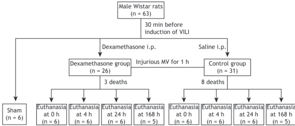
Animals were allocated to one of three groups (Figure 1): dexamethasone, comprising rats that received an
intraperitoneal injection of dexamethasone (6 mg/kg) at 30 min before the induction of VILI(24,25); control,
comprising rats that received an intraperitoneal injection of the same volume of saline, also at 30 min before the induction of VILI; and sham, comprising rats that received neither dexamethasone nor saline, were submitted to normal (non-injurious) ventilation for 10 min, and underwent the same experimental procedures performed in the irst two groups.
The rats were anesthetized with intraperitoneal injections of xylazine (8 mg/kg) and ketamine (80 mg/kg). After conirmation of the depth of anesthesia by paw clamp, the animals were intubated with a 16-gauge intravenous catheter and ventilated in the volume-controlled mode with a small animal ventilator (Inspira ASV; Harvard Apparatus, Holliston, MA, USA). To induce VILI (in the dexamethasone and control group rats), the ventilator parameters were set as follows: tidal volume of 35 mL/kg; respiratory rate of 18 breaths/min; inspiratory to expiratory ratio of 1:2; FiO2 of 1; and positive end-expiratory pressure (PEEP) of 0 cmH2O. After 1 h of this injurious MV, the animals were extubated and kept under observation to recover from the anesthesia.
After VILI induction, animals in the dexamethasone and control groups were euthanized (n = 23 in each group): immediately after VILI induction (0 h, n = 6 in each group); at 4 h after VILI induction (n = 6 in each group); at 24 h after VILI induction (n = 6 in each group); and at 168 h (7 days) after VILI induction (n = 5 in each group). At each of those time points, the animals were again anesthetized with intraperitoneal injections of xylazine (8 mg/kg) and ketamine (80 mg/kg), after which a surgical tracheostomy was performed and a 14-gauge cannula was inserted. An arterial catheter was inserted into the left carotid to obtain blood samples for arterial blood gas analysis (ABL90 FLEX; Radiometer, Copenhagen, Denmark). The rats were then paralyzed with an intra-arterial injection of rocuronium (1 mg/kg) and mechanically ventilated at the following settings: tidal volume of 6 mL/kg; respiratory rate of 80 breaths/ min; inspiratory to expiratory ratio of 1:2; FiO2 of 1; and PEEP of 2 cmH2O. After 10 min of ventilation, an arterial blood gas analysis was performed. Subsequently, a laparotomy was performed; the animals were euthanized by exsanguination and sectioning of the diaphragm. The trachea was clamped at end-expiration, with a PEEP of 2 cmH2O, and the lungs were extracted for further analysis. The animals in the sham group (n = 6) were euthanized 10 min after non-injurious MV.
Measurements
was blinded to the groups, using a conventional light microscope (Carl Zeiss, Hallbergmoos, Germany).
The acute lung injury score, which was the primary outcome measure, was determined by using a weighted scoring system, as described elsewhere.(26)
In brief, 20 random ields at a magniication of ×400 were scored independently. Values of 0, 1, or 2 were used in order to represent the severity based on the following indings: neutrophils in the alveolar space, neutrophils in the interstitial space, hyaline membranes, proteinaceous debris illing the airspaces, and alveolar septal thickening. To generate an acute lung injury score, the sum of the ive variables was weighted according to the relevance ascribed to each one. The resulting score was a continuous value between 0 (normal) and 1 (the most severe injury). In addition, the extent of each acute lung injury score component was calculated on the basis of the sum of the severity values (0, 1, or 2) for the 20 ields analyzed.
To obtain a BAL luid sample, the left lung was washed three times via a tracheal tube with 4 mL of PBS containing 10 nM of EDTA. Each sample was
centrifuged for 10 min at 1,500 rpm to separate it into its cellular and non-cellular components. The samples were diluted in Türk solution (2% acetic acid), and total leukocyte counts were determined in a Neubauer chamber under light microscopy. The cell pellet was resuspended in PBS and stained with May-Grünwald-Giemsa for differential cell counts, which were performed with a minimum of 300 cells.
The lung wet-to-dry weight ratio was determined in the right upper lobe, as previously described.(27) In brief,
the right upper lobe was isolated and weighed (wet weight) using a precise balance (Ohaus Adventurer; Toledo do Brasil, São Bernardo do Campo, Brazil). It was then dried in a microwave at low power (200 W) for 5 min. The drying process was repeated until the difference between two consecutive measurements was less than 0.002 g. The last weight determined was considered the dry weight.
Statistical analysis
The distribution of the data was tested for normality using the Kolmogorov-Smirnov test. Values are Figure 2. Log-rank test comparison of survival probability within the irst 24 h after induction of ventilator-induced lung injury.
Male Wistar rats (n = 63)
30 min before induction of VILI
Dexamethasone i.p. Saline i.p.
Dexamethasone group (n = 26)
Injurious MV for 1 h Control group (n = 31)
3 deaths 8 deaths
Sham (n = 6)
Euthanasia at 168 h
(n = 5) Euthanasia
at 0 h (n = 6)
Euthanasia at 4 h (n = 6)
Euthanasia at 24 h (n = 6)
Euthanasia at 168 h
(n = 5) Euthanasia
at 0 h (n = 6)
Euthanasia at 4 h (n = 6)
Euthanasia at 24 h (n = 6)
Cumulativ
e pro
bability
o
f surv
ival
0 5 10 15 20
Hours 1.0
0.8
0.6
0.4
0.2
0.0
Dexamethasone
Control
p = 0.19
expressed as mean ± standard deviation or median (interquartile range), as appropriate. Data for each group were analyzed by one-way ANOVA followed by the Dunnett’s test or by the Kruskal-Wallis test followed by the Mann-Whitney test, as appropriate. Adjustments for repeated measures were performed with Bonferroni correction. Comparisons between the dexamethasone and control groups at each time point were made by unpaired t-test or the Mann-Whitney test, as appropriate. The log-rank test was used for comparison of survival between the dexamethasone and control groups. Values of p < 0.05 were considered signiicant. All statistical analyses were performed with the Statistical Package for the Social Sciences, version 17.0 (SPSS Inc., Chicago, IL, USA).
RESULTS
Survival
Survival was not initially an objective of this study, and the animals were therefore not followed for a pre-determined period of time speciically for the study of that outcome. However, some deaths were observed in the dexamethasone and control groups during the period between the post-induction anesthesia recovery and the time of euthanasia. Death occurred in 3 (12%) of the 26 animals in the dexamethasone group and in 8 (26%) of the 31 animals in the control group (p = 0.19). Of those 11 deaths, 10 occurred within the irst 6 h after VILI induction, and 1 occurred more than 6 h but less than 24 h after VILI induction (Figure 2).
Lung histology and BAL
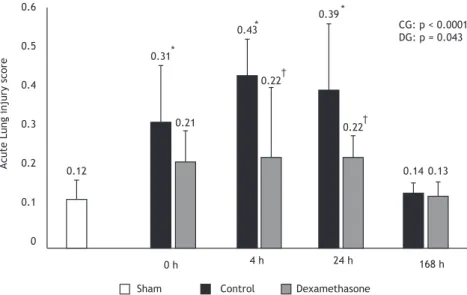
At 0, 4, and 24 h after VILI induction, the rats in the control group showed greater lung injury, as determined by the acute lung injury score, than did those in the
sham group. By 168 h after VILI induction, the mean control group score had decreased and was no different from that observed for the sham group. There were no signiicant differences between the dexamethasone group and the sham group, in terms of the acute lung injury score, at any of the time points evaluated. There was signiicantly less lung injury in the dexamethasone group than in the control group at 4 h and 24 h after VILI induction (Figure 3).
In the analysis of each component of the acute lung injury score, the animals in the control group showed higher numbers of interstitial neutrophils than did those in the sham group, at 0, 4, and 24 h after VILI induction, those numbers being highest at 4 h after VILI induction. No such neutrophil iniltration was seen in the dexamethasone group animals, which showed lower numbers of interstitial neutrophils, compared with the control group, at 4 and 24 h after VILI induction. Also at 4 h and 24 h after VILI induction, there was alveolar neutrophil iniltration in the control group, although not in the sham group. Such iniltration was inhibited by dexamethasone, the animals in the dexamethasone group showing lower numbers of alveolar neutrophils at 4 h and 24 h after VILI induction than did the animals in the control group. In addition, proteinaceous debris was more often observed in the control group rats than in the sham group rats at 0 h and 24 h after VILI induction (Table 1).
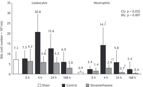
In the control group, the BAL neutrophil counts increased after VILI induction, peaking at 4 h after but returning to levels that were not statistically different from those observed for the sham group at 24 h and 168 h after. The BAL neutrophil counts also increased after VILI induction in the dexamethasone group, although to a lesser degree, and were lower in the dexamethasone group than in the control group
0.6
0.5
0.4
0.3
0.2
0.1
0
0 h 4 h 24 h 168 h
†
†
CG: p < 0.0001 DG: p = 0.043
Dexamethasone Control
Sham Acute Lung injury score 0.12
0.31
0.21
0.43
0.22 0.39
0.22
0.14 0.13 *
*
*
at 4 h and 24 h after VILI induction. Compared with the control group rats, the dexamethasone group rats showed lower numbers of leukocytes in the BAL at 4 h after VILI induction (Figure 4).
Arterial blood gases
Immediately after VILI induction (at 0 h), the PaO2 in the control group was lower than that observed for the sham group, although it showed progressive improvement at each subsequent time point. No such impairment in oxygenation was observed in the dexamethasone group (Table 2).
Lung wet-to-dry weight ratio
No differences in the lung wet-to-dry weight ratio were found between the dexamethasone and control groups.
DISCUSSION
The main inding of this study was that dexamethasone was able to reduce the inlammatory response caused by VILI and consequently reduce the acute lung injury, as well as attenuating the effects that such injury has on oxygenation. We also demonstrated that VILI occurred early after injurious MV and progressively improved after the removal of the insult, an improvement that was maintained throughout the observation period.
In this model of VILI induction, MV with high tidal volumes resulted in immediate lung injury, characterized by elevated acute lung injury scores after 1 h of such injurious ventilation. The score remained elevated at 4 h and 24 h after the ventilation and eventually normalized by 168 h after. These indings are in
accordance with those of previous studies showing that MV with high tidal volumes can induce VILI within the irst 60-90 min. In those studies, the histological alterations peaked within the irst 48 h after the insult and normalized within 7 to 14 days.(23,28)
We studied not only the temporal evolution of VILI but also the impact that the administration of an anti-inlammatory drug has on such injury. The decision to administer dexamethasone 30 min before injurious MV was based on previous experimental studies in which its anti-inlammatory effects on VILI were demonstrated.(24,25) In the present study, the animals
that received dexamethasone presented acute lung injury scores comparable to those of the animals in the sham group at all of the time points analyzed. In addition, the acute lung injury scores were lower in the dexamethasone group than in the control group at 4 h and 24 h after the injurious MV. That milder lung injury was associated with better oxygenation at the end of the MV. Although neither the inlammatory pathways involved in VILI nor the impact of corticos-teroid administration were evaluated, the milder lung injury and the lower pulmonary dysfunction observed among the animals treated with dexamethasone are in accordance with what is already known about the pathophysiology of VILI. It has been shown that MV with high tidal volumes promotes neutrophil chemotaxis and activation in the lungs, by at least two different mechanisms: activation of the transcription factor nuclear factor-kappa B, which mediates the release of certain pro-inlammatory cytokines, such as TNF-α, IL-6, and IL-8(23); and upregulation of CD11b/CD18
induced by the deformation of neutrophils as they pass through pulmonary capillaries compressed by alveoli
Table 1. Acute lung injury scores, by component, in rats submitted to 10 min of protective mechanical ventilation (sham group) or 1 h of injurious mechanical ventilation (dexamethasone and control groups, evaluated over time).a
Component Group
Time point pb
0 h 4 h 24 h 168 h
Interstitial neutrophils Control
Dexamethasone Sham
22.8 ± 6.9* 15.8 ± 6.6 12.3 ± 4.3
35.0 ± 5.2* 15.8 ± 8.4†
-30.3 ± 9.0* 18.2 ± 7.2†
-18.0 ± 3.3 16.8 ± 4.3
-< 0.0001 NS
Alveolar neutrophils Control Dexamethasone Sham
5.5 (9.5) 4 (7.5) 0.5 (2.5)
11.5 (4.8)* 2 (6.5)†
-11 (14.3)* 3 (4)†
-1 (0.5) 1 (1)
-0.001 NS
Hyaline membranes Control Dexamethasone Sham
0 (1) 0 (1.3)
0 (0)
0 (0.3) 0 (0.5)
-0 (1.3) 0 (0.8)
-0 (-0) 0 (0)
-NS NS
Proteinaceous debris Control Dexamethasone Sham
15 (18.3)* 12.5 (7)*
2 (4.3)
9.5 (12.3) 4 (6)
-13 (-13.3)* 9.5 (10.3)
-0 (-0) 0 (1)
-0.001 0.01
Septal thickening Control Dexamethasone Sham
14.5 ± 10.9 12.0 ± 5.5 10.0 ± 7.5
20.0 ± 6.7 18.7 ± 7.8
-16.8 ± 10.0 16.5 ± 5.5
-3.2 ± 2.3 3.2 ± 3.7
-NS NS
overinlated by the high tidal volumes.(29) Corticosteroids
are able to inhibit both pathways, thereby reducing the inlux of neutrophils into the lungs as well as their activation, thus inhibiting or minimizing the injury they induce.(25,30) These corticosteroid effects could explain
our indings, which showed that the treated animals presented lower numbers of neutrophils in the BAL luid, as well as less interstitial/alveolar iniltration by neutrophils and, consequently, milder lung injury.
Most experimental studies of the effects of corticoster-oids on VILI have been conducted in the acute phase of
the injury and have shown that these anti-inlammatory agents are able to decrease the VILI-induced release of local/systemic cytokines, neutrophil iniltration of the lungs, histological alterations, and functional impairment.(23,25,28,29) In the present study, we showed
that, in addition to those acute beneits, dexamethasone maintained the inlammation and lung injury at lower levels throughout the period of recovery after VILI induction.
In another experimental model of VILI, Hegeman et al.(22) showed that dexamethasone inhibits neutrophil
Table 2. Arterial blood gases in rats submitted to 10 min of protective mechanical ventilation (sham group) or 1 h of injurious mechanical ventilation, as well as lung wet-to-dry weight ratio in the latter (dexamethasone and control) groups, which were evaluated over time.a
Variable Group
Time point p
0 h 4 h 24 h 168 h
PaO2 (mmHg) Control Dexamethasone Sham
222 ± 98* 279 ± 76 388 ± 26
274 ± 87 313 ± 131
-387 ± 31 388 ± 71
-335 ± 71 385 ± 21
-0.003 NS
PaCO2 (mmHg) Control Dexamethasone Sham
52 ± 9 51 ± 6 51 ± 12
49 ± 6 44 ± 4
-54 ± 6 52 ± 10
-57 ± 10 51 ± 12
-NS NS
pH Control Dexamethasone Sham
7.25 ± 0.05 7.26 ± 0.02 7.30 ± 0.08
7.28 ± 0.05 7.26 ± 0.04
-7.27 ± 0.04 7.27 ± 0.06
-7.26 ± 0.09 7.27 ± 0.05
-NS NS
HCO3 (mEq/L) Control Dexamethasone Sham
22.6 ± 1.3 21.7 ± 2.5 21.9 ± 1.0
23.0 ± 3.3 19.3 ± 2.6
-23.0 ± 2.0 22.2 ± 2.2
-24.1 ± 2.7 24.3 ± 2.6
-NS NS
Lung W/D weight ratio Control
Dexamethasone
4.9 ± 0.2 4.9 ± 0.3
4.8 ± 0.7 4.7 ± 0.4
4.5 ± 0.2 4.4 ± 0.1
4.5 ± 0.1 4.9 ± 0.5
NS NS NS: non-signiicant; and W/D: wet-to-dry. aData are expressed as mean ± SD. bOverall signiicance of the differences among the time points within each group (one-way ANOVA). *p < 0.05 vs. sham group (Dunnett’s test).
35
30
25
20
15
10
5
0
0 h 4 h
4 h 24 h 168 h
CG: p < 0.032 DG: p = 0.007
Dexamethasone Control
Sham
BAL
(cell number × 10
4/ml)
Leukocytes Neutrophils
†
† 7.3
0 h 7.5 6.2
168 h 6.0
3.0 †
20.8
5.8
24 h 12.6
5.7
*
*
0.9 2.4
1.4 14.1
2.9 5.8
0.7 2.4
0.4
inlux and inlammatory mediator expression, as well as preventing vascular endothelial growth factor expression in the lungs. The authors found such expression to be associated with vascular leakage and with regulation of vascular permeability. However, they did not observe a protective effect of dexamethasone against alveolar-cap-illary barrier dysfunction, inding that glucocorticoid treatment had no effect on protein levels in the BAL luid or on the lung wet-to-dry weight ratio. (22) The
same result in relation to the lung wet-to-dry weight ratio was observed in our study. It has been shown that dexamethasone can increase extravascular lung water in animals exposed to hyperoxia, countering its positive effects on pulmonary injury so that the overall effect is neutral or even deleterious.(31)
We observed a lower mortality rate among the animals treated with dexamethasone than among those in the control group, although the difference was not statistically signiicant. This might have occurred because the number of animals studied was insuficient, given that survival was not an outcome for which we had planned. The study design prevented us from determining the cause of death in the 11 animals that died. Nevertheless, because those deaths occurred within the irst hours after VILI induction, a period during which the levels of lung inlammation and injury were highest, we can hypothesize that they were related to the lung injury. We can also infer that dexamethasone had a positive effect on survival. Another possible hypothesis, based on the results of other studies, is that VILI could have generated a systemic inlammatory response, with hemodynamic instability, that might have been improved by the administration of dexamethasone.(32) This hemodynamic effect was
demonstrated by Nin et al.,(25) who showed, in another
experimental model of VILI, that the administration of dexamethasone attenuated the lung injury and avoided the hemodynamic instability seen among the animals that did not receive pre-treatment.
We chose to ventilate rats at a tidal volume of 35 mL/kg, on the basis of previous studies that have used the same settings to study VILI and the effects of therapeutic strategies.(25,33,34) Although that tidal
volume might seem quite distant from what is employed in clinical practice, it should be borne in mind that, in ARDS, the alveolar area available to ventilation is highly limited (the so-called “baby lung” concept), and the tidal volume (which is set according to the ideal body weight) delivered to those available areas can be extremely high.(35)
Our study has some limitations that should be con -sidered. The analyses carried out at the different time points were performed only with the surviving animals, which constitutes a bias. The results obtained at each time point within the dexamethasone and control groups were compared with those obtained for the sham group, in which the animals were ventilated for only 10 min with a protective strategy. A more precise comparison would have included intubation and extubation of the sham group animals, euthanasia being performed at the same time points as in the other groups.
The design of this study, with induction of VILI followed by extubation and observation for a period of time, does not necessarily correlate with clinical practice. However, it furthers understanding of the course of VILI and of how the use of corticosteroids can alter that course. Although we have demonstrated the temporal evolution of VILI and its inlammatory nature, mainly by showing the capacity of dexamethasone to attenuate the injury, we did not evaluate the pathways involved. In conclusion, we have described the course of VILI induced by high tidal volumes and demonstrated that pre-treatment with dexamethasone is able to attenuate this injury in Wistar rats. The molecular mechanisms and the possible clinical role of corticosteroid therapy in VILI remain to be elucidated.
REFERENCES
1. Dreyfuss D, Saumon G. Ventilator-induced lung injury: lessons from experimental studies. Am J Respir Crit Care Med. 1998;157(1):294-323. http://dx.doi.org/10.1164/ajrccm.157.1.9604014
2. Pinhu L, Whitehead T, Evans T, Grifiths M. Ventilator-associated lung
injury. Lancet. 2003;361(9354):332-40. http://dx.doi.org/10.1016/ S0140-6736(03)12329-X
3. Ventilation with lower tidal volumes as compared with traditional tidal volumes for acute lung injury and the acute respiratory distress syndrome. The Acute Respiratory Distress Syndrome Network. N Engl J Med. 2000;342(18):1301-8. http://dx.doi.org/10.1056/ NEJM200005043421801
4. Amato MB, Barbas CS, Medeiros DM, Magaldi RB, Schettino GP, Lorenzi-Filho GP, et al. Effect of a protective-ventilation strategy on mortality in the acute respiratory distress syndrome. N Engl J Med. 1998;338(6):347-54. http://dx.doi.org/10.1056/ NEJM199802053380602
5. Serpa Neto A, Simonis FD, Schultz MJ. How to ventilate patients without acute respiratory distress syndrome? Curr Opin Crit Care. 2015;21(1):65-73. http://dx.doi.org/10.1097/ MCC.0000000000000165
6. Mechanical Ventilation Committee of the Brazilian Intensive Care Medicine Association; Commission of Intensive Therapy of the Brazilian Thoracic Society. Brazilian recommendations of mechanical
ventilation 2013. Part I. J Bras Pneumol. 2014;40(4):327-63. http:// dx.doi.org/10.1590/S1806-37132014000400002
7. Gattinoni L, Protti A, Caironi P, Carlesso E. Ventilator-induced lung injury: the anatomical and physiological framework. Crit Care Med. 2010;38(10 Suppl):S539-48. http://dx.doi.org/10.1097/ CCM.0b013e3181f1fcf7
8. Slutsky AS, Ranieri VM. Ventilator-induced lung injury. N Engl J Med. 2013;369(22):2126-36. http://dx.doi.org/10.1056/NEJMra1208707
9. Tremblay LN, Slutsky AS. Ventilator-induced injury: from barotrauma
to biotrauma. Proc Assoc Am Physicians. 1998;110(6):482-8.
10. De Prost N, Dreyfuss D. How to prevent ventilator-induced lung injury? Minerva Anestesiol. 2012;78(9):1054-66.
11. Uhlig S, Uhlig U. Pharmacological interventions in ventilator-induced lung injury. Trends Pharmacol Sci. 2004;25(11):592-600. http://dx.doi. org/10.1016/j.tips.2004.09.002
12. Barnes PJ. Corticosteroid effects on cell signalling. Eur Respir J. 2006;27(2):413-26. http://dx.doi.org/10.1183/09031936.06.0012540 4
13. Rhen T, Cidlowski JA. Antiinlammatory action of glucocorticoids--new mechanisms for old drugs. N Engl J Med. 2005;353(16):1711-23. http://dx.doi.org/10.1056/NEJMra050541
respiratory distress syndrome? Respir Care. 2010;55(1):43-55.
15. Thompson BT. Glucocorticoids and acute lung injury. Crit Care Med. 2003;31(4 Suppl):S253-7. http://dx.doi.org/10.1097/01. CCM.0000057900.19201.55
16. Agarwal R, Nath A, Aggarwal AN, Gupta D. Do glucocorticoids decrease mortality in acute respiratory distress syndrome? A meta-analysis. Respirology. 2007;12(4):585-90. http://dx.doi.org/10.1111/ j.1440-1843.2007.01060.x
17. Lamontagne F, Briel M, Guyatt GH, Cook DJ, Bhatnagar N, Meade M. Corticosteroid therapy for acute lung injury, acute respiratory distress syndrome, and severe pneumonia: a meta-analysis of randomized controlled trials. J Crit Care. 2010;25(3):420-35. http:// dx.doi.org/10.1016/j.jcrc.2009.08.009
18. Meduri GU, Marik PE, Chrousos GP, Pastores SM, Arlt W, Beishuizen A, et al. Steroid treatment in ARDS: a critical appraisal of the ARDS network trial and the recent literature. Intensive Care Med. 2008;34(1):61-9. http://dx.doi.org/10.1007/s00134-007-0933-3
19. Tang BM, Craig JC, Eslick GD, Seppelt I, McLean AS. Use
of corticosteroids in acute lung injury and acute respiratory distress syndrome: a systematic review and meta-analysis. Crit Care Med. 2009;37(5):1594-603. http://dx.doi.org/10.1097/ CCM.0b013e31819fb507
20. Peter JV, John P, Graham PL, Moran JL, George IA, Bersten A. Corticosteroids in the prevention and treatment of acute respiratory distress syndrome (ARDS) in adults: meta-analysis. BMJ. 2008;336(7651):1006-9. http://dx.doi.org/10.1136/ bmj.39537.939039.BE
21. Steinberg KP, Hudson LD, Goodman RB, Hough CL, Lanken PN, Hyzy R, et al. Eficacy and safety of corticosteroids for persistent acute respiratory distress syndrome. N Engl J Med. 2006;354(16):1671-84. http://dx.doi.org/10.1056/NEJMoa051693
22. Hegeman MA, Hennus MP, Cobelens PM, Kavelaars A, Jansen NJ, Schultz MJ, et al. Dexamethasone attenuates VEGF expression and inlammation but not barrier dysfunction in a murine model of ventilator-induced lung injury. PLoS One. 2013;8(2):e57374. http:// dx.doi.org/10.1371/journal.pone.0057374
23. Nin N, Lorente JA, de Paula M, El Assar M, Vallejo S, Peñuelas O, et al. Rats surviving injurious mechanical ventilation show reversible pulmonary, vascular and inlammatory changes. Intensive Care Med. 2008;34(5):948-56. http://dx.doi.org/10.1007/s00134-007-0959-6
24. Nin N, Lorente JA, Fernández-Segoviano P, De Paula M, Ferruelo A, Esteban A. High-tidal volume ventilation aggravates sepsis-induced multiorgan dysfunction in a dexamethasone-inhibitable manner. Shock. 2009;31(4):429-34. http://dx.doi.org/10.1097/ SHK.0b013e318188b720
25. Nin N, Peñuelas O, de Paula M, Lorente JA, Fernández-Segoviano
P, Esteban A. Ventilation-induced lung injury in rats is associated with organ injury and systemic inlammation that is attenuated by dexamethasone. Crit Care Med. 2006;34(4):1093-8. http://dx.doi. org/10.1097/01.CCM.0000205663.92384.E7
26. Matute-Bello G, Downey G, Moore BB, Groshong SD, Matthay MA, Slutsky AS, et al. An oficial American Thoracic Society workshop report: features and measurements of experimental acute lung injury in animals. Am J Respir Cell Mol Biol. 2011;44(5):725-38. http:// dx.doi.org/10.1165/rcmb.2009-0210ST
27. Peterson BT, Brooks JA, Zack AG. Use of microwave oven for determination of postmortem water volume of lungs. J Appl Physiol Respir Environ Exerc Physiol. 1982;52(6):1661-3.
28. Curley GF, Contreras M, Higgins B, O’Kane C, McAuley DF, O’Toole D, et al. Evolution of the inlammatory and ibroproliferative responses during resolution and repair after ventilator-induced lung injury in the rat. Anesthesiology. 2011;115(5):1022-32. http://dx.doi. org/10.1097/ALN.0b013e31823422c9
29. Ohta N, Shimaoka M, Imanaka H, Nishimura M, Taenaka N, Kiyono
H, et al. Glucocorticoid suppresses neutrophil activation in ventilator-induced lung injury. Crit Care Med. 2001;29(5):1012-6. http://dx.doi. org/10.1097/00003246-200105000-00027
30. Held HD, Boettcher S, Hamann L, Uhlig S. Ventilation-induced chemokine and cytokine release is associated with activation of nuclear factor-kappaB and is blocked by steroids. Am J Respir Crit Care Med. 2001;163(3 Pt 1):711-6. http://dx.doi.org/10.1164/ ajrccm.163.3.2003001
31. Ramsay PL, Smith CV, Geske RS, Montgomery CA, Welty SE. Dexamethasone enhancement of hyperoxic lung inlammation in rats independent of adhesion molecule expression. Biochem Pharmacol. 1998;56(2):259-68. http://dx.doi.org/10.1016/S0006-2952(98)00138-5
32. Imai Y, Parodo J, Kajikawa O, de Perrot M, Fischer S, Edwards V, et al. Injurious mechanical ventilation and end-organ epithelial cell apoptosis and organ dysfunction in an experimental model of acute respiratory distress syndrome. JAMA. 2003;289(16):2104-12. http:// dx.doi.org/10.1001/jama.289.16.2104
33. Dreyfuss D, Basset G, Soler P, Saumon G. Intermittent positive-pressure hyperventilation with high inlation positive-pressures produces pulmonary microvascular injury in rats. Am Rev Respir Dis. 1985;132(4):880-4.
34. Eyal FG, Hamm CR, Parker JC. Reduction in alveolar macrophages
attenuates acute ventilator induced lung injury in rats. Intensive Care Med. 2007;33(7):1212-8. http://dx.doi.org/10.1007/s00134-007-0651-x