Immunohistochemical analysis of apoptosis and
cell proliferation in lungs of premature infants with
chronic lung disease (bronchopulmonary dysplasia)
Análise imuno-histoquímica da apoptose e proliferação celular em pulmões de prematuros
com doença pulmonar crônica (displasia broncopulmonar)
Sandra Mara Witkowski; Lúcia de Noronha; Cristina T. Okamoto; Carlos F. Oldenburg Neto; Tammy Almeida; Seigo Nagashima; João A. Bahr
Pontifícia Universidade Católica do Paraná (PUCPR), Paraná, Brazil.
First submission on 21/06/16; last submission on 29/06/16; accepted for publication on 03/11/16; published on 20/12/16
ABSTRACT
Introduction: The pathophisiology of chronic lung disease (CLD), clinically known as bronchopulmonary dysplasia is not clear. It is believed that protective mechanisms, such as the release of inlammatory mediators and the activation of apoptotic and/or proliferative processes are activated in the lung tissue of premature infants in an attempt to repair tissue injury caused by exposure to oxygen and mechanical
ventilation. Objective: Assess the presence of apoptosis and cell proliferation in the lungs of premature infants with CLD, exposed to oxygen and/or mechanical ventilation, by analyzing the proteins expression: proliferating cell nuclear antigen (PCNA), phosphatase and tensin homolog (PTEN), B-cell lymphoma 2 (Bcl-2), tumor necrosis factor receptor family member (Fas), fas-associated protein with death domain (FADD), tumor necrosis factor receptor type 1-associated death domain protein (TRADD), Caspase 3 and Caspase 8. Material and methods: We analyzed 32 infants autopsies at gestational age of less than 34 weeks exposed to oxygen therapy. The study was divided into three groups: “classic” CLD, “new” CLD and “without” CLD. Immunohistochemical analysis was performed. Results and discussion: A
higher proliferation rate was observed in infants with CLD suggesting that longer exposure to mechanical ventilation may stimulates cell proliferation. The PTEN and Caspase 8 expressions were higher in the “new” CLD group, compared to the “without” CLD group, indicating that the “new” CLD form is more susceptible to apoptosis. Conclusion: Apoptosis and cell proliferation are involved in the pathophisiology of CLD. The “new” CLD form is more susceptible to apoptosis, while cell proliferation is more evident in the groups with CLD.
Key words: apoptosis; pharmacological biomarkers; cell proliferation; bronchopulmonary dysplasia; neonatology.
INTRODUCTION
The pathophysiology of bronchopulmonary dysplasia (BPD) can be the key for prevention of this disease, research with cell cycle proteins appear to be very promising for elucidating this pathology.
Apoptosis and cell proliferation are controlled physiological tissue repair processes regulated by mediators and proteins. Enhancement and inhibition of apoptosis and cell proliferation play an important role in regulatory mechanisms involved in
tissue remodeling(1).
In an attempt to repair tissue injury in the lungs of premature infants exposed to oxygen and mechanical ventilation, protective mechanisms, such as the release of inlammatory mediators and the activation of apoptotic and/or proliferative processes,
are triggered(2-4). These mechanisms seem to be part of the
pathophysiology of BPD.
BPD is a chronic lung disease (CLD) affecting mainly low-weight newborns (particularly those weighing less than 1500 g) that frequently receive mechanical ventilation and/or oxygen therapy. Neonates are clinically deined as having BPD/CLD if they still need oxygen supplementation at 28 days of life (or respiratory support at 36 weeks corrected age)(5). However, the pathological
alterations of BPD/CLD can be observed at an earlier age, before 28 days of life, and they are based on the presence of the classic characteristics of exacerbated inlammatory process, over-distended acini, atelectasis, ibrosis, squamous metaplasia and airway and vascular smooth-muscle hypertrophy in the “classic” form. With the use of antenatal steroids, exogenous surfactant and less aggressive mechanical ventilation in recent decades, another form of BPD, known as “new” BPD, have emerged to replace the “classic” form. The fundamental histological characteristic of “new” BPD/CLD is reduced alveolarization. The mechanisms involved in this abnormal alveolar development are not yet well
understood(6-8).
As cell proliferation and apoptosis can adversely affect lung-tissue homeostasis, the efforts to elucidate the pathophysiology of “new” BPD/CLD have involved research on the cell-cycle proteins, since this type of BPD/CLD appears to be the result of interrupted distal lung growth(9, 10).
Studies in animal models and cell cultures show that apoptosis increases in the lungs of premature infants exposed to oxygen and mechanical ventilation(11-15). Although few studies have been
carried out on humans(10, 16).
The aim of this study is to investigate the expression of the proteins involved in the cell-cycle [proliferating cell nuclear antigen (PCNA), phosphatase and tensin homolog (PTEN), B cell lymphoma 2 (Bcl-2), death receptor (Fas), Fas-associated protein with death domain (FADD), tumor necrosis factor receptor type 1-associated death domain protein (TRADD), aspartic acid protease 3 (Caspase 3) and cysteine-aspartic acid protease 8 (Caspase 8)] in lung autopsy samples from premature infants that required assisted ventilation, with pathological evidence of “classic” or “new” CLD, and compare them to the expression of the same proteins in newborns without pathological evidence of CLD.
MATERIAL AND METHODS
All 466 autopsy reports on infants who died in the neonatal period were reviewed by the Anatomic Pathology Service, Hospital de Clínicas (HC), Universidade Federal do Paraná (UFPR), between 1991 and 2007 (post exogenous surfactant era). The ethics review board of this hospital reviewed and approved the study (Register number 1099.138/2005-08; approved on 30 August 2005). Only premature infants with a gestational age between 25-34 weeks who were submitted oxygen therapy were included in the study. Premature infants with congenital
abnormalities, chronic intrauterine diseases or meconium aspiration syndrome, as well as those whose medical records or samples were inadequate, were excluded. Thirty-two cases were included in this study.
The medical records were analyzed to collect the data related to the clinical events such as gender, gestational age, birth weight, Apgar score at the irst e ifth minute, pregnancy hypertension event, gestational diabetes, chorioamnionitis, longer time with amniotic sac disruption, asphyxia, antibiotic therapy, surfactant therapy, necrotizing enterocolitis, bronchopneumonia, pulmonary hemorrhage, pulmonary hypertension, intracranial hemorrhage, sepsis, corrected age postpartum and time spent on mechanical ventilation and oxygen therapy.
Morphometric analysis
The formalin-ixed parafin-embedded lung tissue samples were reexamined and classiied into one of the three groups according to the histopathological and morphometric changes (without considering the clinical data): A) the histopathological indings of “classic” CLD (n = 11); B) the histopathological and morphometric indings of “new” CLD (n = 5); and C) “without”
histopathological and morphometric criteria for CLD (n = 16).
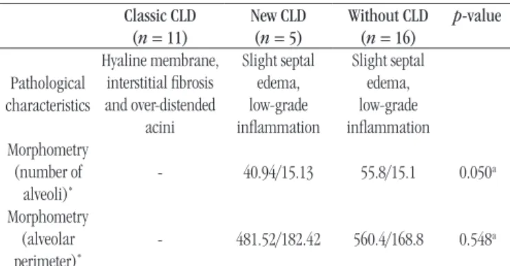
As the alterations in alveolar formation could be very subtle, the samples in groups B and C were analyzed only to conirm the existence of the morphometric criteria of “new” CLD in the group B(17-20), data are shown in Table 1.
Olympus BX 50 microscope (Olympus Optical Co. LTDA, Japan) linked to a Dino eye camera and a computer were used to obtain 10 medium power ield photomicrographs (200×) per case (the total area of medium power ield is 475.439 square micrometers). These photomicrographs (200×) were subjected to the morphometric analysis, using Image Pro Plus software. All alveoli in each medium power ield were counted and their perimeters were measured (micrometers). Mean values (number of alveoli and perimeter) for each patient were used for statistical
analysis.
Immunohistochemistry staining
Tissue microarrays were collected from lung samples from all the cases and analyzed immunohistochemically (four samples for each case with 3 mm diameter each), since this is the most suitable technique to analyze protein expression in this type of
material(17, 21).
TABLE 1 − Pathological data for the groups Classic CLD
(n = 11)
New CLD (n = 5)
Without CLD (n = 16)
p-value
Pathological characteristics
Hyaline membrane, interstitial ibrosis and over-distended
acini
Slight septal edema, low-grade inlammation
Slight septal edema, low-grade inlammation Morphometry
(number of
alveoli)*
- 40.94/15.13 55.8/15.1 0.050a
Morphometry (alveolar perimeter)*
- 481.52/182.42 560.4/168.8 0.548a
CLD: chronic lung disease; *: mean µm2/standard deviation; a: nonparametric
Mann-Whitney test, p < 0.05.
apoptosis, PTEN is associated with susceptibility to apoptosis, Fas, FADD, TRADD, Caspase 3 and Caspase 8 are proteins involved in apoptosis(22-25).
Samples were deparafinized with warm xylol (37ºC), dehydrated in a graded alcohol series and rehydrated with water. Methyl alcohol and H2O2 were used for the irst endogenous peroxidase blocking, and distilled water and H2O2
for the second. The samples were then incubated overnight with the following primary antibodies: PCNA, PTEN, anti-Bcl-2, anti-Fas, anti-FADD, anti-TRADD, anti-Caspase 3 and anti-Caspase 8. Anti-PCNA is a rat monoclonal antibody, clones PC10, DAKO™, DakoCytomation, Hostrup, Denmark. Anti-PTEN is a rat monoclonal antibody, 28H6, NOVOCASTRA™, Newcastle, United Kingdom. Anti-Bcl-2 is a rat monoclonal antibody, clones 124, DAKO™, DakoCytomation, Hostrup, Denmark. Anti-Fas is a rat monoclonal antibody, clones GM30, 1:160, NOVOCASTRA™, Newcastle, United Kingdom. Anti-FADD is a rabbit polyclonal antibody, ab55399, 1:30, ABCAM™, Cambridge, United States of America. Anti-TRADD is a rat monoclonal antibody, clones 18A11, 1:50, DAKO™, DakoCytomation, Hostrup, Denmark. Anti-Capase 3 is a rabbit polyclonal antibody, clone 3CSP03, 1:200, BIOSYSTEMS™, Pleasanton, Canada. Anti-Caspase 8 is a rabbit polyclonal antibody, 1:100, ABR™, Colorado, United States of America. The secondary antibody was incorporated to a dextran polymer for samples incubation for 30 minutes. DAKO ADVANCED™ HRP SYSTEM, DakoCytomation, Inc from CA, USA was used to reveal immunoreactivity, and the slides were counterstained with Mayers’ hematoxylin.
Septal cells (ibroblasts, endothelial cells and mononuclear cells) and alveolar cells (pneumocytes) were considered positive when exhibiting the brown nuclear staining (PTEN and PCNA) or perinuclear (Bcl-2), or citoplasmatic staining for Caspase 3,
Caspase 8, TRADD and FADD or membrane staining for Fas as expected for each antibody. An Olympus BX50 microscope (Olympus Optical Co, Ltd., Japan) with a 40× objective, was used
to examine the slides.
The staining was interpreted as follows: for PCNA, ten high-power ields for each four samples of each case were examined at a magniication of 400×. The total number of positive cells in the high-power ields for the septa and alveoli were counted, and the mean number of positive cells for all four samples was calculated (the diameter of high-power ield is 106 micrometers). For PTEN and Bcl-2, a score was assigned according to the degree of positive staining as well as a pattern of staining. For each of the four samples of each case a degree of positive staining was assigned as follows: 0 for negative samples, 1 for weakly positive staining, and 2 for strongly positive staining. The total score for each case were added to provid the total sum scores for the four samples. The pattern of staining was scored as follows: 1 for focal, 2 for multifocal and 3 for diffuse. The scores for each case were determined by adding the scores for each of the four samples(26).
For Fas, FADD, TRADD, Caspase 3 and Caspase 8 the immunostained slides were observed using an optical microscope Olympus® BX50 (Tokyo, Japan), coupled to a Dino eye video
camera enhanced by image analysis software Image Pro Plus™ (Maryland, USA). For each sample, 12 photomicrographs were taken in high-power ield [(HPF) = 400×], with a total area of 115,226.1 µm2 and with 1024 × 768 pixels each.
The positive control HPF photomicrography was chosen as the “mask”, which contained adequate levels of positive tissue immunoexpression signal. The mask was then superimposed to the samples photomicrographs. Based on the ideal positive tissue immunoexpression signal obtained from the mask, the image analysis software Image Pro Plus(TM) identiied the positive
areas in the samples and is able to transform these results into positive tissue immunoexpression area per square micron (µm2).
The area in µm2 obtained with this method was divided by the
constant 115,226.1 µm2, which is the total area of the HPF
observed, thus generating a percentage value for the positive tissue immunoexpression area for each HPF. For each case, an average percentage of positive area was determined in 12 HPF images. TRADD was not submitted to morphometric analysis because most cases were negative for this protein.
The observer did not have prior knowledge to which group the samples belong.
Kruskal-Wallis, and Fisher were used for statistical analysis. The group “without” CLD was used as the control group for the Dunnett’s test. The signiicance level was set at p < 0.05. For
the Fisher test the signiicance level was correct for Bonfarroni
(p < 0.017). Analysis of covariance (Ancova) was tested using
corrected age and birth weight as covariates.
RESULTS
Clinical data
The “classic” CLD group received oxygen therapy for the longest period of time (21.36 days) than the other groups. The clinical proile of the study population is shown in Tables 2, 3
and 4.
Immunohistochemical data
There was a high PCNA tissue immunoexpression, in septal cells in the “classic” form of the disease (p = 0.057). PTEN
(p = 0.015) and Caspase 8 (p = 0.010) tissue immunoexpression
was greater in the “new” CLD group. FADD expression was greater in the group without CLD (p < 0.001). All cases were analyzed
immunohistochemically for the proteins, and the results are shown in Table 5 andFigure.
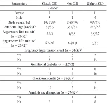
TABLE 2 − Clinical date for the groups
Parameters Classic CLD New CLD Without CLD Gender
Female 6 4 11
Male 5 1 5
Birth weight* (g) 1021/289 1340/398 959/358 Gestational age (weeks)*b 32/3.3 33.4/3.1 28.8/2.6 Apgar score first minute*
(n = 29/32)† 2.6/2 6/3.5 3.5/2.7 Apgar score fifth minute*
(n = 29/32)† 6.2/2.6 8.4/1.9 5/3.5 Pregnancy hypertension event (n = 31/32)†
Yes 3 3 2
No 8 2 13
Gestational diabetes (n = 32/32)†
Yes 0 1 0
No 11 4 16
Chorioamnionitis (n = 32/32)†
Yes 1 0 2
No 10 5 14
Amniotic sac disruption (n = 27/32)†
Yes 1 1 4
No 9 4 8
CLD: chronic lung disease; *:mean/standart deviation; †: number of cases; a:
Kruskal-Wallis nonparametric test (p < 0.05); b:p = 0.005.
TABLE 3 − The postnatal clinical factors for the groups
Parameters Classic CLD (n = 11)
New CLD (n = 5)
Without CLD
(n = 16) p-value Patent ductus arteriosus
Yes 10 2 2 0.0003**
No 1 3 14
Antibiotic therapy
Yes 10 4 8 > 0.017‡
No 1 1 8
Surfactant therapy
Yes 7 0 1 0.0089
No 4 5 15
Necrotizing enterocolitis
Yes 7 2 1 0.002‡
No 4 3 15
Bronchopneumonia
Yes 6 0 1 0.009‡
No 5 5 15
Pulmonary hemorrhage
Yes 3 1 1 > 0.017‡
No 8 4 15
Pulmonary hypertension
Yes 2 1 0 > 0.017‡
No 9 4 16
Intracranial hemorrhage
Yes 4 2 1 > 0.017‡
No 7 3 15
Sepsis
Yes 11 4 3 0.001‡
No 0 1 13
Corrected gestational age (weeks)*
32/3.3 33.4/3.1 28.8/2.6 0.005§
Survival time (days)*
23.4/12.3 12.4/5.5 1.4/1.6 < 0.001§
Asphyxia
Yes 6 1 9
> 0.017‡
No 5 4 7
CLD: chronic lung disease;*mean/ standart deviation; **: Pearson test; ‡: Fisher test, p >
0.017 (adjusted for Bonferroni); §:Kruskal-Wallis nonparametric test (p < 0.05).
TABLE 4 − Use of oxygen by the groups
Classic CLD New CLD Without CLD
Days of mechanical ventilation* 12.90/9.23 3.4/3.91 1.33/1.69 Days of CPAP* 5.6/7.66 4.4/3.2 1.91/1.63 Days of oxygen tent* 3.8 1.8 0 Days of oxygen catheter* 0.27 0.8 0
FiO2Max (%)* 95/12.24 85/30 98.75/7.07 PIP Max (mmHg)* 21.16/3.37 20/0 23.18/6.98
PEEP Max (mmHg)* 4.7 5 5.05
Days of oxygen therapy/survival time* 21.36/12.26 10.4/2.79 1.4/1.6
DISCUSSION
BPD is a chronic lung disease, characterized by an impaired lung function due to a reduced inal alveolar number and vascular growth. It is the most common sequelae of ventilation premature
neonates(27).
Various studies are currently being carried out to elucidate the pathogenesis of this condition. It is known to be multifactorial, with a genetic predisposition, prolonged use of oxygen at high concentrations, insuficient surfactant, exposure to mechanical ventilation (leading to volutrauma, biotrauma and barotrauma) and pre or postnatal infection as the main etiologic factors(8).
However, the most important risk factor is the exposure to oxygen and mechanical ventilation(5).
From the pathological point of view, a injury and repair process, with early alveolar and interstitial inlammation and ibrosis, characterizes “classic” CLD. “New” CLD, on the other hand, as observed by Coalson et al. (1999) in baboon lungs, is characterized by alveolar hypoplasia, variable saccular wall ibrosis and minimal airway disease. Coalson et al. (1995) also
reported decreased alveolarization and internal alveolar surface area. These indings were conirmed by other authors in human, sheep, lamb and rabbit lungs(17-20, 28-30).
During lung development there is a natural equilibrium between apoptosis and cell proliferation(31). Loss of this equilibrium may result
in chronic lung pathologies in newborns as a result of impaired vascular and alveolar growth. The pathophysiology of alveolar hypoplasia, which is present in the “new” CLD, has not been fully elucidated to date. It is known that apoptosis and cell proliferation are implicated in this process and that pulmonary exposure to oxygen and cyclic stretching in mechanical ventilation can trigger changes in the cell-cycle, particularly changes in apoptosis(10). Fewer alveoli
and decreased alveolar perimeter were found in the “new” CLD group compared to the group “without” CLD, conirming impaired alveolar growth in neonates with this pathology.
The number of females is greater than males. Neonates in the “new” CLD group had heavier weight and more mature at birth than in the other two groups (mean weight 1,340 g and mean corrected age 33.4 weeks). The group “without” CLD was more premature at birth (mean 28.8 weeks). The presence of maternal pathology such as hypertensive disorders of pregnancy and gestational diabetes did not inluence the incidence of CLD.
From the point of view of infection (chorioamnionitis and membranes rupture for more than 18 hours), comparing the groups, there were no statistically signiicant differences. However the postnatal infection (bronchopneumonia, necrotizing enterocolitis and sepsis) was greater in the “classic” CLD group,
TABLE 5 − Immunohistochemical expression of proteins PCNA, PTEN, Bcl-2, Fas, FADD, Caspase 3 and Caspase 8 in each group
Protein
Classic CLD (n = 11)
m/SD
New CLD (n = 5) m/SD
Without CLD (n = 16)
m/SD
p-value§
PCNA septal (n = 26/32)* 69.1/55.8 61.5/38.1 24.2/40.5 0.057 PCNA alveoli (n = 26/32)* 4.59/3.63 3.25/1.55 3.21/5.55 0.246 Bcl-2 degree 2.18/1.83 1.8/1.79 0.94/1.61 0.236 Bcl-2 pattern 2.18/1.83 2.18/1.83 0.88/1.45 0.196 PTEN degree* (n = 29/32)* 4.67/2 7.5/1 3.63/2.39 0.015 PTEN pattern* (n = 29/32)* 5.78/3.15 8.75/3.95 5/3.86 0.2
Fasa 0.032/2289.2 0.039/4650.9 0.030/2483.3 0.967 FADDa 0.028/2476.3 0.024/1073.5 0.104/3425.1 < 0.001 Caspase 3a 0.012/777.3 0.012/1060.5 0.014/1235 0.993 Caspase 8a 0.031/589 0.046/951.7 0.032/766.3 0.010
*: number of positive cells; a: immunopositive area (%/SD); §:Kruskal-Wallis
nonparametric test (p < 0.05).
PCNA: proliferating cell nuclear antigen; PTEN: phosphatase and tensin homolog; Bcl-2: B cell lymphoma 2; Fas: tumor necrosis factor receptor family member; FAAD: fas-associated protein with death domain; CLD: chronic lung disease; m: mean; SD: standard deviation.
FIGURE − Immunohistochemical reaction
A, B and C – immunohistochemical reaction for PTEN. A) “classic” CLD; B) “new” CLD; C) “without” CLD groups cases. There are a few septal and alveoli positive cells in the “without” CLD group, and much more positive septal and alveoli cells in the “new” and the “classic” CLD groups (400×).
D, E and F – Immunohistochemical reaction for Caspase 8. D) “classic” CLD; E) “new” CLD; F) “without” CLD groups cases. There are more positive cells in the “new” CLD group (400×).
G, H and I – Immunohistochemical reaction for FADD. G) “classic” CLD; H) “new” CLD; I) “without” CLD groups. There are more positive cells in the “without” CLD group (400×).
PTEN: phosphatase and tensin homolog; CLD: chronic lung disease; FAAD: fas-associated protein with death domain.
A
D
G
B
E
H
C
F
showing that infection could increase the risk of developing CLD, or because these neonates might be more exposed to oxygen and mechanical ventilation. The presences of pathologies such as pulmonary hemorrhage, pulmonary hypertension and intracranial hemorrhage have no statistically signiicant differences in the groups.
The length of oxygen therapy use and the time of survival were almost a like. The lungs of premature infants were in different stages of development during the repair/remodeling processes. In the group “without” CLD, the length of time of oxygen therapy and the survival time (mean 1.4 days) were shorter than the other two groups. This could explain the reason why this group has not shown any pathological features of CLD (Table 4).
The “classic” CLD group received oxygen and presented longer survival (21.36 days) compared to the other groups but was, therefore, more exposed to high peak inspiratory pressure and positive expiratory pressure, showing that the oxygen and the agressive mechanical ventilation was involved in CLD development. On the other hand, the “classic” group had more time for cell differentiation, a process that is characteristic for this phase.
It is important to bear in mind that we use only pathological criteria to deine these three groups, without considering the clinical data.
Statistically signiicant differences were found when expression of the proteins was analyzed.
PCNA is a nuclear protein which acts as an accessory factor of deoxyribonucleic acid (DNA) polymerase delta, it is required for DNA replication and repair, and consequently for cell replication(22).
Its expression is increased in cells which are proliferating. If PCNA is reduced or not present in a cell, apoptosis will take place(32). In
this study there was a higher proliferation index, as evidenced by a high PCNA tissue immunoexpression, in septal cells in the “classic” form of the disease compared to the group “without” CLD (p = 0.057), showing a statistical tendency (Table 5). The pathological features of the “classic” form showed an obliterative bronchiolitis, an interstitial ibrosis that is compatible with proliferative activity. These indings may be associated with the ability of a pulmonary cell to proliferate in response to mechanical
strain(33, 34), knowing that the “classic” form was submitted to
mechanical ventilation for more days than the group “without” CLD. Regarding the exposure to oxygen, some studies with cell cultures showed that hyperoxia inhibits cell proliferation(13, 14),
while another study say the opposite(35).
There was no difference in proliferation indices between the “new” CLD and “classic” CLD group, and between the “new” CLD and the group “without” CLD. There were no statistically signiicant differences in the degree of positivity for PCNA in alveolar cells between the groups, which were all positive for this
marker as cell proliferation is normally present in the canalicular and saccular stages of lung development, corresponding to the 22nd to 36th weeks of gestation(10).
PTEN is a lipoprotein phosphatase that plays an important role in cell proliferation and apoptosis by negatively regulating the cell-cycle and suppressing growth(36). PTEN induces cell death
by increasing the tumor suppressor activity of p53 and reducing cell proliferation by blocking the phosphoinositol 3 kinase/ Akt signaling pathway, with a consequent reduction in Ki67, a protein associated with cell proliferation(23). The greater the
number of cells expressing this protein in a particular tissue, the more likely the tissue would have a higher apoptotic index(36).
In this study, PTEN tissue immunoexpression was greater in the “new” CLD group (degree and pattern score tending to be strong and diffuse staining) than in the group “without” CLD. Caspase 8 immunoexpression was also greater in the “new” CLD group. Caspase 8 is a protein that belongs to the apoptotic pathways. These indings were statistically signiicant and could indicate that the “new” form of the disease presents more cells susceptible to apoptosis than the group “without” CLD, consistent with the decreased alveolarization observed in this phase of the disease (Figure and Table 5)(6). Conirming these indings, previous
studies in animal models have shown that there is increased apoptosis in lungs exposed to oxygen, which are susceptible to barotrauma, volutrauma and biotrauma(11, 12). A study by
Hargitai in 2001, also found a high apoptotic index in alveolar and bronchial cells in lungs with BPD.
There were no statistically signiicant differences in PTEN tissue immunoexpression between the “classic” CLD group and the group “without” CLD, compatible with data in literature indicating that “classic” CLD is more associated with inlammatory response than with the apoptosis processe(30).
There was no statistically signiicant difference in Bcl-2 tissue immunoexpression between the groups. Bcl-2 is an anti-apoptotic protein associated with the regulation of apoptosis, it is of special importance as it is one of the proteins that determine which cells will undergo apoptosis and in which apoptosis it will
be inhibited(24).
In the present study, Bcl-2 expression was higher in “classic” CLD than in the group “without” CLD (Table 5), but this inding has not been statistically signiicant. However, this result might be related to the better long-term survival of the group with “classic” CLD, which allowed more time for cell differentiation and consequently led to increased Bcl-2 expression, indicating higher resistance to apoptosis.
the adapter protein Fas-associated death domain, FADD, and the initiator cysteine protease Caspase 8. Subsequent activation of the effector caspases through mitochondria dependent or independent pathways results in activation of Caspase 3, the key effector caspase. Activated Caspase 3 cleaves a variety of substrates, including DNA repair enzymes, cellular and nuclear structural proteins, endonucleases, and many other cellular constituents, culminating in effective cell death(37-39).
FADD is an adaptor molecule that mediates cell apoptotic signals. It can allow recruiting Caspase 8 or Caspase 10 to the activated Fas receptor. The resulting aggregate called the death-inducing signaling complex (DISC) performs Caspase 8 proteolytic activation. Active Caspase 8 initiates the subsequent cascade of caspases mediating apoptosis, and then FADD binding to Fas receptor is degraded. The antibody used in this study can bind Fas into FADD portion, in free FADD. Thus in this study, FADD expression was higher in the group without CLD. This may indicate that apoptosis is lower or did not happen in the group without CLD (FADD-Fas was not degraded) comparing to other groups (Figure)(40).
For immunoexpression of the other proteins involved in cell apoptosis (Fas and Caspase 3) there were no statistically signiicant differences in the groups. In the literature, the function of Caspase 3 is not clear, Caspase 3 could be involved in the pathophisiology of oxygen and ventilation inducing apoptosis(10).
CONCLUSION
Apoptosis was involved in the pathophysiological of CLD. The process of lung tissue lesion appears to be related to an imbalance between inlammatory response, apoptosis and cell proliferation that affects alveolar formation and pulmonary vascular growth(2-4).
Increased apoptosis (“new” form) and cell proliferation in lungs with CLD were observed in this study.
FINANCIAL DISCLOSURE
ACKNOWLEDGEMENTS
The other authors have indicated they have no inancial relationships relevant to the disclure of this article.
ETHICAL CONDUCT OF RESEARCH
The authors state that they have obtained appropriate institutional review board approval or have followed the principles outlined in the Declaration of Helsinki for all human or animal experimental investigations. The ethics review board of the HC-UFPR reviewed and approved the study (Register number 1099.138/2005-08; approval 30 August 20).
RESUMO
Introdução: A fisiopatologia da doença pulmonar crônica, clinicamente conhecida como displasia broncopulmonar (DBP), ainda é incerta. Acredita-se que mecanismos de proteção, como liberação de mediadores inflamatórios e ativação de processos apoptóticos e/ou proliferativos, são acionados no tecido pulmonar de prematuros na tentativa de reparar os danos teciduais causados pela exposição ao oxigênio e à ventilação mecânica. Objetivo: Avaliar a existência de apoptose e proliferação celular em pulmões de neonatos prematuros com DBP, expostos ao oxigênio e/ou à ventilação mecânica, por meio do estudo da expressão das proteínas: antígeno nuclear de proliferação celular (PCNA), homólogo da fosfatase e tensina (PTEN), linfoma de células B 2 (Bcl-2), membro da família de receptor do fator de necrose tumoral (Fas), proteína de domínio de morte associada ao Fas (FADD), proteína do domínio de morte associada ao receptor do fator de necrose tumoral (TRADD), Caspase 3 e Caspase 8. Material e método: Foram analisadas 32 autópsias de recém-nascidos, com idade gestacional inferior a 34 semanas, expostos ao oxigênio. O estudo foi dividido em três grupos: DBP “clássica”, DBP “nova” e “sem” DBP; realizou-se estudo imuno-histoquímico. Resultados e discussão: Um índice de proliferação mais elevado foi observado nos recém-nascidos com DBP, sugerindo que o maior tempo de exposição à ventilação mecânica pode estimular a proliferação celular. A expressão das proteínas PTEN e Caspase 8 foram maiores no grupo da DBP “nova” em relação ao grupo sem DBP, indicando que a DBP “nova” é mais suscetível à apoptose. Conclusão: A apoptose e a proliferação celular estão envolvidas na fisiopatologia da DBP, sendo a apoptose mais evidente no grupo com DBP “nova”.
REFERENCES
1. Pedreira PR, Garcia-Prieto E, Albaiceta GM, Taboada YF. Respuesta inlamatória y apoptosis em La lesión pulmonar aguda. Med Intensiva. 2006; 30: 268-75.
2. Speer CP. Inlammation and bronchopulmonary dysplasia. Semin Neonatol. 2003; 8: 29-38.
3. Speer CP. Inlammation and bronchopulmonary dysplasia: a continuing story. Semim Fetal Neonatal Med. 2006; 11: 354-62. 4. Turato G, Zuin R, Saetta M. Pathogenesis and pathology of COPD. Respiration. 2001; 68: 117-28.
5. Jobe H, Bancalari A, Bancalari E. Bronchopulmonary dysplasia. Am J Respir Crit Care Med. 2001; 163: 1723-9.
6. Shawn K, Conway A, Conway S. Aberrant signaling pathways of the lung mesenchyme and their contributions to the pathogenesis of bronchopulmonary dysplasia. Birth Defects Res A Clin Mol Teratol. 2012; 94: 3-15.
7. Tapia JL, Agost D, Alegria A, et al. Bronchopulmonary dysplasia: incidence, risk factors and resource utilization in a population of South American very low birth weight infants. J Pediatr. 2006; 82: 15-20. 8. Bhandari A, Bhandari V. Bronchopulmonary dysplasia an update. Indian J Pediatr. 2007; 74: 73-7.
9. Shaw GM, O’Brodovich HM. Progress in understanding the genetics of bronchopulmonary dysplasia. Seminars in Perinatology. 2013; 37: 85-93.
10. May M, Ströbel P, Preisshofen T, Seidenspinner S, Marx A, Speer CP.
Apoptosis and proliferation in lungs of ventilated and oxygen-treated preterm infants. Eur Respir J. 2004; 23: 113-21.
11. Barazzone C, Horowitz S, Donati YR, Rodriguez I, Piguet PF. Oxygen toxicity in mouse lung: pathways to cell death. Am J Respir Cell Mol Biol. 1998; 19: 573-81.
12. McGrath-Morrow SA, Stahl J. Apoptosis in neonatal murine lung exposed to hyperoxia. Am J Respir Cell Mol Biol. 2001; 25: 150-5. 13. Hussain N, Wu F, Christian C, Kresch MJ. Hyperoxia inhibits fetal rat lung ibroblast proliferation and expression of procollagens. Am J Physiol. 1997; 273: 726-32.
14. Rancourt RC, Staversky RJ, Keng PC, O’Reilly MA. Hyperoxia inhibits proliferation of Mv1Lu epithelial cells independent of TGF-β
signaling. Am J Physiol. 1999; 277: 1172-8.
15. Mokres LM, Parai K, Hilgendorff A, et al. Prolonged mechanical ventilation with air induces apoptosis and causes failure of alveolar septation and angiogenesis in lungs of newborn mice. Am J Physiol Lung Cell Mol Physiol. 2010; 298: 23-35.
16. Hargitai B, Szabó V, Hajdú J, et al. Apoptosis in various organs of preterm infants: histopathologic study of lung, kidney, liver, and brain of ventilated infants. Pediatr Res. 2001; 50: 110-4.
17. Okamoto CT, Silva LLG, Noronha L, Bahr JA. Análises histopatológica e morfométrica no diagnóstico da “nova” displasia broncopulmonar e comparação clinicopatológica com a forma clássica da doença. J Bras Patol Med Lab. 2009; 45: 155.
18. Coalson JJ, Winter V, Lemos RA. Decreased alveolarization in baboon survivors with bronchopulmonary dysplasia. Am J Respir Crit Care Med. 1995; 152: 640-6.
19. Coalson JJ, Winter VT, Siler-Khodr T, Yoder BA. Neonatal chronic lung disease in extremely immature baboons. Am J Respir Crit Care Med. 1999; 160: 1333-46.
20. Mascaretti RS, Mataloun MMGB, Gibelli MACB, et al. Preterm rabbits exposed to prolonged hyperoxias as a model for the study of bronchopulmonary dysplasia. Pediatr Res. 2003; 53: 436.
21. Cronin M, Pho M, Dutta D, et al. Measurement of gene expression in archival parafin-embedded tissues: development and performance of 92- gene reverse transcriptase-polymerase chain reaction assay. Am J Pathol. 2004; 164: 35-42.
22. Gary R, Kim K, Cornelius HL, Park MS, Matsumoto Y. Proliferating
cell nuclear antigen facilitates excision in long-patch base excision repair. J Biol Chem. 1998; 274: 4354-63.
23. Weng LP, Brown JL, Eng C. PTEN induces apoptosis and cell cycle arrest through phosphoinositol-3-kinase/Akt-dependent and independent pathways. Hum Mol Genet. 2001; 10: 237-42.
24. Adams JM, Cory S. The Bcl-2 protein family: arbiters of cell survival. Science. 1998; 281: 1322-6.
25. De Paepe ME, Gundavarapu S, Tantravahi U, et al. Fas-ligand-induced apoptosis of respiratory epithelial cells causes disruption of postcanalicular alveolar development. Am J Pathol. 2008; 173: 42-56.
26. Allred DC, Harvey JM, Berardo M, Clarck GM. Prognostic and predictive factors in breast cancer by immunohistochemical analysis. Mod Pathol. 1998; 11: 144-68.
27. Sánchez Luna M, Moreno Hernando J, Bolet Mussons F, et al. Displasia broncopulmonar: deiniciones y clasiicación. An Pediatr. 2013; 79: 4. 28. Grisai D, Tassone E, Dedja A, et al. L-citrulline prevents alveolar and vascular derangement in a rat model of moderate hyperoxia-induced lung injury. Lung. 2012; 190: 419-30.
29. Grisai D, Pozzobon M, Dedja A, et al. Human amniotic luid stem cells protect rat lungs exposed to moderate hyperoxia. Pediatr Pulmonol. 2013; 48: 1070-80.
30. Rebello CM, Mascaretti RS. A nova displasia broncopulmonar. In: Manual de atualizações em Neonatologia. Rio de Janeiro: Panmericana; 2004. p. 87-132.
31. Weaver TE, Whitsett JA. Function and regulation of expression of pulmonary surfactant-associated proteins. Biochem J. 1991; 15: 249-64.
32. Paunesku T, Mittal S, Protić M, et al. Proliferating cell nuclear antigen (PCNA): ringmaster of the genome. Int J Radiat Biol. 2001; 77: 1007-21.
33. Maniscalco WM, Watkins RH, O’Reilly MA, Shea CP. Increased epithelial cell proliferation with chronic lung disease. Am J Physiol Lung Cell Mol Physiol. 2002; 283: 991-1001.
35. Gao Y, Xue XD, Li JY, Wang N. Expression and roles of CDK4 and p21 in lung tissues of premature rats with hyperoxia-induced chronic lung disease. CJCP. 2007; 9: 595-600.
36. Di Cristofano A, Pandoli PP. The multiple roles of PTEN in tumor suppression. Cell. 2000; 100(4): 387-90.
37. Nagata S. Fas ligand-induced apoptosis. Annu Rev Genet. 1999; 33: 29-55.
38. Sharma K, Wang RX, Zhang LY, et al. Death the Fas way: regulation and pathophysiology of CD95 and its ligand. Pharmacol Ther. 2000; 88: 333-47.
39. Walczak H, Krammer PH. The CD95 (APO-1/Fas) and the TRAIL (APO-2L) apoptosis systems. Exp Cell Res. 2000; 256: 58-66.
40. Anti-FADD ab55399, ABCAM™, Cambridge, United States of America.
CORRESPONDING AUTHOR
Lúcia de Noronha e/ou Sandra Mara Witkowski