The spectrum of non alcoholic fatty liver disease in morbidly obese patients. Prevalence
and associate risk factors
1Suerda Guiomar FeijóI, José Milton de Castro LimaII, Maria Aparecida Alves de OliveiraIII, Régia Maria Vidal PatrocínioIV, Luis
Gonzaga Moura-JuniorV, Antônio Borges CamposVI, José Wellington Oliveira LimaVII, Lúcia Libanez Bessa Campelo BragaVIII
IMD, MSc, Gastroenterologist, University Hospital Walter Cantideo, Ceara Federal University (UFC), Brazil. Conception of the study, acquisition and
interpretation of data, manuscript writing.
IIPhD, Associate Professor, Division of Clinical Gastroenterology, Department of Internal Medicine, UFC, Fortaleza-CE, Brazil. Acquisition of data,
critical revision.
IIIFellow PhD degree, Postgraduate Program in Surgery, UFC, Fortaleza-CE, Brazil. Acquisition and interpretation of data. IVMD, Department of Pathology and Legal Medicine, UFC, Fortaleza-CE, Brazil. Histological analysis, critical revision. VMD, MSc. Assistant Professor, Department of Surgery, Ceara State University, Brazil. Surgical procedures, acquisition of data. VIPhD, Associate Professor, Department of Surgery, UFC, Fortaleza-CE, Brazil. Surgical procedures, acquisition of data. VIIPhD, Department of Pathology and Legal Medicine, UFC, Fortaleza-CE, Brazil. Statistical analysis, interpretation of data.
VIIIPhD, Full Professor,Department of Internal Medicine, UFC, Fortaleza-CE, Brazil. Conception and design of the study, manuscript writing, critical
revision.
ABSTRACT
PURPOSE: To determine the prevalence of non alcoholic fatty liver disease (NAFLD) and non alcoholic steatohepatitis (NASH) in morbidly obese patients undergoing bariatric surgery and to identify risk factors associated with the disease spectrum.
METHODS: Liver biopsy was performed in 60 patients who underwent bariatric surgery, after other causes of liver disease were excluded. Clinical, biochemical and histological features were evaluated.
RESULTS: NAFLD was detected in ifty-seven patients (95%) of the sample and forty patients (66.7%) of the total sample met the
criteria for NASH. Perisinusoidal ibrosis was only found in three (7.5%) patients with NASH. The γGT was an independent predictive
factor associated with the degree of hepatic steatosis. The variables such as dyslipidemia and ALT were independently associated with
the presence of Mallory’s corpuscles with the following values, respectively, OR 0, 05, 95% CI 0.002 to 0.75, P = 0.031 and OR 10, 99, 95% CI 1.44 to 83.93, P = 0.021.
CONCLUSIONS: Non alcoholic fatty liver disease seems to be an obese-related condition with approximately half of asymptomatic
morbidly obese patients having histological NASH. The γGTwas an independent predictor of the degree of steatosis.
Introduction
Non alcoholic fatty liver disease (NAFLD) is considered one of the most common liver diseases, affecting a wide spectrum of patients worldwide. The estimated prevalence in the general
population is 10 to 24% and 70% in obese patients1,2
. Given
the obesity epidemic, it is thought to become one of the most important liver pathology in the future. Increased levels of insulin,
triglycerides, low HDL, type 2 diabetes glucose intolerance are
considered important risk factors.
Approximately 60% of patients with NAFLD have
metabolic syndrome3.There is a wide spectrum of liver histology in NAFLD, ranging from steatosis to steatohepatitis (NASH),
advanced ibrosis and cirrhosis4
. NASH is characterized by zone 3 macrovesicular steatosis, inlammatory changes, ibrosis and
may ultimately lead to cirrhosis and hepatocelular carcinoma
in the absence of alcohol use. NASH histological indings resolve or remit in approximately 80% of patients after bariatric
surgery5. Several studies have been conducted to evaluate the real prevalence of NASH and NAFLD in severely obese patients that underwent bariatric surgery, when liver biopsy was performed.
The prevalence varied from 27 to 98%, which may be explained by recent modiications in histological classiication6.
It is of great importance to predict and prevent the development of advanced liver diseases in obese patients. Determining the prevalence of NASH in those patients and further understanding the role of certain predictor factors is of interest, since it would aid in identifying patients at high risk. Furthermore,
patients undergoing bariatric surgery would greatly beneit from
strategies to allow detection of NASH and closer follow-up given they are at increased risk for the development of cirrhosis, hepatocellular and end-stage liver disease.
Therefore, the aim of this study was to prospectively determine the prevalence of NASH in obese patients undergoing bariatric surgery from Northeastern of Brazil and to identify risk factors involved in the development of NASH.
Methods
The study was approved by the Ethics Committee for Clinical Investigation at Ceara Federal University of Brazil. Informed written consent was obtained from every patient. Seventy eight patients from the Nucleus of Obese of state of Ceara scheduled to perform gastric bypass, after consultation with the surgeon, anesthetist and dietitians, were invited to participate in the study. Eighteen failed to meet the selection criteria. The study
was composed of sixty consecutive patients with obesity class II
or III, deined as a BMI> 35kg/m2 that were submitted to bariatric gastric bypass surgery type. Intraoperative ine-needle liver biopsy was performed in all patients using 16 gauge biopsy needle-22mm
(Bard - Max Core).
Exclusion criteria included: daily alcohol consumption > 30 g in men or > 20 g in women7at the time of evaluation or for a
period longer than two years at any time, hepatitis B or C, previous
history or laboratory evidence for a speciic liver disease, had
undergone extensive small bowel resection or jejuno-ileal or were on parenteral nutrition. Additionally, patients were excluded if on tamoxifen, amiodarone, corticosteroids, high doses of estrogen, methotrexate and cyclins in the last six months or for a prolonged period of time.
Serology for hepatitis C (anti HCV), hepatitis B (HBsAg, anti HBc IgM, anti HBc IgG, anti HBs), autoantibodies (antinuclear antibodies, anti smooth muscle and anti mitochondrial) was performed. Iron study (ferritin and tranferrin saturation index),
serum levels of AST, ALT, γGT, alkaline phosphatase, total
bilirubin and fractions, protein fractions, and prothrombin time, ceruloplasmin, uric acid and TSH were evaluated. Additionally,
lipid proile (HDL, LDL, VLDL and triglycerides) and glycemic proile (fasting glucose, glycated hemoglobin A1c). An assessment
of past and present alcohol consumption was done.
Patients were diagnosed with diabetes mellitus (DM) type
2 or impaired fasting glucose, according to previous classitication8,
with hypertension when they had a resting blood pressure greater
than or equal to 140/90 mmHg in two measurements. Patients were
considered dyslipedemic based on the criteria of the Brazilian Society of Cardiology. The diagnosis of metabolic syndrome followed the criteria established by the International Diabetes
Federation 2005 9. Alcohol consumption was classiied into four
categories: 0, no consumption or sporadic, 1, and 10 g / day; 2 between 10-20g/dia; 3 between 20-30g/dia.Waist circumference (WC) was considered at risk when greater than 102cm in men and> 88cm in women and waist-quandril (WHR) was assessed by
measuring waist circumference divided by hip circumference10.
Histological evaluation
The material was ixed in parafin and stained: H&E;
periodic acid-Schiff after diastase (PAS-d), Perl’s and Masson’s trichrome, and examined by a single pathologist (RP), without prior knowledge of clinical and laboratory data of patients. All
steatosis on biopsy, which was graded on a scale of 0 to 3 according
to the criteria of Brunt et al.11. NASH diagnosis was deined as the presence of at least three of the four initial histological
indings: 1) predominant macrovesicular steatosis in zone 3, 2) neutrophilic lobular inlammation 3), hepatocellular ballooning, 4) perisinusoidal ibrosis, according to the criteria of Brunt et al.11. The degree of steatosis was assessed on a scale of 0 to 3.
Zero, indicating no: 1, mild (<33% of parenchyma involved) 2, moderate (33% to 66% of parenchyma involved), and 3, severe (> 66% of parenchyma involved). The degree of lobular inlammation was also graded on a scale of 0 to 3. Zero: no inlammation; 1: 1 to 2 inlammatory foci; 2: 2 to 4 inlammatory foci; 3: > 4 inlammatory foci. The stage classiication required an assessment of the degree of perisinusoidal ibrosis / portal on a scale of 1 to 4. Stage 1, perisinusoidal ibrosis / pericellular in zone 3, focal or extensive; 2 ibrosis, perisinusoidal / pericellular in zone 3 and portal ibrosis, focal or extensive, 3, perisinusoidal ibrosis / pericellular ibrosis in zone 3 and portal w / ibrosis bridge, focal or extensive; 4, cirrhosis with or without perisinusoidal ibrosis. The presence of portal ibrosis in the absence of perisinusoidal ibrosis was not considered for the stage of NASH but was studied
separately. Thus, histological variables described above, constitute
the system of grade and stage of NASH: Grades on a scale of 1 to 3 and stage scale of 1 to 411.
Statistical analysis
Results are expressed as mean ± SD and percentages. Comparisons of quantitative variables were made using the Student’s t-test. Categorical variables and proportions were tested by the qui square and Fisher exact test. Multivariate analysis was tested using logistic regression. A p-value <0.05 was considered as
statistically signiicant All variables with a P value of 0.25 or less
in univariate analyses were included in the full logistic regression model, according to recommendations of the Hosmer-Lameshow
goodness-of-it-test. Odds ratio (OR) and 95% conidence intervals
(CI) were used as an estimated of the risk. Analysis was performed
with SPSS computer software (version 11.5).
Results
Seventy-eight patients were invited to participate in the study, one was excluded for presenting positive marker for hepatitis C during the evaluation, four decided not to carry out with surgery, two had inadequate biopsy sample and eleven did not undergo biopsy for various technical reasons. A total of 60 patients,
(73% female and 27% male) were considered for analysis.
Laboratory and demographic evaluation
Patients (mean age ± SD, 38.4 ± 11.6,) had a mean ± SD BMI of 44.2 kg/m2 ± 5.5, mean ± SD CC 125.8 ± 15.2 cm and mean ± SD of the sample WHR 0.93 ± 0.11. Thirty-four (56.6%) were hypertensive, twenty (33%) were type 2 diabetic or had changes in fasting glucose, thirty-one (51.7%) had altered lipid proile and thirty-nine (65%) fell within the metabolic syndrome. Thirty-eight (63.3%) were abstainers, and only one (1.67%) consumed between 20 - 30 g alcohol / day. The liver function tests together (AST, ALT,
GGT, total bilirubin and fractions and alkaline phosphatase) were
normal in seven patients (45%). γGT was raised in twenty-three (38.3%), ALT in twenty (33.3%), AST in eight (13.33%). The mean ratio of AST / ALT was 0.76 (range, 0.27 to 1.61), with the ratio AST / ALT> 1 detected in six (10%). Twenty-one (35%)
had slightly elevated levels of ferritin and transferrin saturation index normal in all.
Histological data
Liver biopsy results are summarized in Table 1. Fifty-seven patients (95%) had hepatic steatosis. Twenty-four (42.1%) had moderate to severe steatosis. Forty (66.6%) met all histologic
criteria for NASH as described by Brunt et al.11
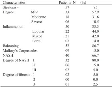
TABLE 1 - Hepatic histologic characteristics in 60 morbidly obese patients.
Characteristics Patients N (%)
Steatosis - 57 95
Degree Mild 33 57.9
Moderate 18 31.6
Severe 06 10.5
Inlammation 50 83.3
Lobular 22 44.0
Mixed 21 42.0
Portal 07 14.0
Balooning 52 86.7
Mallory’s Corpuscules: 09 15.0
NASH 40 66.7
Degree of NASH I 32 80.0
II 06 15.0
III 02 5.0
Degree of ibrosis 1 02 5.0
2 00 0.0
Factors associated with the degree of hepatic steatosis
Table 2 shows the association of demographic,
anthropometric and laboratory variables with the degree of hepatic
steatosis (I versus II and III degree of hepatic steatosis). γGT was the only variable signiicantly and independently associated with the degree of steatosis ( OR 0.04, 95% CI 0.003 to 0.63, P = 0.022)
TABLE 2 - Crude and adjusted odds ratios (OR) with
95% conidence interval (CI) for the degree of steatosis according
to clinical and biological variable
Variables
Crude Odds Ratio (OR)
Adjusted Odds Ratio (OR)
OR 95% IC P OR 95% IC P
Age 1.04 0.99 – 1.09 0.075 0.94 0.86 – 1.04 0.273
Gender 0.31 0.09 – 1.03 0.057 8.96 0.35 - 230, 0.186
Diabetes 2.26 0.74 – 6.84 0.151 0.65 0.07 – 5.59 0.693
BMI 2.52 0.97 – 6.56 0.059 0.26 0.04 – 1.81 0.174
WC 1.03 0.99 – 1.07 0.075 1.01 0.92 – 1.11 0.820
HDL-C 0.93 0.87 – 0.99 0.036 1.07 0.97 – 1.19 0.186
Triglycerides 1.00 0.99 – 1.01 0.099 1.00 0.99- 1.01 0.720
Uric acid 4.50 1.36 – 14.83 0.013 0.19 0.02 – 1.74 0.140
AST 5.17 0.94- 28.35 0.059 1.17 0.1 – 12.75 0.985
ALT 14.50 3.77– 55.74 0.000 0.15 0.02 – 1.07 0.059 γGT 5.21 1.65 – 16.41 0.050 0.04 0.00 – 0.63 0.022
Ferritin 6.30 1.89 – 20.92 0.003 0.28 0.04 – 1.84 0.184
Factors associated with histological features present in NAFLD
In univariable analysis Mallory corpuscles were
signiicantly associated with type 2 diabetes (p = 0.036), dyslipidemia (p = 0.017), glucose (p = 0.008), HbA1c (p = 0.027), ALT (p = 0.004) and ferritin (p = 0.040). After subjecting these
values to the logistic regression variables dyslipidemia and ALT
were conirmed as factors independently associated with presence
of Mallory’s corpuscles with the following values, respectively,
OR 0.05, 95% CI 0.002 to 0.75, P = 0.031 and OR 10.99, 95% CI 1.44 to 83.93, P = 0.021. Age was the only variable that was
independently associated with the presence of ballooning, OR
0.89, 95% CI 0.79 to 0.99, P = 0.029.
Factors associated with NASH
In the univariable analysis, the factors that were
signiicantly associated with NAFLD were: triglycerides (P = 0.029); group consisting of type 2 diabetes and those with impaired fasting glucose (P = 0.044), fasting glucose greater than 110mg/ dL (p = 0.034), ALT changes (p = 0.044). The logistic regression analysis identiied no factor independently associated with NASH.
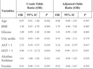
Factors associated with ibrosis
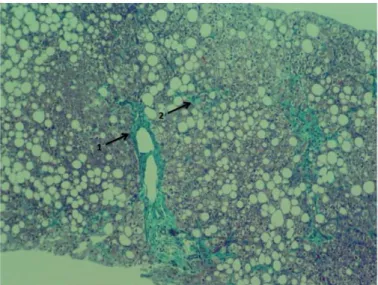
Zone 3 perisinusoidal ibrosis was detected in three cases (7.5%) while the isolated portal ibrosis was seen in seven (11.6%). Figure 1The univariable analysis for factors associated with perisinusoidal ibrosis was compromised by the low prevalence of perisinusoidal ibrosis in the sample. However, taking into account that in morbid obesity, portal ibrosis alone
has received attention12, all type of ibrosis (perisinusoidal, portal,
perisinusoidal and portal) were analyzed as a group, and portal
ibrosis also been analyzed separately. Only the elevation of γ GT showed a signiicant association with the irst group (p = 0.035). After logistic regression analysis, γ GT did not remain as a signiicant independent factor. However, there was a trend towards signiicancy (OR 5.69, 95% CI 0.99 to 32.73, P = 0.051) Table 3.
FIGURE 1 - Masson staining of the liver(x100) shows portal ibrosis (1)
TABLE 3 - Crude and adjusted odds ratios with 95%
conidence interval (CI) for ibrosis according to clinical and biological variable
Variables
Crude Odds Ratio (OR)
Adjusted Odds Ratio (OR)
OR 95% IC P OR 95% IC P
Age 0.97 0.91 – 1.04 0.382 0.98 0.89 – 1.07 0.597
BMI 1.50 0.47 – 4.78 0.496 1.79 0.46 – 7.02 0.403
Glicose 1.00 0.99 – 1.02 0.340 1.01 0.99 – 1.02 0.402
Uric acidic 0.22 0.03 – 1.85 0.161 0.19 0.02 – 2.13 0.179
ALT > 1 2.33 0.59 – 9.27 0.229 2.16 0.34 – 13.97 0.417
GGT > 1 4.96 1.13 – 21.72 0.034 5.69 0.99 – 32.73 0.051
Alcaline
fosfatase 1.01 1.00 – 1.02 0.161 1.01 0.99 – 1.02 0.529
Ferritin 0.41 0.08 – 2.13 0.287 0.32 0.04 – 2.67 0.293
Discussion
The present prospective study evaluated liver biopsies of patients that underwent bariatric surgery. The prevalence of
NAFLD in the sample in this study was 95%, and among patients with NAFLD, twenty-four (42.1%) scored higher (Grade II and
III).
The γGTwas the only laboratory parameter that showed independent association with the degree of liver steatosis after logistic regression analysis. Interestingly. AST did not correlate with the degree of steatosis even in the univariate analysis and
an AST / ALT ratio below 1 was found in 90% of cases. These indings suggest that increased levels of γ GT are not associated
with alcohol consumption. Recent studies have demonstrated an association of GT levels with NAFLD. However, few studies have
evaluated the frequency of such indings13
. Additionally, increased
levels of γ GT have been associated with oxidative stress14
and insulin resistance syndrome15. Our indings highlight the
importance of monitoring γ GT levels in patients with NAFLD. Among the histological indings in NAFLD, the
presence of corpuscles Mallory was independently associated with dyslipidemia and ALT changes. Free fatty acids, acting at the intracellular level may result in the formation of corpuscles underlying Mallory16. The hepatocyte ballooning showed an independent association with age and a trend towards statistical
signiicance with type 2 diabetes. The trend towards an independent association of type 2 diabetes with hepatocyte ballooning as
well as and dyslipidemia and ALT in the presence of Mallory’s
corpuscles may support the idea of type 2 diabetes, dyslipidemia
and ALT as factors associated with NAFLD. The ballooning of hepatocytes and presence of Mallory’s corpuscles are now crucial for the framework of NASH in the spectrum of NAFLD13.It has been suggested that the mechanisms of pathogenesis of steatosis and ballooning are closely linked to insulin resistence, and their long-term evolution may be predicted by early improvement in insulin resistence17.
The prevalence of NASH in this study was 66.7%,
similarly with what was reported from Belo Horizonte Southeastern
of Brazil 57%18, lower than found in Ribeirao Preto also in
Southeastern of Brazil 89%19 and in Greek 98%20, however it is
higher than that reported from Dixon et al. 25% 5 and Beymer et al. 33%21 in the USA. In this study we did not ind any signiicant
association between NASH and gender unlike others studies that found higher prevalence of NASH in men22,23.
In the univariable analysis, diabetes mellitus and changes
in fasting glucose were signiicantly associated with NASH,
as well as laboratory variables ALT, triglyceride and glucose
levels >110mg/dL. However, none of these variables remained signiicant after the adjusted analysis. The BMI, WHR and waist circumference did not show statistical signiicance as factors
associated with NASH in our study, in accordance with several studies2,5.
We found a lower rate of ibrosis in this asymptomatic population (12%) than is commonly identiied and no evidence
of cirrhosis. One explanation may include referral bias in other published reports, but the exclusion criteria that applied to preexisting liver disease may also be related. Another possibility could be due to the fact that the mean age of the patient is lower
than the one reported from the studies that found higher ibrosis
rates since it has been reported that age could be a predictive factor
for ibrosis in several studies2.
The role of isolated portal ibrosis, despite being reported
in patients with NAFLD, has not been fully understood. Abrams
et al.12 found isolated portal ibrosis in 30.3% of 195 liver biopsies
in obese morbid patients and hyperglycemia. Our indings in the isolated portal ibrosis highlight the importance of this type
of in the group of morbidly obese patients13. The two groups
of ibrosis studied were not associated with any of the variables evaluated. It has been described a signiicant association between AST, ALT and γ GT elevation and NASH/ibrosis. However, high
Conclusions
Non alcoholic fatty liver disease seems to be an obese-related condition with approximately half of asymptomatic
morbidly obese patients having histological NASH. The γGT was
an independent predictor of the degree of steatosis.
References
1. Angulo P. GI epidemiology: nonalcoholic fatty liver disease. Aliment Pharmacol Ther. 2007;25:883-9.
2. Clark JM. The epidemiology of nonalcoholic fatty liver disease in adults. J Clin Gastroenterol. 2006;40(3 Suppl 1):S5-10.
3. Blackburn GL, Mun EC. Effects of weight loss surgeries on liver disease. Semin Liver Dis. 2004;24(4):371-9.
4. Wang JT, Liu YL. Non-alcoholic fatty liver disease: the problems we are facing. Hepatobiliary Pancreat Dis Int. 2003;2(3):334-7. 5. Dixon JB, Bhatal PS, Obrien PE. Nonalcoholic fatty liver disease:
predictors of nonalcoholic steatohepatitis and liver ibrosis in severely obese. Gastroenterology. 2001;121:91-100.
6. Machado M, Marques-Vidal P, Cortez-Pinto H. Hepatic histology
in obese patients under going bariatric surgery. J Hepatol. 2006;45(4):600-6.
7. Neuschwander-Tetri BA, Brunt EM, Wehmeier KR, Oliver D, Bacon BR. Improved nonalcoholic steatohepatitis after 48 weeks of treatment with the PPARγ ligand rosiglitazone. Hepatology. 2003;38:1008-17.
8. Report of the expert committee on the diagnosis and classiication
of diabetes mellitus.Expert Committee on the Diagnosis and
Classiication of Diabetes Mellitus. Diabetes Care. 2003;26 Suppl 1:S5-20.
9. Ford ES. Prevalence of the metabolic syndrome deined by the
International Diabetes Federation among adults in the U.S. Diabetes
Care. 2005;28(11):2745-9.
10. Cronk C, Roche AF. Race and soc-speciic reference data for triceps and subcapsular skinfolds and weight/stature. Am J Clin Nutr. 1998;35:347-54.
11. Brunt EM, Janney CJ, Di Bisceglie AM, Neuschwander-Tetri BA,
Bacon BR. Non-alcoholic steatohepatitis: a proposal for grading and
staging the histologic lesions. Am J Gastroenterol. 1999;94:2467-74.
12. Abrams GA, Kunde SS, Lazenby AJ, Clements RH. Portal ibrosis
and hepatic steatosis in morbidly obese subjects: spectrum of
nonalcoholic fatty liver disease. Hepatology. 2004;40:475-83. 13. Sanyal AJ. Aga: technical review on nonalcoholic fatty liver disease.
Gastroenterology. 2002;123:1705-25.
14. Lee DH, Ha MH, Kim JH, Christiani DC, Gross MD, Steffes M, Blomhoff R, Jacobs DR Jr. Gamma-glutamyltransferase and diabetes a 4 year follow-up study. Diabetologia. 2003;46:359-64. 15. Yokoyama H, Moriya S, Homma Y, Ogawa T. Association between
gamma-glutamyl transpeptidase activity and status of disorders constituting insulin resistance syndrome. Alcohol Clin Exp Res.
2003;27:225-55.
16. Wanless IR, Bargman JM, Oreopoulus DG, Vas SI. Subcapsular
steatonecrosis in response to peritoneal insuline delivery: a clue to the pathogenesis of steatonecrosis in obesity. Mod Pathol.
1989;2:69-74.
17. Mathurin P, Hollebecque A, Arnalsteen L, Buob D, Leteurtre E,
Caiazzo R, Pigeyre M, Verkindt H, Dharancy S, Louvet A, Romon
M, Pattou F. Prospective study of the long-term effects of bariatric surgery on liver injury in patients without advanced disease.
Gastroenterology. 2009;137(2):532-40.
18. Lima ML, Mourão SC, Diniz MT, Leite VH. Hepatic histopathology
of patients with morbid obesity submitted to gastric bypass. Obes Surg. 2005;15(5):661-9.
19. Júnior WS, Nonino-Borges CB. Clinical predictors of different grades of nonalcoholic fatty liver disease. Obes Surg. 2012;22(2):248-52. 20. Stratopoulos C, Papakonstantinou A, Terzis I, Spiliadi C, Dimitriades
G, Komesidou V, Kitsanta P, Argyrakos T, Hadjiyannakis E.
Changes in liver histology accompanying massive weight loss after
gastroplasty for morbid obesity. Obes Surg. 2005;15(8):1154-60. 21. Beymer C, Kowdley KV, Larson A, Edmonson P, Dellinger
EP, Flum DR. Prevalence and predictors of asymptomatic liver disease in patients undergoing gastric bypass surgery. Arch Surg.
2003;138:1240-44.
22. Zelber-Sagi S, Nitzan-Kaluski D, Halpern Z, Oren R. Prevalence
of primary non-alcoholic fatty liver disease in a population-based study and its association with biochemical and anthropometric
measures. Liver Int. 2006;26:856–63.
23. Browning JD, Szczepaniak LS, Dobbins R, Nuremberg P, Horton
JD, Cohen JC, Grundy SM, Hobbs HH. Prevalence of hepatic
steatosis in an urban population in the United States: impact of
ethnicity. Hepatology. 2004;40:1387-95.
24. Mofrad P, Contos MJ, Haque M, Sargeant C, Fisher RA, Luketic VA, Sterling RK, Shiffman ML, Stravitz RT, Sanyal AJ. Clinical and
histologic spectrum of nonalcoholic fatty liver disease associated
with normal ALT values. Hepatology. 2003;37(6):1286-92.
Correspondence:
Prof. Dra Lucia L.B.C. Braga Departamento de Medicina Interna Instituto de Biomedicina - UFC
Rua Nunes de Melo, 1315
60430-270 Porangabussu Fortaleza – CE Brasil Tel.: (55 85)3223-6982
Fax: (55 85)3281-5212
Received: July 11, 2013 Review: Sept 10, 2013 Accepted: Oct 14, 2013 Conlict of interest: none
Financial source: none
1Research performed at Nucleus of Obese, State of Ceara and Division