Iranian Journal of Basic Medical Sciences
ijbms.mums.ac.ir
Effects of gamma oryzanol on factors of oxidative stress and
sepsis‐induced lung injury in experimental animal model
Elmira Zolali
,, Parina Asgharian , Hamed Hamishehkar , Maryam Kouhsoltani , Hajhir
Khodaii , Hadi Hamishehkar *
Iranian Evidence Based Medicine Center of Excellence, Tabriz University of Medical Sciences, Tabriz, Iran Student Research Committee, Faculty of Pharmacy, Tabriz University of Medical Sciences, Tabriz, Iran Drug Applied Research Center, Pharmaceutics Department, Tabriz University of Medical Sciences, Tabriz, Iran Department of Oral and Maxillofacial Pathology, Faculty of Dentistry, Tabriz University of Medical Sciences, Tabriz, Iran
Drug Applied Research Center, Clinical Pharmacy Pharmacotherapy Department, Tabriz University of Medical Sciences, Tabriz, Iran A R T I C L E I N F O A B S T R A C T
Article type:
Short communication Objective (s):sepsis that finally leads to organ damage or even death. Gamma oryzanol GO is one of the major There is corroborating evidence to substantiate redox imbalance and oxidative stress in bioactive components in rice bran has been considered to function as an antioxidant. The present study was carried out to evaluate the antioxidant activity of gamma oryzanol invitro and its efficacy in
sepsis.
Materials and Methods: To induce sepsis, cecal ligation and puncture CLP method was performed on
the rats. A study group of forty male Wistar rats were divided into the following groups: sham group; CLP group; mg/kg GO‐ treated CLP group and mg/kg GO‐ treated CLP group. GO was administered with an oral gavage hr prior to inducing sepsis. Tissue and blood samples were collected hr after CLP to prepare tissue sections for histopathological study and assay the oxidative stress biomarkers including: SOD Superoxide Dismutase , TAC total antioxidant capacity , MDA Malondialdehyde , MPO Myeloperoxidase and PAI‐ Plasminogen Activator Inhibitor‐ . Data are given as mean ± SD. The ANOVA with Tukey posthoc test was used to determine the differences
between groups andP< . was considered as statistical significance.
Results: TAC level increased in GO‐ treated CLP groups P< . . Inflammation score of lung tissue and
MPO activity were significantly lower in GO treated CLP group P< . .
Conclusion:It seems that GO has a protective effect on lung inflammation and improves the body
redox capacity during sepsis. Article history:
Received: Mar , Accepted: Oct , Keywords: CLP
Gamma oryzanol Oxidative stress Sepsis
►Please cite this article as:
Zolali E, Asgharian P, Hamishehkar H, Kouhsoltani M, Khodaii H, Hamishehkar H. Effects of gamma oryzanol on factors of oxidative stress and sepsis‐induced lung injury in experimental animal model. Iran J Basic Med Sci ; : ‐ .
Introduction
Sepsis is the condition of the presence of an infection with evidences of a systemic response that
is called the systemic inflammatory response syndrome
SIRS and is one of the most important reasons that
results in admitting patients to intensive care units ICUs in the world . Following the invasion of microorganism, macrophages and neutrophils immigrate to the site of inflammation and release free radicals to destroy pathogens. Free radicals that are released following activation of immune system lead to peroxidation of lipids, proteins and DNA; moreover, reactive oxygen species ROS produced by macro‐ phage cause lung tissue endothelium damage and
acute respiratory distress syndrome ARDS . The
imbalance between ROS production and its degradation by endogenous antioxidants during
sepsis causes free radicals overload and production of inflammatory mediators . There is increasing evidence that oxidative stress following free radical overload plays an essential role in the development of multi organ dysfunction MOD . Antioxidants can control the sepsis‐induced inflammation and tissue damage by directly scavenging free radicals or enhancing endogenous antioxidant defence system
, . Various studies established that the
administration of a natural antioxidant in the septic animal showed a remarkable positive effect on improving the sepsis manifestations , .
Gamma oryzanol GO , with the structure shown in Figure , is a part of unsaponifiable matter of crude rice bran oil, which is achieved in the milling process of rice . It has been demonstrated that GO is a mixture of ferulic acid esters of phytosterol
*Corresponding author: Hadi Hamishehkar. Faculty of Pharmacy, Tabriz University of Medical Sciences, Daneshgah St. Tabriz‐Iran. email:
Figure1. Chemical structures of four major components of gamma oryzanol
and triterpene alcohols. Cycloartenyl ferulate, ‐ methylene cycloartanyl ferulate, sitosteryl ferulate and campesteryl ferulate are the major components of GO . It is demonstrated that GO, a natural product, is a powerful scavenger of diphenyl picrylhydrazyl DPPH , hydroxyl, and superoxide radicals . It is able to protect cells against lipid peroxidation in oxidative status. It has also been reported that GO poses anti‐inflammatory effects. A report showed that GO treatment could suppress colitis and mucosal inflammation in mice by inhibiting the transcription of NFκB, interleukin IL‐ and IL‐ . Furthermore, it is shown that GO had the potential to attenuate the amounts of total cholesterol, very low‐density lipoprotein VLDL and low‐density lipoprotein LDL , and to
enhance plasma high‐density lipoprotein HDL in
hypercholesterolemic hamsters . In another
study, in which the hypocholesterolemic activity of GO had been investigated, it is demonstrated that the cholesterol‐lowering effects of GO is due to inhibition of ‐hydroxy‐ ‐methylglutaryl‐coenzyme A HMG‐ CoA reductase activity and preventing from cholesterol uptake by intestine epithelial cells .
In order to create a condition similar to sepsis, we used cecal ligation and puncture CLP model on the test group of rats to evaluate the antioxidant effects of GO in animal model of sepsis. Furthermore, we theorized that GO, a known antioxidant, may prevent acute lung injury ALI during sepsis; therefore, investigation of the microscopic evidence on lung tissues of septic rats was performed.
Materials
and
Methods
Animals
rats. The animals were housed in standard polypropylene cages, four per cage, under a : hr light/dark schedule at an ambient temperature of ± °C. Animals had free access to food and water. All experiments performed under ethical guidelines for the care and use of laboratory animals of Tabriz University of Medical Sciences, Tabriz, Iran.
The rats were divided into the four groups: I sham group, rats were subjected to laparotomy without any other manipulation; II CLP group, rats were subjected to CLP without any treatment; III
mg/kg GO‐ treated CLP group; and IV mg/kg GO‐ treated CLP group.
GO powder was received as a gift from Tsuno Rice Fine Chemicals Co., Ltd. Wakayama, Japan . GO is a white or yellowish white, odorless, crystalline powder with the purity ≥ % and is oil soluble. It was dissolved in olive oil and prepared freshly on the day of the experiment. As regards the maximum plasma level of GO is nearly hr after oral
administration , it was administered with an oral
gavage hr before CLP. The rats had free access to food and water after surgery, until they were sacrificed.
Treatmentprotocol
Since, daily dosage of GO in humans is
mg/day , and mg/kg body weight were
selected to be administered with an oral gavage in
rats, based on the body surface area .
Surgicalprocedure
A CLP model was used for induction of polymicrobial sepsis on the rats. Each rat was anesthetized by administering ketamine hydrochloride
expose the cecum. The cecum was isolated and ligated with / silk ligature, just distal to the ileocecal valve. One puncture wound was made piercing the cecum with an ‐gauge needle, the cecum was repositioned into the peritoneal cavity. Finally, closure of the abdominal incision with sterile / silk sutures was performed. Laparotomies performed on the sham‐ operated group with no perforation of the cecum. Pre‐
warmed normal saline ml/ g body weight was
administered subcutaneously for fluid resuscitation post‐operatively.
SpecimenCollection
All four groups were anaesthetized after hr and ml of blood drawn by cardiac puncture. Three ml of the blood was extracted and transferred to a plastic vial and centrifuged for min at
rpm/min. The plasma separated and stored at ‐ °C
until analyse. The ml whole‐blood sample was transferred to plastic vial and stored at ‐ °C until analyse. The lungs were surgically removed and washed in an ice‐cold saline solution. One specimen
of lung tissue was stored at ‐ °C and retained for biochemical analyses; the second specimen of
lung tissue fixed in % formalin solution for
histopathological analyses.
Measurement of tissue myeloperoxidase (MPO)
activity
Myeloperoxidase activity was assayed according to the Bradley et al method . The lung tissue
specimen was homogenized in . % hexadecyl trimethyl ammonium bromide HTAB solution prepared with potassium phosphate buffer, mM;
pH= for min, at rpm. The homogenates
were sonicated for sec, frozen and thawed
times, then centrifuged at rpm, in °C for
min. μl of supernatant was added to . ml of o‐
Dianisidine dihydrochlorid and hydrogen peroxide solution. After five min, the reaction was stopped by adding . ml of hydrochloric acid . M . Lastly, the absorbance was measured spectophotometrically at nm and concentrations expressed as MPO Unit/mg tissue.
Measurementofplasmaandtissuemalondialdehyde
(MDA)
MDA levels in the lung tissue and plasma were
measured according to the Olgen etal method .
Tissues were mixed in . % KCl to achieve a % W/V homogenate. One ml of each supernatant and plasma was added to a mixture of O‐ phosphorous acid % and thiobarbituric acid . % in an aqueous solution. The reaction mixture was heated for min up to °C and then was cooled to room
temperature. Three ml of n‐BuOH butanol was
added and centrifuged. The absorbance of the
n‐BuOH butanol phase was measured spectro‐
photometrically at nm, and MDA levels reported
as nmol MDA/mg tissue and nmol MDA/ml plasma.
Measurement of tissue superoxide dismutase
(SOD), whole blood total antioxidant capacity
(TAC) and plasma plasminogen activator
inhibitor‐1(PAI‐1)
SOD activity in lung tissue, TAC level of whole blood and PAI‐ level of plasma were measured according to the manufacturer’s instructions and guidelines using assay kits Randox Laboratories Ltd., Crumlin,UK for SOD and TAC, Glory science Co., Ltd, USA for PAI‐ . SOD activity was reported as Unit/mg protein, TAC level was reported as mmol/l and PAI‐ level was reported as ng/ml.
Histopathologicalexaminationoflungtissue
Lung tissues samples were fixed in % formalin, and after fixation they were embedded in paraffin. Five to six µm sections were taken from tissue samples and stained with hematoxylin‐eosin HE . After staining, sections were examined under light microscopy and then scored based on the degree of inflammation and neutrophil infiltration by an experienced observer blinded to treatment. The severity of tissue injury was scored as follows: : normal; : mild; : moderate; and : severe.
Dataanalysis
Data are expressed as the mean ± SD. Kolmogorov–Smirnov test demonstrated that all data in each group distributed normally. The one‐way analysis of variance ANOVA with Tukey post hoc
test was used to determine the differences in the oxidative stress plasma and tissue extract levels.
P< . was accepted as the statistically significant
difference.
Results
Thelevelsofoxidativestressfactorsinlungtissue
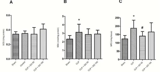
There was no significant difference between groups in tissue SOD enzyme activity P> .
Figure .
The lung tissue level of MDA was significantly higher in the CLP group as compared to sham group
P< . . MDA level has a relative decrease in GO‐
treated CLP groups as compared to CLP group, but it was not statistically significant P> . Figure .
Figure2. The parameters of lung tissue oxidative stress measured hr after cecal ligation and puncture n= . A SOD activity. B MDA level. C MPO activity. CLP, cecal ligation and puncture; GO , Gamma oryzanol mg/kg peroral. hr before CLP; GO , Gamma oryzanol mg/kg p.o. hr before CLP. * ‐indicates P< . compared to sham group. # ‐indicates P < . compared to control group.
Values are expressed as mean±SD
Thelevelsofoxidativestressfactorsinplasmaand
wholeblood
The level of TAC in whole blood samples was significantly lower in CLP group as compared to sham group. However, a significant increase occurred in the GO‐ treated CLP groups as compared to CLP group
P< . Figure .
There was no significant difference between the groups in plasma levels of PAI and MDA P> .
Figure .
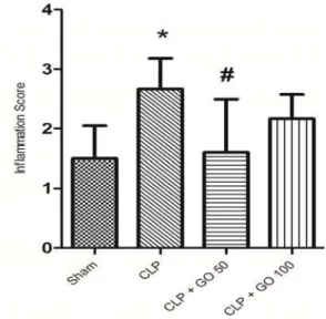
Histopathologicalresultsforlungtissues
There was a significant difference between sham group and CLP group in terms of inflammation scores
P< . Figure , . The mean inflammation score in
sham group was . and . in the CLP group. On the contrary, the results of histopathological study showed significant decrease in lung tissue damage in GO mg/kg‐ treated CLP group compared to CLP group
P< . . Inflammation scores had a slight decrease in
the GO mg/kg‐ treated CLP group compared to CLP
group, but it was not statistically significant P> . .
Figure3.Thewhole blood and parameters of plasma oxidative stress measured hr after cecal ligation and puncture n= . A TAC
level. B PAI level. C MDA level. CLP, cecal ligation and puncture; GO , gamma Oryzanol mg/kg p.o. hr before CLP; GO , gamma oryzanol mg/kg p.o. hr before CLP. * ‐indicates P < . compared to sham group. # ‐indicates P< . compared to control group.
Figure4.The lung tissue inflammation scores measured hr after cecal ligation and puncture n= . CLP, cecal ligation and puncture; GO , gamma oryzanol mg/kg p.o. hr before CLP; GO , gamma oryzanol mg/kg p.o. hr before CLP. * ‐indicates P< . compared to sham group, # ‐indicates P< .
compared to control group. Values are expressed as mean±SD
Discussion
Sepsis causes progressive damage in multiple
organs and if progress to septic shock with a significant drop in blood pressure can lead to death. Treatment with antibiotics alone is not an adequate treatment plan to increase the possible survival rate of the septic patient. Considering this fact, it is thought that the use of antioxidants may have an important role in the treatment of septic patients .
GO, as a natural product, has a potent antioxidant
properties . The result of an animal study
showed that GO has protective effects on liver injury
induced by chronic ethanol administration .
Furthermore, there is scientific evidence reporting its immunomodulatory properties and the effects of GO in stimulating immune responses in experimental
animal models of immunity .
Oxidative stress influences the molecular mechanisms that control inflammation and directly
cause tissue damage. This type of tissue damage is believed to be one of the most important
mechanisms those results in multiple organ failure
MOF in septic patients .The lung is the primary
organ that is affected initially and most severely
in intra‐abdominal sepsis . Under normal
physiological conditions, there is a balance between the levels of antioxidants and oxidants in the lungs,
and a disruption in this balance is considered to be one of the primary events that can cause an
inflammatory response in the lungs during a septic
infection . The accumulation and activation of
neutrophilsinthelungsisanimportantearly event
in the development ofan ALI in the experimental
Figure5. The histopathological view of lung section. A Sham group: B CLP cecal ligation and puncture group: C gamma oryzanol mg/kg‐ treated CLP group: D gamma oryzanol mg/kg‐ treated CLP group. Inflammatory cell infiltration arrow indicated ; Rb: respiratory bronchiole. All magnifications: ×
animal models of sepsis . The measurement of
MPO activity as a marker of active neutrophil accumulation in lung tissue showed that MPO
activity enhanced in the lungs of septic animals .
Decreasing level of MPO activity by treatment with GO mg/kg may indicate that GO suppresses the severity of sepsis, and it may have lung protective effects in CLP model of sepsis. Furthermore, the results of histopathological findings demonstrated that GO prevented damage to the lung tissue. Severe sepsis has been associated with microvascular thrombosis, which is mediated by plasminogen activators PAs . In disseminated intravascular coagulation DIC syndrome, intravascular activation of coagulation occurs that may delay sufficient blood supply to the organs. PAI‐ that is secreted by the endothelial cells increases in inflammatory conditions and causes fibrosis and thrombosis in
otherwise healthy tissues . Sepsis‐induced DIC
might result in a poor prognosis; therefore, early diagnosis and treatment may improve the
outcome of patients with sepsis . Findings of a study showed that PA and PAI levels were increased during sepsis and caused fibrin remaining in microvasculature, which led to inadequate organ
perfusion . However, PAI‐ level as a marker for
coagulopathy did not rise in the CLP group in this study as compared to the sham group.
Superoxide anion O._ is a free radical that is used by SOD and is converted to hydrogen
there is no compensatory elevation in the peroxidases, the excess amount of H O is converted to hydroxyl radicals OH., which is suggested to be
the most dangerous radical . Itis demonstrated
that SOD activity is decreased in lung tissue during sepsis . In this regard, it was illustrated that SOD activity decreased hr after inducing sepsis as a result of neutrophils activation and production of superoxide in response to the inflammation . On the other hand, the results of a study
demonstrated that CLP‐ induced sepsis caused an elevation in SOD activity of CLP group in response to overproduction of mitochondrial superoxide and expression of inflammatory mediators. However, in the present study SOD did not rise in CLP group as compared to the sham group.
MDA is the product of oxidative stress and is formed during the destruction of cellular membrane phospholipids. When phospholipids such as arachidonic acid AA are attacked by hydroxyl radical OH•, lipid endoperoxide is formed and undergoes spontaneous breakage, which causes MDA
production . Oxidized lipids and proteins play an
important role in damaging cell membranes and are
associated with septic mortality . Previous
studies have shown elevated MDA levels following lipid peroxidation in rats with CLP‐ induced sepsis
, . In the present study, measurement of MDA in
the tissue extract showed significant increase in the CLP group as compared to the sham group, but it was not significant in the plasma samples.. Therefore it seems that measurement of MDA in the tissue comparing it with plasma will predict sepsis more precisely.
In addition, the results of our study demonstrated that treatment with GO decreased MDA tissue levels, but depression was not significant.
The elevation of oxidants and free radicals and suppression of antioxidants in plasma samples of patients with sepsis has been established previously . The study of Gadek etal in patients with sepsis‐
induced ALI demonstrated that administration
of antioxidants prevented lung injury by reducing pro‐inflammatory mediators; moreover, it is shown that patients who developed SIRS had a lower
antioxidant status . On the other hand, some
investigators noticed that TAC level was elevated in
severe septic patients , . In the present study,
TAC level was lower in the CLP group as compared to the sham group, and treatment with GO was able to increase the TAC level significantly in septic rats.
Based on the results of the present study and evidence‐based approaches, the difference between
the effects of mg/kg GO and mg/kg GO is not
interpretable. However, trends of results shows that
mg/kg GO may be more effective than mg/kg
GO. Regarding that antioxidant level of plasma samples is increased in patients with septic shock
and taking care in antioxidant‐related therapy during sepsis is important and also very challenging topic.
Limitations
of
study
As regards the present study was a pilot study to assess the effects of GO in sepsis and because of low sample size and lack of survival study due to ethical limitations, dose‐response effects of GO may not be interpretable.
Conclusion
In conclusion, it appears that GO has a protective effect on the lungs during an acute phase of sepsis. It attenuated lung inflammation and neutrophils infiltration in septic rats. GO improved the body redox capacity during the acute sepsis induced by CLP to overcome oxidative stress.
Acknowledgment
The authors would like to acknowledge Amir M Vatankhah for his helpful technical assistance Drug Applied Research Center . This is a report of a database from thesis entitled "Evaluation of gamma oryzanol effects on factors of oxidative stress in animal model of sepsis" registered in Drug Applied Research Center and was financially supported by
grant no. / from the Drug Applied Research
Center of Tabriz University of Medical Sciences, Tabriz, Iran.
References
. Nguyen HB, Rivers EP, Abrahamian FM, Moran GJ,
Abraham E, Trzeciak S, etal. Severe sepsis and septic
shock: review of the literature and emergency department management guidelines. Ann Emerg Med
; : ‐e .
. Chabot F, Mitchell J, Gutteridge J, Evans T. Reactive oxygen species in acute lung injury. Eur
Respir J ; : ‐ .
. Gutteridge JMC, Mitchell J. Redox imbalance in the
critically ill. Br Medical Bull ; : ‐ .
. Bian K, Murad F. Diversity of endotoxin‐induced nitrotyrosine formation in macrophage‐endothelium‐
rich organs. Free Radic Biol Med ; : ‐ .
. Ritter C, Andrades ME, Reinke A, Menna‐Barreto S, Moreira JCF, Dal‐Pizzol F. Treatment with N‐ acetylcysteine plus deferoxamine protects rats against oxidative stress and improves survival in
sepsis. Crit Care Med ; : ‐ .
. Zolali E, Hamishehkar H, Maleki‐Dizaji N,
Zolbanin NM, Ghavimi H, Kouhsoltani M, et al.
Selenium effect on oxidative stress factors in septic
rats. Adv Pharm Bull ; : ‐ .
. Toklu HZ, Tunali Akbay T, Velioglu‐Ogunc A,
Ercan F, Gedik N, Keyer‐Uysal M, etal. Silymarin, the
antioxidant component of silybum marianum, prevents sepsis‐induced acute lung and brain injury. J
Surg Res ; : ‐ .
. Ohara K, Kiyotani Y, Uchida A, Nagasaka R,
Maehara H, Kanemoto SH, etal. Oral administration
of γ‐aminobutyric acid and γ‐oryzanol prevents stress‐induced hypoadiponectinemia. Phytomed
; : ‐ .
. Lakkakula NR, Lima M, Walker T. Rice bran stabilization and rice bran oil extraction using ohmic
heating. Bioresour Technol ; : ‐ .
. Xu Z, Godber JS. Purification and identification of components of γ‐oryzanol in rice bran oil. J Agr Food
Chem ; : ‐ .
. Juliano C, Cossu M, Alamanni MC, Piu L. Antioxidant activity of gamma‐oryzanol: mechanism of action and its effect on oxidative stability of
pharmaceutical oils. Int J Pharm ; : ‐ .
. Islam M, Murata T, Fujisawa M, Nagasaka R, Ushio
H, Bari A, et al. Anti‐inflammatory effects of
phytosteryl ferulates in colitis induced by dextran
sulphate sodium in mice. Br J Pharmacol ;
: ‐ .
. Wilson TA, Nicolosi RJ, Woolfrey B, Kritchevsky D. Rice bran oil and oryzanol reduce plasma lipid and lipoprotein cholesterol concentrations and aortic cholesterol ester accumulation to a greater extent than ferulic acid in hypercholesterolemic hamsters. J
Nutr Biochem ; : ‐ .
. Mäkynen K, Chitchumroonchokchai C, Adisa‐ kwattana S, Failla M, Ariyapitipun T. Effect of gamma‐ oryzanol on the bioaccessibility and synthesis of
cholesterol. Eur Rev Med Pharmacol Sci ; : ‐ .
. Kim JS, Lee J‐S, Chang P‐S, Lee HG. Optimization,
in vitro release and bioavailability of γ‐oryzanol‐
loaded calcium pectinate microparticles reinforced
with chitosan. New Biotechnol ; : ‐ .
. Ghatak SB, Panchal SJ. Investigation of the immunomodulatory potential of oryzanol isolated from crude rice bran oil in experimental animal
models. Phytother Res ; : ‐ .
. Reagan‐Shaw S, Nihal M, Ahmad N. Dose translation
from animal to human studies revisited. FASEB J ;
: ‐ .
. Rittirsch D, Huber‐Lang MS, Flierl MA, Ward PA. Immunodesign of experimental sepsis by cecal
ligation and puncture. Nat Protoc ; : ‐ .
. Bradley PP, Priebat DA, Christensen RD, Rothstein G. Measurement of cutaneous inflammation: estimation of neutrophil content with an enzyme marker. J Invest
Dermatol ; : ‐ .
. Olgen S, Coban T. Antioxidant evaluations of novel N‐H and N‐substituted indole esters. Biol
Pharm Bull ; : ‐ .
. Atli M, Erikoglu M, Kaynak A, Esen HH, Kurban S. The effects of selenium and vitamin E on lung tissue
in rats with sepsis. Clin Investig Med ; :E ‐
E .
. Hiramitsu T, Armstrong D. Preventive effect of antioxidants on lipid peroxidation in the retina.
Ophthalmic Res ; : ‐ .
. Chotimarkorn C, Ushio H. The effect of trans‐ ferulic acid and gamma‐oryzanol on ethanol‐induced
liver injury in C BL mouse. Phytomed ;
: ‐ .
. Babayigit H, Kucuk C, Sozuer E, Yazici C, Kose K, Akgun H. Protective effect of β‐glucan on lung injury
after cecal ligation and puncture in rats. Intensive
Care Med ; : ‐ .
. Guo R‐F, Ward PA. Role of oxidants in lung injury
during sepsis. Antioxid Redox Signal ; : ‐
.
. Chow C‐W, Herrera Abreu MT, Suzuki T, Downey GP. Oxidative stress and acute lung injury. Am J
Respir Cell Mol Biol ; : ‐ .
. Mimuro J. Type plasminogen activator
inhibitor: its role in biological reactions. Rinsho
Ketsueki, Jpn J Clin Hematol ; : ‐ .
. Madoiwa S, Nunomiya S, Ono T, Shintani Y,
Ohmori T, Mimuro J, et al. Plasminogen activator
inhibitor promotes a poor prognosis in sepsis‐ induced disseminated intravascular coagulation. Int J
Hematol ; : ‐ .
. Robbie L, Dummer S, Booth N, Adey G, Bennett B. Plasminogen activator inhibitor and urokinase‐type plasminogen activator in plasma and leucocytes in
patients with severe sepsis. Br J Haematol ;
: ‐ .
. Halliwell B, Gutteridge J. Free Radicals in Biology
and Medicine. th ed. Oxford and New York:
Clarendon Press; : ‐ .
. Ozturk E, Demirbilek S, Begec Z, Surucu M,
Fadillioglu E, Kırımlıoglu H, etal. Does leflunomide
attenuate the sepsis‐induced acute lung injury?
Pediatr Surg Int ; : ‐ .
. Andrades M, Ritter C, de Oliveira MR, Streck EL, Fonseca Moreira JC, Dal‐Pizzol F. Antioxidant treatment reverses organ failure in rat model of sepsis: role of antioxidant enzymes imbalance, neutrophil infiltration, and oxidative stress. J Surg
Res ; :e ‐e .
. Lorente L, Martín MM, Abreu P, Domínguez‐
Rodriguez A, Labarta L, Díaz C, etal. Sustained high
serum malondialdehyde levels are associated with severity and mortality in septic patients. Crit Care
; :R .
. Koksal G, Sayilgan C, Aydin S, Oz H, Uzun H. Correlation of plasma and tissue oxidative stresses in
intra‐abdominal sepsis. J Surg Res ; : ‐ .
. Goode HF, Cowley HC, Walker BE, Howdle PD, Webster NR. Decreased antioxidant status and increased lipid peroxidation in patients with septic shock and secondary organ dysfunction. Crit Care
Med ; : ‐ .
. Gadek JE, DeMichele SJ, Karlstad MD, Pacht ER,
Donahoe M, Albertson TE, et al. Effect of enteral
feeding with eicosapentaenoic acid, gamma‐linolenic acid, and antioxidants in patients with acute
respiratory distress syndrome. Crit Care Med ;
: ‐ .
. Chi C‐H, Shiesh S‐C, Lin X‐Z. Total antioxidant capacity and malondialdehyde in acute abdominal
pain. Am J Emerg Med ; : ‐ .
. Pascual C, Karzai W, Meier‐Hellmann A,
Oberhoffer M, Horn A, Bredle D, etal. Total plasma
antioxidant capacity is not always decreased in
sepsis. Crit Care Med ; : ‐ .
. Tsai K, Hsu T‐G, Kong C‐W, Lin K‐C, Lu F‐J. Is the endogenous peroxyl‐radical scavenging capacity of plasma protective in systemic inflammatory disorders in