2
Rev Dor. São Paulo, 2014 jan-mar;15(1):2-5
ABSTRACT
BACKGROUND AND OBJECTIVES:here are many non invasive treatment modalities for internal temporomandibu-lar joint derangements described in the literature, including counseling, drug therapy, physical therapy and interocclusal devices. However, some patients become refractory to con-servative treatments and procedures such as arthrocentesis, arthroscopy and temporomandibular joint surgery are indi-cated. Viscosupplementation is a less invasive, low cost ap-proach with good short and long term results. his study aimed at discussing viscosupplementation to treat internal temporomandibular joint alterations with results after four months of follow-up.
METHODS: Participated in the study 55 patients with re-ducing and non rere-ducing disc displacement and osteoarthritis refractory to conservative treatments who were submitted to sodium hyaluronate iniltrations. here has been statistically signiicant pain improvement for all groups.
RESULTS:Patients with non reducing disc displacement and osteoarthritis had signiicant mouth opening improvement. Such results were constant along the four months of follow-up.
CONCLUSION: Viscosupplementation with sodium hyal-uronate may be considered a good alternative to function-ally reestablish temporomandibular joint in the short term in patients with internal alterations refractory to conservative treatments.
Keywords: Hyaluronic acid, Temporomandibular joint, Treatment.
Viscosupplementation as a treatment of internal derangements of the
temporomandibular joint: retrospective study*
Viscossuplementação como tratamento das alterações internas da articulação
temporomandibular: estudo retrospectivo
Daniel Bonotto1, Eduardo Machado1, Rafael Schlogel Cunali1, Paulo Afonso Cunali1
*Received from Federal University of Paraná, Curitiba, PR, Brazil.
1. Federal University of Paraná, Curitiba, PR, Brazil.
Submitted in May 16, 2014.
Accepted for publication in January 27, 2014. Conlict of interests: none.
Correspondence to: Paulo Afonso Cunali
Rua Cel. Napoleão Marcondes França, 360 80040-270 Curitiba, PR, Brasil. E-mail: [email protected]
© Sociedade Brasileira para o Estudo da Dor
RESUMO
JUSTIFICATIVA E OBJETIVOS: As formas de tratamento consideradas não invasivas para as alterações internas das articu-lações temporomandibulares descritas na literatura são muitas, incluindo aconselhamento, farmacoterapia, isioterapia e dis-positivos interoclusais. No entanto, alguns pacientes tornam-se refratários aos tratamentos contornam-servadores, tornam-sendo indicados procedimentos como artrocentese, artroscopia e cirurgias das articulações temporomandibulares. A viscossuplementação é uma abordagem pouco invasiva, de baixo custo e com bons resultados em curto e médio prazo. O objetivo deste estudo foi discutir a viscossuplementação no tratamento das alterações internas da articulação temporomandibular com os resultados depois de quatro meses de acompanhamento.
MÉTODOS: Cinquenta e cinco pacientes com deslocamento de disco com redução, deslocamento de disco sem redução e osteoartrite refratários a tratamentos conservadores foram submetidos a iniltração com hialuronato de sódio. Foi obser-vada melhora estatisticamente signiicativa para dor nos três grupos.
RESULTADOS: Pacientes com deslocamento de disco sem redução e osteoartrite apresentaram aumento signiicativo da abertura bucal. Estes resultados se mantiveram constantes ao longo dos quatro meses de acompanhamento.
CONCLUSÃO:A viscossuplementação com hialuronato de sódio pode ser considerada uma boa alternativa no reestabe-lecimento funcional da articulação temporomandibular em curto prazo em pacientes com alterações internas refratárias a tratamentos conservadores.
Descritores: Ácido hialurônico, Articulação temporoman-dibular, Tratamento.
INTRODUCTION
Among temporomandibular disorders (TMD), the derange-ment of the condyle-disc complex derives from the collapse of the normal rotational function of the disc on the condyle. Usually this situation occurs with elongation of the discal collateral ligaments and the inferior retrodiscal lamina. his group of articular TMD includes reducing and non-reducing disk displacement. hese disorders are, many times, associat-ed with inlammatory alterations such as synovitis, capsulitis
ORIGINAL ARTICLE
3
Viscosupplementation as a treatment of internal derangements of the temporomandibular joint: retrospective study
Rev Dor. São Paulo, 2014 jan-mar;15(1):2-5
and retrodiscitis or degenerative alterations like osteoarthrosis and osteoarthritis1.
Generally the primary protocol to control TMD prioritizes the simplest measures, which are reversible and less invasive1.
However since intracapsular dysfunctions are often a result of pathologies of the articular surface, that is, of existing structural alterations, the conservative treatment sometimes proves to be inefective. Several forms of treatment for inter-nal dysfunctions of the temporomandibular joint (TMJ) are supported by the literature: functional rest, non-steroid anti-inlammatory drugs, oral splint, physical therapy support ex-ercises, intra-articular corticosteroid injection, arthrocentesis, arthroscopy, open joint surgery for TMJ, among others. Viscosupplementation with intra-articular injection of sodi-um hyaluronate (SH) – the sodisodi-um salt of the hyaluronic acid (HA) – was irst used as a treatment for traumatic arthritis on racehorses2, subsequently used in humans to treat
osteoar-thritis in large joints such as knees, hips and shoulder. At In 1979, sodium hyaluronate started to be indicated for internal TMJ alterations3, and since then some studies have tried to
assess the efectiveness of the technique, as well as to establish a protocol to its utilization.
A multicenter randomized double blind and placebo-con-trolled study with 121 patients has presented promising re-sults4. A group of 80 patients received SH injections (35 had
reducing disk displacement (RDD), 8 presented non-reduc-ing disk displacement (NRDD), and 37 with degenerative alterations of TMJ), while 41 patients received injection with saline solution (15 with RDD, 6 with NRDD and 20 with degenerative alterations of TMJ). Results showed that for pa-tients with RDD, joint sounds were subjectively reduced in both groups, without statistically signiicant diference, but the degree and importance of the mandibular deviation im-proved signiicantly in the SH group. Patients with NRDD treated with SH presented an improvement of mouth open-ing in the irst ive weeks when compared to the group treated with placebo, however, statistically there was no signiicant diference. Regarding pain assessment with visual analogue scale (VAS), results indicated that the group treated with SH obtained signiicant improvement in comparison to placebo group.
Other study5 performed a retrospective study comparing the
efectiveness of intra-articular injection of SH to the absence of treatment in patients with disk NRDD. A group of 60 patients with NRDD was submitted weekly to an injection of 1ml of SH during 5 weeks. A second group of 76 patients diagnosed with NRDD was only monitored without receiv-ing any treatment (control group). Durreceiv-ing a period of two years, patients were examined monthly regarding mandibular movement range and joint pain. After this period, 82.3% of patients from the SH group presented an improvement (de-ined by the authors as mouth opening range over 35mm and absence of joint pain) against 64.7% from the control group, indicating statistically signiicant diference. Furthermore, it was observed that patients from the SH group presented fast remission of the symptomatology when compared to
untreat-ed patients, concluding that SH appears to be an efective method for the treatment of NRDD.
Other authors have assessed the efficacy of intra-articular injection of SH in 38 patients presenting RDD by a ran-domized placebo-controlled clinical trial6. Patients from
SH group received two SH injections in the upper com-partment of the affected TMJ while control group patients received saline solution injection. SH group presented sta-tistically significant improvement for all evaluated aspects, while the placebo group presented significant improve-ment only for pain. It was concluded that SH injection is an efficient therapeutic option for the treatment of RDD in a six month period6.
Comparison of injections of corticosteroid (CO) and sodium hyaluronate (SH) in 33 patients with arthralgia and RDD un-responsive to conservative treatments was also performed. In that controlled double-blind study, 18 patients received two iniltrations with 0.5mL SH 1% with a two weeks interval, while 15 patients received corticosteroid injection (0.5mL of bethametasone). Evolution was assessed using a questionnaire regarding pain, functional limitation, articular sounds and symptoms persistence, and a clinical evaluation. VAS indi-cated signiicant improvement, with a reduction of the initial algic condition of 30% for SH group and of 40% for the CO group7.
A randomized controlled clinical trial with 67 patients with RDD, NRDD or degenerative alteration of TMJ compared injections of sodium hyaluronate and corticosteroid. The work group received 0.5mL of SH associated to 1ml bupivacaine 1% once a week totalizing from three to four injections. Control group received 0.5mL of prednisolone 2.5% with 1ml of bupivacaine 1% once a week, total of three or four injections. During 5 weeks monitoring period both groups presented significant improvement of pain and function, with no statistically significant difference between them8.
A randomized double-blind clinical trial evaluated 41 pa-tients with rheumatoid arthritis in the temporomandibular joints, dividing them in three groups: 14 individuals were treated with intra-articular SH injections; 14 with corticoste-roid injections; 13 with saline solution injection. Monitoring period was of four weeks, and an improvement of symptoms and in the clinical indexes of dysfunction was observed in all groups. Better results were observed in the groups treated with SH and CO9.
One study reports that 6 patients (7.5% of the sample) pre-sented reactions such as discomfort and edema at the injec-tion site4. Other author claims that 13 patients (37.1% of
the sample) who received SH injection complained of pain during the procedure and within three days, 3 patients (8.5% of the sample) presented acute malocclusion at the injection side and muscular strength reduction8.
4
Bonotto D, Machado E, Cunali RS and Cunali PA Rev Dor. São Paulo, 2014 jan-mar;15(1):2-5
METHODS
his is a retrospective study performed by assessment of medi-cal records of patients treated at the TMD and Orofacial Pain Clinic of the Federal University of Paraná (UFPR) and at pri-vate clinic. Fifty ive patients with articular TMD diagnosis – RDD, NRDD and osteoarthritis (OA) – and unresponsive to conservative treatments, such as occlusal splints and mandible exercises, received viscosupplementation on the afected TMJ. Diagnostic clinical criteria on RDC/TMD were followed by two specialists in TMD and orofacial pain throughout the as-sessment of all patients. he technique described by Bonotto, Custodio and Cunali10 for sodium hyaluronate 1mL
iniltra-tion was utilized in every procedure. Two specialists in TMD and orofacial pain performed all procedures during the period from February 2006 to March 2011. Both examiners were calibrated to data collection, and executed the technique. All patients have received from one to three iniltrations of SH with at least 10 days between them. hroughout the post-operative period patients were instructed to continue with routine conservative treatment, oral splint and/or mandibular exercises. Non-steroid anti-inlammatory drug was prescribed for the three days subsequently to the procedure.
Patients’ evolution regarding temporomandibular pain com-plains, was assessed using a VAS before and 4 months after the procedure. To evaluate alteration concerning mandibular function, measurements of interincisal opening were also per-formed with the same interval. he same professionals that had executed previous procedures performed all assessments. In order to compare average mouth opening and average in-dexes of the VAS, before and after treatment, the Wilcoxon signed rank test was applied. A 95% conidence interval was considered.
his study was approved by the Ethics Committee, Federal University of Paraná under number 1245.170.11.2010.
RESULTS
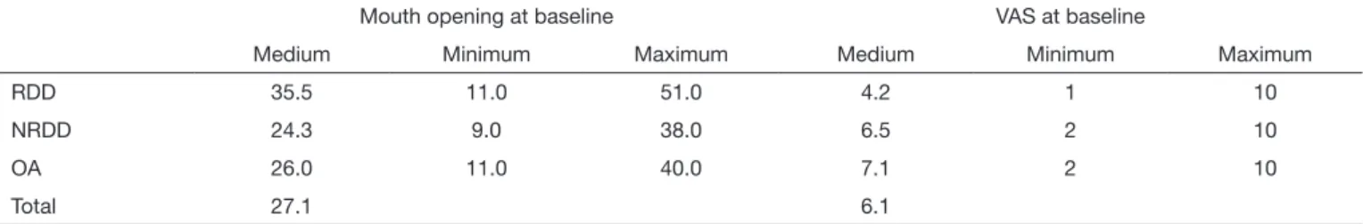
From the 55 evaluated patients, 46 were females (83.64%) and 9 were males (16.36%). Average age was 32.98±15.84 years. After clinical assessment using RDC/TMD criteria, di-agnosis was RDD for 21.8% (12 patients), NRDD for 54.5% (30 patients) and OA for 23.6% (13 patients). Table 1 shows mouth opening and VAS data at baseline.
Figure 1 indicates patient’s evolution for mandibular function before and after the viscosupplementation treatment. Statistic
signiicant improvement of the mandibular function was ob-served in patients with RDD and OA (p<0.001).
Figure 2 shows the improvement for temporomandibular dis-orders pain complaint, which was statistically signiicant for all groups: RDD, NRDD and OA.
Figure 1. Interincisal opening average pre and post treatment for the three groups
RDD: reducing disc displacement; NRDD: non-reducing disc displacement; OA: osteoarthritis.
*Statistical difference was observed in patients with NRDD and OA using Wilco-xon signed rank test (p<0.001).
RDD RDD
50
40
30
20
10
50
40
30
20
10
Befor
e tr
eatment (mm)
After tr
eatment (mm)
NRDD OA NRDD* OA*
RDD RDD
10
8
6
4
2
0
10
8
6
4
2
0
V
AS befor
e tr
eatment
V
AS after tr
eatment
NRDD OA NRDD* OA*
Figure 2. TMJ pain intensity measured by visual analog scale (0-10 scale) before and after treatment for all groups
RDD: reducing disc displacement; NRDD: non-reducing disc displacement; OA: osteoarthritis
*Statistically signiicant difference was observed for all groups at Wilcoxon signed rank test (p<0.001).
Table 1: Mouth opening (in millimeters) and visual analogue scale (0-10 scale) at baseline for the three groups
Mouth opening at baseline VAS at baseline
Medium Minimum Maximum Medium Minimum Maximum
RDD 35.5 11.0 51.0 4.2 1 10
NRDD 24.3 9.0 38.0 6.5 2 10
OA 26.0 11.0 40.0 7.1 2 10
Total 27.1 6.1
5
Viscosupplementation as a treatment of internal derangements of the temporomandibular joint: retrospective study
Rev Dor. São Paulo, 2014 jan-mar;15(1):2-5
Complaints of mild discomfort in the irst 48 hours were re-ported by 9% of patients, while 7.2% of them have experi-enced an open bite in the injection side.
DISCUSSION
here is no precise indication for viscosupplementation in the literature, however there seems to be a consensus on its uti-lization in cases of internal symptomatic alteration of TMJ, specially in the presence of limited range of movement. Dur-ing the monitorDur-ing of the reported cases, viscosupplementa-tion demonstrated to be an eicient treatment to control pain in patients with RDD, NRDD and OA. his result is con-sonant with those presented by several authors4,6-9.
Further-more, viscosupplementation improved mandibular function of patients with limited mouth opening caused by RDD and OA, corroborating others5,9.
Results regarding improvement of mandibular function ob-served in this study may be considered expressive, since they refer to patients who did not respond to conservative treat-ment. However it must be highlighted that this is a retrospec-tive study based on chart review with short term follow-up and that the same examiners have performed both treatment and postoperative assessment procedures. herefore some obliquity may be considered in the interpretation of results. Maintenance of conservative treatment during the follow-up period may have contributed to improvement of patients. However, these patients were refractory to conservative ther-apy alone. Furthermore, in clinical practice viscosuplementa-tion should be associated with conservative treatment. HA is a mucopolysaccharide acid and an essential component of animal tissues. HA is composed by multiple alternating units of D-glucuronic acid and N-acetylglucosamine, form-ing highly viscous gelatinous solution due to its elevated hy-drophilicity11. It is the major component of the synovial luid
and plays an important role in the articular tissues lubrication due to its high molecular weight11. Inlammatory and
degen-erative alteration of the joints reduces the concentration and molecular weight of HA11,12.
SH injection increases the concentration and molecular weight of HA at the synovial luid, associated to the relief of pain13. By clearing the adherence zones between articular disk
and the mandibular fossa, the articular mobility is enlarged allowing a better circulation of the synovial luid.
It was veriied the presence of Prostaglandin E2 and Leukotri-ene B4 in the synovial luid of patients with TMD suggesting that these mediators are among the factors able to generate joint pain14. It is also suggested that the analgesic efect of
viscosupplementation may occur by blocking receptors and endogenous algic substances in the synovial tissues.
A strictly mechanic mechanism by the interruption of trauma caused by mechanic block of the disk or of both adherence zones was also suggested4, what could explain the efects of
therapy in medium and long term, because although the in-jected HA is kept on the joint only for a few days the results last for months15,16.
Only two articles reported side efects of the technique, which seem to be brief and self-limiting4,8. During the follow-up of
the cases reported in this study, no severe side-efect was ob-served. Most common complaints were mild soreness, edema and open bite at the injection side. However in all cases side-efects were self-limiting conirming other authors’ indings8.
According to results of this study, viscosupplementation can be considered an eicient alternative for the management of pain and function improvement in patients with RDD, NRDD and OA refractory to conservative treatments.
CONCLUSION
After the monitoring of clinical cases it is possible to conclude that viscosupplementation with SH may be an interesting proposal to reduce TMJ pain and improve mouth opening. Controlled clinical trials with signiicant samples and longer monitoring period are required to evaluate real efectiveness of viscosupplementation technique and to establish an objec-tive protocol.
REFERENCES
1. Okeson JP, de Leeuw R. Diferential diagnosis of temporomandibular disorders and other orofacial pain disorders. Dent Clin North Am. 2011;55(1):105-20.
2. Butler J, Rydel NW, Balazs EA. Hyaluronic acid in synovial luid. VI. Efect of intra--articular injection of hyaluronic acid on the clinical symptoms of arthritis in track horses. Acta Vet Scand. 1970;11(12):139-55.
3. Kopp S, Wenneberg B. Efects of occlusal treatment and intraarticular injec-tions on temporomandibular joint pain and dysfunction. Acta Odontol Scand. 1981;39(2):87-96.
4. Bertolami CN, Gay T, Clark GT, Rendell J, Shetty V, Liu C, et al. Use of sodium hya-luronate in treating temporomandibular joint disorders: a randomized, double-blind, placebo controlled clinical trial. J Oral Maxillofac Surg. 1993;51(3):232-42. 5. Sato S, Oguri S, Yamaguchi K, Kawamura H, Motegi K. Pumping injection
of sodium hyaluronate for patients with non-reducing disc displacement of the temporomandibular joint: two years follow-up. J Craniomaxillofac Surg. 2001;29(2):89-93.
6. Hepguler S, Akkoc YS, Pehlivan M, Ozturk C, Celebi G, Saracoglu A, et al. he eicacy of intra-articular sodium hyaluronate in patients with reducing displaced disc of the temporomandibular joint. J Oral Rehabil. 2002;29(1):80-6.
7. Kopp S, Wenneberg B, Haraldson T, Carlsson GE. he short-term efect of intra--articular injections of sodium hyaluronate and corticosteroid on temporomandibular joint pain and dysfunction. J Oral Maxillofac Surg. 1985;43(6):429-35.
8. Shi ZD, Yang F, He ZX, Shi B, Yang MZ. [Comparative study on efects of sodium hyaluronate and prednisolone injecions on experimental temporomandibular joint os-teoarthritis of rabbits]. Zhongguo Xiu Fu Chong Jian Wai Ke Za Zhi. 2002;16(1):5-10. (Chinese).
9. Kopp S, Akerman S, Nilner M. Short-term efects of intra-articular sodium hyalu-ronate, glucocorticoid, and saline injections on rheumatoid arthritis of the temporo-mandibular joint. J Craniomandib Disord. 1991;5(4):231-8.
10. Bonotto D, Custodio L, Cunali P. Viscossuplementation to treat internal temporo-mandibular joint disorders. Case reports. Rev Dor. 2011;12(3):313-7.
11. Radin El, Paul L. Joint lubrication with artiicial lubricants. Arthritis Rheum. 1971;14(1):126-9.
12. Listrat V, Ayral X, Patarnello F, Bonvarlet JP, Simonnet J, Amor B, et al. Arthroscopic evaluation of potential structure modifying activity of hyaluronan (Hyalgan) in osteo-arthritis of the knee. Osteoosteo-arthritis Cartilage. 1997;5(3):153-60.
13. Swann DA, Radin EL, Nazimiec M, Weisser PA, Curran N, Lewinnek G. he role of hyaluronic acid in joint lubrication. Ann Rheum Dis. 1974;33(4):318-26. 14. Quinn JN, Bazan NG. Identiication of protaglandin E2 and leukotriene B4 in the
sinovial luid of painful, dysfunctional temporomandibular joints. J Oral Maxillofac Surg. 1990;48(9): 968-71.
15. Sato S, Ohta M, Ohki H, Kawamura H, Motegi K. Efect of lavage with injection of sodium hyaluronate for patients with nonreducing disk displacement of the temporomandibular joint. Oral Surg Oral Med Oral Pathol Oral Radiol Endod. 1997;84(3):241-4.