DOI: 10.1590/0004-282X20160119 ARTICLE
Red blood cells in cerebrospinal fluid as
possible inhibitory factor for enterovirus
RT-PCR
Presença de hemácias no líquido cefalorraquidiano como possível fator inibitório da
RT-PCR para detecção de enterovirus
Sérgio Monteiro de Almeida1,2,3, Sônia Mara Raboni1, Meri Bordignon Nogueira1, Luine R. Renaud Vidal1
Despite its high sensitivity, polymerase chain reaction (PCR) can still provide false-negative results. The presence of hemoglobin in body fluid samples has been considered as an important inhibitory factor for PCR. Other factors include the quality of samples submitted to the lab, small recovery rates of genetic material from the sample during processing, low specificity of primers, optimization of reagents, failure to use nuclease-free materials, or
the presence of a series of PCR inhibitory factors in the samples. Various body fluids such as saliva, urine, serum, stool, amniotic fluid, and cerebrospinal fluid (CSF) contain intrinsic agents that can inhibit PCR1,2,3.
Accidental introduction of red blood cells (RBC) in CSF is a frequent complication during CSF puncture, mainly in newborns and in young children. It occurs more frequently
among less experienced physicians. he CSF samples with
1Universidade Federal do Paraná, Hospital de Clínicas, Seção de Virologia, Curitiba PR, Brasil;
2Faculdades Pequeno Príncipe, Curitiba PR, Brasil;
3Instituto de Pesquisa Pelé Pequeno Príncipe, Curitiba PR, Brasil.
Correspondence: Sérgio Monteiro de Almeida; Hospital de Clínicas – UFPR, Seção de Virologia, Setor Análises Clínicas; Rua Padre Camargo, 280; 80060-240 Curitiba PR, Brasil; E-mail: [email protected]
Conflict of interest: There is no conlict of interest to declare.
Received 31 December 2015; Received in inal form 13 June 2016; Accepted 14 July 2016. ABSTRACT
The presence of hemoglobin in samples are considered an important inhibitory factor for polymerase chain reaction (PCR). The aim of this study was to examine the inluence of red blood cells (RBC)s in cerebrospinal luid (CSF) as an inhibitory factor to reverse transcription polymerase chain reaction (RT-PCR) for enteroviruses (EV). Forty-four CSF samples from patients showing characteristics of viral meningitis were assessed for EV by RT-PCR. Viral RNA extracted with guanidine isothyocianate buffer and virus detection was performed by in-house nested PCR. Positivity for EV RT-PCR was higher in CSF samples without RBCs than in samples with RBCs: 13(26%) and 36(9.2%), p = 0.001. In the group with positive EV RT-PCR, the mean + SD CSF RBC was 37 ± 183 cell/mm³; the group with negative results had 580 + 2,890 cell/mm³ (p = 0.007). The acceptable upper limit for CSF RBCs that could not inluence RT-PCR was 108 cells/mm³. CSF samples with negative results for EV RT-PCR have more erythrocytes.
Keywords: enterovirus; cerebrospinal luid; reverse transcriptase polymerase chain reaction; hemoglobins; erythrocytes; meningitis
RESUMO
A presença de hemoglobina em amostras de luidos corporais é considerada um fator inibitório importante da reação em cadeia da polimerase (PCR). O objetivo deste estudo era examinar a inluencia de hemácias no líquido cefalorraquidiano (LCR) como um fator inibitório da RT-PCR para enterovirus (EV). Quatrocentos e quarenta amostras de LCR de pacientes com características de meningite viral foram avaliados para enterovirus por RT-PCR. RNA viral foi extraído com tampão de isotiocianato de guanidina e a detecção viral foi feita com nested PCR in-house. A positividade do EV RT-PCR no LCR foi maior nas amostras de LCR sem hemácias do que as amostras com hemácias: 13 (26%) e 36 (9,2%), respectivamente (p = 0,001). No grupo com resultados EV RT-PCR positivo, a media ± DP do número de hemácias no LCR foi 37 ± 183 cell/mm³ e no grupo com resultados negativos foi 580 ± 2.890 cell/mm³ (p = 0,007). O limite superior aceitável de hemácias no LCR para não inibir o resultado do PCR foi 108 cells/mm³. As amostras de LCR com resultados negativos para RT-PCR EV tem mais eritrócitos em comparação com amostras com resultados positivos.
puncture accidents are frequently referred to laboratories, especially in teaching hospitals, and could be responsible for the large number of false-negative PCR results for CSF, thus limiting the value of its interpretation3.
To the authors’ knowledge, there are no studies that analyze the effect of RBCs on the positivity for enterovirus (EV) reverse transcription polymerase chain reaction (RT-PCR) in CSF; this is the first study that tries to establish an acceptable maximum number of RBCs in CSF that does not influence the positivity of PCR. The aims of this study are to determine the influence of red blood cell presence in CSF as an inhibitory factor to the RT-PCR reaction for EV, and to establish an acceptable upper limit for the number of RBCs in CSF that does not influence RT-PCR positivity.
METHODS
Four hundred and forty CSF samples were collected by lumbar puncture from patients with clinically suspected meningitis; CSF samples were referred to the virology laboratory in less than 24 hours on ice and were stored in an RNA/DNase-free and sterile tube at -70°C until molecular
analysis. he study was approved by the Ethical and
Research Committee on Human Beings from the Hospital de Clínicas, UFPR.
Inclusion criteria – CSF samples were included in the study based on the biochemical and cytological characteristics of viral meningitis in CSF, which are as follows: white blood cells
(WBCs) ≥ 5 cells/mm3 with a predominance of lymphocytes;
normal CSF glucose levels (> 45 mg/dL); and CSF lactate level < 3.5 mmol/L4.
Exclusion criteria – [1] Clinical diagnosis of encephalitis,
deined as acute onset (< 3 weeks), the presence of
fever (> 38°C), and signs or symptoms that suggested brain parenchyma involvement (consciousness and/or personality alterations, seizures or focal neurological signs).
[2] Samples with CSF biochemical and cytological indings indicative of bacterial meningitis: WBCs > 5 cells/mm3 with
predominance of neutrophils, low CSF glucose levels (<
45 mg/dL), CSF lactate > 3.5 mmol/L. [3] Samples stored
improperly, i.e., non-refrigerated samples or those sent
improperly to the virology section. [4] Identiication of an
etiologic agent other than enterovirus in CSF.
Cellular and biochemical CSF characteristics
Total CSF protein was quantiied by the sulphosalicylic
acid turbidometric method and CSF glucose levels were
assessed using the enzymatic method. he total cell counts of WBCs and RBCs were quantiied using a Fuchs Rosenthal chamber. For diferential cell counts, CSF samples were
concentrated in a cytospin and the slides were stained by the May Grünwald-Giemsa technique.
Extraction of viral RNA/DNA
Extraction of viral RNA was performed according to a previously described protocol5. Briely, 200 µL of lysis bufer
(guanidine isothyocianate [GuSCN], Invitrogen (USA) 4 M),
0.5% N-lauroylsarcosine salt solution (Fluka [USA]), 1 mM dithiotreitol (DTT) (Invitrogen [USA]), 25 mM sodium citrate (Sigma [USA]), 20 µg/tube of glycogen (Sigma
[USA]), and 100 copies of plasmid with pseudo rabies virus
used as internal control were added to 50 µL of CSF. After
vortexing, the tube was incubated at room temperature
for 10 minutes. To this, 250 µL of isopropyl alcohol (-20ºC)
was added, followed by vortexing and centrifugation for
10 minutes at 17,000 rpm at 4°C. he cell pellet was washed with 500 µL of ethanol 70%, and centrifuged as described. he supernatant was discarded and the open tube was incubated at 56°C for 10 minutes. he dried pellet was
resuspended in ultrapure water and stored at -70°C.
Reverse transcription
Viral RNA was transcribed to obtain cDNA as described previously6. he master mix was prepared in a volume of
17.5 µL/tube. he mix contained 2.5 mM deoxynucleotide triphosphates (dNTPs), 5X bufer (Invitrogen, USA), 0.1M dithiothreitol (DTT) (Invitrogen, USA), 0.25 µL of RNase Out (Invitrogen, USA), and 0.25 µL of the reverse transcriptase enzyme Superscript II (Invitrogen, USA). To 10 µL of the extracted product, 0.5 µL of the enterovirus antisense primer was added. After two minutes at 94ºC, the sample was cooled on ice, and 17.5 µL of the mix was added to the tube, which was incubated at 45°C for one hour.
Nested PCR enterovirus – he PCR for enterovirus was
based on ampliication of the 5’-UTR region of the gene,
which is a highly conserved region in most enterovirus serotypes7. For the irst PCR, the primer sequences used were
EV1-Reverse 5’GAAACACGGACACCAAAGTAGTCG3’ and EV1-Forward 5’CGGTACCTTTGTRCGCCTGTTTTA3’. he ampliication was carried out for two rounds. For the irst PCR, the master mix contained 10 mM Tris-HCL (pH 8.8), 3.5 mM MgCl2, 2.5 mM KCl, 1.5 U of Taq polymerase, and 0.5
µL of each external primer in a concentration of 10 pmol, and the inal volume was adjusted with ultrapure water to 22.5 µL, after which 2.5 µL of cDNA was added. he ampliication
was performed using an Eppendorf thermocycler with one cycle at 94°C for two minutes, followed by 40 cycles:
94°C for 30 seconds, 48ºC for 30 seconds, and 72°C for two minutes, followed by an extension at 72ºC for 10 minutes.
For the second PCR, the primer sequences used were as
follows: EV2 - Reverse 5’GGATTAGCCGCATTCAGGG3’; EV2 – Forward 5’CAAGCACTTCTGTTTCCCCG3’. he
master mix for the second PCR was similar to that described
above and the volume of Taq polymerase used was 0.25 µL. he irst ampliication product had 0.5 µL added to 24.5 µL of the master mix. he second ampliication was
of 94°C for two min, followed by 30 cycles of 94°C for 30 s,
52°C for 30 s, 72°C for one min, followed by an extension at 72°C for 10 minutes. he ampliication products were analyzed on a 1% agarose gel. he expected product size for the enterovirus amplicon was 306–316 bp and that for the
internal control, pseudo rabies virus, was 147 bp.
he CSF samples were analyzed by RT-PCR for enterovirus
detection; the primers used for this study targeted a portion
of the 5’-NCR, a highly conserved portion in diferent enterovirus serotypes. he degenerate primer was designed
based on the sequences of 13 serotypes of enterovirus (Polio1,
Polio2, Polio3, CB1, CB3, CB4, CB5, CA9, CA16, CA21, CA24, CB1, CB3, CB4, CB5, Echo12, and Enterovirus 70).
Statistical analysis
he samples were divided into the following groups:
1. EV RT-PCR negative (n = 391) and EV RT-PCR positive (n = 49); 2. Without RBCs in the CSF (n = 49) and with RBCs in the CSF (n = 391).
Comparisons between groups were made using chi-square tests, Fisher’s exact tests, and non-parametric methods as
appropriate. he acceptable upper limit of RBCs in CSF that could not inluence the enterovirus PCR was determined by
the receiver operating characteristic (ROC) curve8.
Results were considered statistically signiicant at the 5%
alpha level.
RESULTS
The mean ± SD age in the EV RT-PCR negative group
was 6.5 ± 7.4 years while that in the EV RT-PCR positive group was 6 ± 5 years (p > 0.05). The EV RT-PCR negative group had 234 male subjects (59%) and the EV RT-PCR positive group had 27 male (56%) subjects (p = 0.54).
The two groups were comparable in gender, age, CSF biochemistry characteristics (total protein (TP) and glucoses) and CSF WBC number. The percentage of lymphocytes was higher in the EV RT-PCR negative group, with statistical significance; although in both groups there was a predominance of lymphocytes in accordance with the diagnosis of lymphocytic meningitis (Table 1).
Forty-nine (11%) samples showed amplicons consistent with the expected size (306-316 bp in agarose gel stained with ethidium bromide), while 391 (89%) samples were negative.
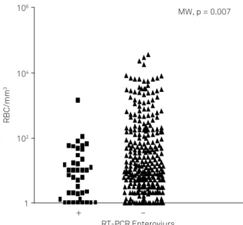
In the group with negative RT-PCR findings for EV, the
RBC number (mean ± SD) was 580 ± 2890 cells/mm3 and in
the group with positive RT-PCR findings, the RBC number was 37 ± 183 cells/mm3 (MW p = 0.007). The cytological
and biochemical characteristics of CSF in both groups are shown in Table 1 and Figure 1.
In the group without RBCs in the CSF, 13 (26.5%) samples
were positive for EV RT-PCR, and in the group with RBCs
in the CSF, 36 (9.2%) samples were positive (X2 p = 0.001;
Table 1. CSF cell and biochemistry characteristics in groups
with positive and negative enterovirus RT-PCR.
Results presented as mean ± SD. EV RT-PCR: enterovirus reverse transcriptase-PCR; RBC: red blood cell; WBC: white blood cell; TP: total protein.
Variable EV RT-PCR Negative
EV RT-PCR
Positive p
N 391 49
RBC cell/mm3 580 ± 2890 37 ± 183 0.007
WBC cell/mm3 163 ± 271 170 ± 279 0.799
Neutrophils (%) 6 ± 17 34 ± 33 0.340
Lymphocytes (%) 81 ± 90 63 ± 31 0.035
Monocytes (%) 17 ± 33 11 ± 23 0.821
TP (mg/dL) 52 ± 32 54 ± 44 0.556
Glucose (mg/dL) 68 ± 55 71 ± 33 0.234
Figure 1. Relationship between the presence of RBCs in CSF
samples and the positivity of RT-PCR to enterovirus.
RT-PCR Enteroviurs
RBC/mm
3
106
104
102
1
MW, p = 0.007
+ –
Figure 2. Percentage of enterovirus RT-PCR positivity in groups
with and without RBC in CSF samples.
CSF
No RBC With RBC
% positivity RT-PCR
30
25
20
15
5
0 10
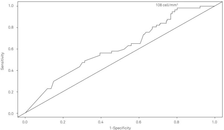
Figure 2). he acceptable upper limit of RBCs in CSF that could not inluence EV RT-PCR was calculated by the ROC
curve to be 108 cells/mm3 (Figure 3).
Considering this cutof to stratify the number of CSF RBCs, 47 (96%) of CSF samples in the group with a positive
EV RT-PCR had CSF RBCs under or equal to 100 cell/mm3,
whereas in the group with a negative EV RT-PCR, 312 (80%) of
CSF samples had RBCs beyond this value (p = 0.003; Table 2).
In the EV RT-PCR negative samples, 35 (9%) of samples had
more than 1000 CSF cells/mm3; in the EV RT-PCR positive
samples, only one (2%) sample had this number.
DISCUSSION
In this study, RT-PCR for enterovirus was 2.3 times more positive in the group without RBCs in the CSF than in the group with RBCs. According to the ROC curve, the upper limit
for RBCs in CSF that could not inluence the results was 108
RBC/mm3. his is considered to be equivalent to a small CSF
puncture accident. However, the ROC curve calculated is not ideal due to its closeness with the diagonal line and the small area under the curve (AUC). Stratifying the number of CSF RBCs in both groups, the values are seen to be in accordance with the value provided by the ROC curve.
Traumatic CSF puncture is the accidental introduction of RBCs during CSF collection, and is a frequent complication of CSF puncture. Heme products from the breakdown of erythrocytes may inhibit the PCR, but modest CSF xanthochromia, high protein levels, or high WBC counts do not have a negative impact on CSF PCR testing.
Several other blood constituents are related to the inhibition of PCR, such as heme, hemoglobin, lactoferrin, heparin, and IgG, resulting in the generation of false-negative results9. All these factors, mainly heme and
Receiver operating characteristic (ROC) curve of the studied groups. The acceptable upper limit for RBCs in CSF that could not inluence enterovirus PCR was 108 cell/mm3 AUC= 0.61. The area under the curve (AUC) serves as a single measure, independent of prevalence, which summarizes the discriminative ability of
a test across the full range of cut-offs. The greater the AUC, the better the test. A perfect test will have an AUC of 1.0, while a completely useless test (one whose curve falls on the diagonal line) has an AUC of 0.5. Youden’s index (J), is the difference between the true positive rate and the false positive rate. Maximizing this index reveals, from the ROC curve, an optimal cut-off point independently from the prevalence. According to its deinition, J is the vertical distance between the ROC curve and the irst bisector (or chance line).
Figure 3. The ROC curve of the studied group.
0.0 0.2 0.4 0.6 0.8 1.0
1-Specificity
Sensitivity
1.0
0.8
0.6
0.4
0.2
0.0
108 cell/mm3
Table 2. CSF RBC counts (cell/mm3) in the groups with
enterovirus RT-PCR positive and negative.
Variable
EV RT-PCR Negative EV RT-PCR Positive P
n % n %
0 36 9.2 13 26 0.001
0.6–5 159 41.0 16 36 0.65
5–120 70 18.0 9 17 1.00
21–100 47 12.0 9 17 0.25
101–500 34 8.7 1 2 0.16
501–1.000 10 2.6 0 0 0.04
hemoglobin, are present in CSF samples with puncture
accidents. hese factors may cause competitive inhibition,
i.e., binding of an inhibitory factor to an active site of the
DNA polymerase, thereby inhibiting ampliication.
In general, endogenous polymerase inhibitors are very
rarely present in CSF compared to other body luids or tissues.
Nevertheless, false-negative results occur in CSF analysis as well. Factors that contribute to low sensitivity, include low viral load, delay in CSF processing or rapid clearance due to a robust host neutralizing antibody response. False negative results may also occur in the presence of endogenous polymerase inhibitors; especially heme products from
artiicial blood contamination, which may inhibit PCR, and
this should be kept in mind in cases of unexpected negative results, as in suspected herpes simplex encephalitis10. here
is no validation for extraction and PCR methods with CSF, and even for the volume of CSF that needs to be used.
Heparin and hemoglobin are natural components of blood. A number of publications have shown that hemoglobin
has a great inhibitory efect on PCR and similar efects have
been reported with heparin as well11,12,13. Usually, heparin
occurs in insuicient quantities in the blood to be detectable
as an anticoagulant.
Hemoglobin is a multichain protein that serves as the oxygen-carrying protein of red blood cells. Hemoglobin is made up of four polypeptides or globin chains: two identical α-chains and two identical β-chains. The globin chains of hemoglobin interact and are connected to each other by the heme group, which contains an iron ion (Fe2+) in the center. The heme group with the iron ion has been shown to be involved in inactivating several DNA polymerases in PCR reactions9.
An earlier experiment determined that a concentration of 1 mg ml-1 hemoglobin or of 0.013 mg ml-1 heparin had
a signiicant inhibitory efect on PCR ampliication.
Concentrations of 10 mg ml-1 hemoglobin in water and
1.3 mg ml-1 of heparin in water were therefore selected as
suitable inhibitor concentrations for all the tests throughout
the study. hese concentrations were 1 and 2 orders of
magnitude, respectively, higher than the concentrations
showing the PCR inhibition efect14.
In this study, we used an in-house made bufer for RNA
extraction that may explain the high rate of RBC interference in the PCR positive results. We suggest the use of commercial kits for extraction, although studies comparing these assays in CSF are necessary.
Enterovirus is an RNA virus. RNA is more labile than double-stranded DNA, and is more susceptible to inhibitory
factors than DNA viruses such as herpesviruses. he impact
of RBCs in CSF must therefore be evaluated in DNA viruses10.
Almost 90% of acute viral meningitis cases are caused by
enteroviruses such as coxsackievirus and echovirus, which have several serotypes15, followed by the herpesviridae
family of viruses16.
he implication of many inhibiting factors in PCR results
is well known. In CSF samples collected less than three
days after the onset of neurological symptoms, only 12%
samples showed positive results. A higher positivity was
observed on the fourth or ifth day1,17. In our study, most CSF
samples were collected on the second day (median 12 h)18,
which could explain the high number of negative samples. Additionally, the elevated number of RBCs due to traumatic lumbar puncture in the CSF samples, as well as the presence
of hemoglobin, probably had an inhibitory efect generating
false-negative results. We excluded CSF samples with a predominance of neutrophils from this analysis, as the aim
of this study was lymphomonocytary meningitis. his could
have had little impact on the number of positive PCR samples as the samples included in this study were collected with a mean of 12 h.
Although the PCR technique is highly sensitive due to the
million-fold ampliication of the genomic material present in
the tested sample, the exact sensitivity in particular clinical situations is not known for many organisms due to the lack of a gold standard for comparison. For some organisms, the sensitivity is low, leading to false-negative results. Factors that might contribute to the low sensitivity ( false negatives) include low viral load due to delay in obtaining the CSF specimen for testing, or rapid clearance due to robust host neutralizing antibody responses. False-negative tests may also occur if endogenous polymerase inhibitors that interfere with PCR are present in the CSF sample10.
A variant of classical PCR, called nested PCR, was utilized for the analysis of specimens in which very few viral particles are presumed to be present, such as CSF, with the
goal of substantially increasing the sensitivity and speciicity of the PCR. In nested PCR, the irst PCR is followed by an additional ampliication with a second set of primers that
are complementary to sequences internal to the sequence
targeted by the irst set of primers. he replicating virus and viral nucleic acid do not persist indeinitely in infected
patients, particularly in immunocompetent patients who
mount an efective neutralizing antibody response19.
However, most CSF testing is performed in the clinical setting of suspected meningitis or meningoencephalomyelitis within one to two days following the onset of neurologic symptoms, at a time that the yield from PCR testing is likely to be at its peak.
Positive CSF PCR test results have been noted for up to four weeks after onset of clinical symptoms, depending on the pathogen20.
he strength of this study is in the substantial number of
cases with suspicion of acute lymphocytic meningitis that were analyzed.
he present study is not without limitations; the results
must be viewed carefully as other less-frequent viruses that could be related to meningitis were not investigated, such as
Other causes of lymphomonocytary meningitis, although less prevalent, could be associated with other infectious agents such as Mycobacterium tuberculosis, Treponema pallidum, Cryptococcus neoformans, Listeria monocytogenes, Brucella spp., Mycoplasma spp., neurocysticercosis, leptospirosis or non-infectious conditions including autoimmune diseases and carcinomatous meningitis, must also be considered4.
he main limitation of this study is the lack of an optimal
gold standard; thus, this is a descriptive comparative study. In conclusion, the presence of RBCs in CSF with a number greater than 108 cell/mm3 may interfere with the
positivity of EV RT-PCR, causing false-negative results.
Cerebrospinal luid samples with negative results for EV PCR
have greater numbers of erythrocytes in compared with the samples showing positive results. We stress the importance of observing technical precepts when collecting CSF in order to reduce the number of CSF puncture accidents for greater
efectiveness of RT-PCR in diagnosis and treatment.
his study was performed with an in-house extraction using guanidine isothyocianate (GuSCN) bufer. hese
results are valid for EV and for the in-house extraction kit. We cannot extend the results to DNA viruses such as herpes
simplex virus type 1 or 2 that are major causes of sporadic
encephalitis. More studies need to be conducted to evaluate the impact of RBCs in CSF using commercial kits and for DNA viruses.
References
1. Davies NWS, Brown LJ, Gonde J, D Irish, R O Robinson, A V Swan et al. Factors influencing PCR detection of viruses in cerebrospinal fluid of patients with suspected CNS infections. J Neurol Neurosurg Psychiatry. 2005;76(1):82-7. doi:10.1136/jnnp.2004.045336
2. Ochert AS, Boulter AW, Birnbaum W, Johnson NW, Teo CG. Inhibitory effect of salivary luids on PCR: potency and removal. PCR Methods Appl. 1994;3(6):365-8. doi:10.1101/gr.3.6.365
3. Spreer A. Detection of infectious agents. In: Deisenhammer F, Sellebjerg F, Teunissen CE, Tumani H, editors. Cerebrospinal luid in clinical neurology. New York: Springer; 2015. p. 131-42.
4. Almeida SM, Nogueira MB, Raboni SM, Vidal LRR. Laboratorial diagnosis of lymphocytic meningitis. Braz J Infect Dis. 2007;11(5):489-95. doi:10.1590/S1413-86702007000500010
5. Casas I, Powell L, Klapper P, Cleator G. New method for the extraction of viral RNA and DNA from cerebrospinal luid for use in the polymerase chain reaction assay. J Virol Methods. 1995;53(1):25-36. doi:10.1016/0166-0934(94)00173-E
6. Casas I, Tenorio A, Echevarría JM, Klapper PE, Cleator GM. Detection of enteroviral RNA and speciic DNA of herpesviruses by multiplex genome ampliication. J Virol Methods. 1997;66(1):39-50. doi:10.1016/S0166-0934(97)00035-9
7. Casas I, Pozo F, Trallero G, Echevarría JM, Tenorio A. Viral diagnosis of neurological infection by RT multiplex PCR: a search for entero and herpes viruses in a prospective study. J Med Virol.1999;57(2):145-51. doi:10.1002/(SICI)1096-9071 (199902)57:2<145::AID-JMV10>3.0.CO;2-N
8. Akobeng AK. Understanding diagnostic tests 3: receiver operating characteristic curves. Acta Paediatr. 2007;96(5):644-7. doi:10.1111/j.1651-2227.2006.00178.x
9. Lodish H, Berk A, Kaiser CA, Krieger M, Scott MP, Bretscher et al. Regulating the eukaryotic cell cycle. In: James D, Lodish H, Baltimore D. Molecular cell biology. 4th ed. New York: W. H. Freeman; 2000. p. 51-104.
10. Debiasi R, Tyler K. Molecular methods for diagnosis of viral encephalitis. Clin Microbiol Rev. 2004;17(4):903-25. doi:10.1128/CMR.17.4.903-925.2004
11. Al-Soud W, Rådström P. Puriication and characterization of PCR-inhibitory components in blood cells. J Clin Microbiol. 2001;39(2):485-93. doi:10.1128/JCM.39.2.485-493.200
12. Akane A, Matsubara K, Nakamura H, Takahashi S, Kimura K. Identiication of the heme compound copuriied with deoxyribonucleic acid (DNA) from bloodstains, a major inhibitor of polymerase chain reaction (PCR) ampliication. Forensic Sci. 1994;39(2):362-72. doi:10.1520/JFS13607J
13. Satsangi J, Jewell DP, Welsh K, Bunce M, Bell JI. Effect of heparin on polymerase chain reaction. Lancet. 1994;343(8911):1509-10. doi:10.1016/S0140-6736(94)92622-0
14. Perch-Nielsen IR, Bang DD, Poulsen CR, El-Ali J, Wolff A. Removal of PCR inhibitors using dielectrophoresis as a selective ilter in a microsystem. Lab Chip. 2003;3(3):212-6. doi:10.1039/b304549h
15. Brown B, Oberste MS, Maher K, Pallansch MA. Complete genomic sequencing shows that polioviruses and members of human
enterovirus species C are closely related in the noncapsid coding region. J Virol. 2003;77(16):8973-84. doi:10.1128/JVI.77.16.8973-8984.2003
16. Chadwick DR, Lever AML. The impact of new diagnostic methodologies in the management of meningitis in adults at a teaching hospital. QJM. 2000;95(10):663-70. doi:10.1093/qjmed/95.10.663
17. Puchhammer-Stöckl E, Presterl E, Croÿ C, Aberle S, Popow-Kraupp T, Kundi M et al. Screening for possible failure of herpes simplex virus PCR in cerebrospinal luid for the diagnosis of herpes simplex encephalitis. J Med Virol. 2001;64(40:531-6. doi:10.1002/jmv.1082
18. Vidal LR, Almeida SM, Messias-Reason IJ, Nogueira MB, Debur MC, Pessa LF et al. Enterovirus and herpesviridae family as etiologic agents of lymphomonocytary meningitis, Southern Brazil. Arq Neuropsiquiatr. 2011;69(3):475-81. doi:10.1590/S0004-282X2011000400013
19. Tang YW, Mitchell PS, Espy MJ, Smith TF, Persing DH. Molecular diagnosis of herpes simplex virus infections in the central nervous system. J Clin Microbiol. 1999;37(7):2127-36.