Official Journal of the Brazilian Psychiatric Association Volume 34 • Number 4 • December/2012
Psychiatry
Revista Brasileira de Psiquiatria
Abstract
Objective: Mounting evidence suggests that the limbic system is pathologically involved in cases of psychiatric comorbidities in temporal lobe epilepsy (TLE) patients. Our objective was to develop a conceptual framework describing how neuropathological and connectivity changes might contribute to the development of psychosis and to the potential neurobiological mechanisms that cause schizophrenia-like psychosis in TLE patients. Methods: In this review, clinical and neuropathological indings, especially brain circuitry of the limbic system, were examined together to enhance our understanding of the association between TLE and psychosis. Finally, the importance of animal models in epilepsy and psychiatric disorders was discussed. Conclusions: TLE and psychiatric symptoms coexist more frequently than chance would predict. Damage and deregulation among critical anatomical regions, such as the hippocampus, amygdala, thalamus, and the temporal, frontal and cingulate cortices, might predispose TLE brains to psychosis. Studies of the effects of kindling and injection of neuroactive substances on behavior and electrophysiological patterns may offer a model of how limbic seizures in humans increase the vulnerability of TLE patients to psychiatric symptoms.
Psychiatric comorbidities in temporal lobe epilepsy:
Possible relationships between psychotic disorders
and involvement of limbic circuits
Ludmyla Kandratavicius,
1,3Cleiton Lopes-Aguiar,
1Lézio Soares Bueno-Júnior,
1Rodrigo Neves Romcy-Pereira,
2Jaime Eduardo Cecilio Hallak,
1,3João Pereira Leite
11Faculdade de Medicina de Ribeirão Preto, Department of Neurosciences and Behavior, Universidade de São Paulo, Brazil 2Brain Institute, Universidade Federal do Rio Grande do Norte, Natal, Brazil
3National Institute of Science and Technology in Translational Medicine (Instituto Nacional de Ciência e Tecnologia - INCT-TM; CNPq)
Submitted on March 9, 2012; accepted on April 23, 2012
DESCRIPTORS:
Temporal Lobe Epilepsy; Psychosis; Psychiatric Comorbidities; Neuropathology; Limbic Circuits.
SPECIAL ARTICLE
Corresponding author: Ludmyla Kandratavicius, PhD Faculdade de Medicina de Ribeirão Preto, Department of Neurosciences and Behavior, Universidade de São Paulo. Av Bandeirantes 3900, CEP 14049-900, Ribeirao Preto, SP, Brazil.
Phone: (+55 16) 3602 2796; Fax: (+55 16) 3633 0866. E-mail: [email protected] 1516-4446 - ©2012 Elsevier Editora Ltda. All rights reserved.
Comorbidades psiquiátricas na epilepsia do lobo temporal: Possíveis relações entre desordens psicóticas e comprometimento de circuitos límbicos
Resumo
Objetivo: Existem cada vez mais evidências de que o sistema límbico está envolvido na patologia das comorbidades psiquiátricas em pacientes com epilepsia do lobo temporal (ELT). Nosso objetivo foi elaborar um desenho conceitual descrevendo como aspectos neuropatológicos e de conectividade podem contribuir para o desenvolvimento de psicose em pacientes com ELT.
Métodos: Nesta revisão, achados clínicos e neuropatológicos, e especialmente os aspectos da circuitaria límbica, foram examinados em conjunto para auxiliar nossa compreensão sobre a associação entre ELT e psicose. Achados em modelos animais de epilepsia e esquizofrenia também foram levados em consideração. Conclusões: ELT e comorbidades psiquiátricas coexistem com maior frequência que o predito pela associação ao acaso. Dano e desregulação entre estruturas anatômicas críticas, como hipocampo, amígdala, tálamo, e córtices temporal, frontal e cingulado podem predispor o cérebro com ELT à psicose. Estudos sobre efeitos comportamentais e eletrofisiológicos do abrasamento elétrico e injeções de substâncias neuroativas em modelos animais podem oferecer pistas sobre como crises límbicas em humanos aumentam a vulnerabilidade de pacientes com ELT a sintomas psiquiátricos.
DESCRITORES:
Epilepsia do lobo temporal; Psicose; Esquizofrenia; Comorbidades psiquiátricas; Neuropatologia; Circuitos límbicos.
Background
Temporal lobe epilepsy (TLE) is a focal neurological condi-tion, in which seizures are able to spread and compromise a whole set of limbic structures and neighboring cortices.1 In
contrast, various forms of psychosis are not clearly related to any consistent pathological localization. Instead, they have been attributed to neurochemical and ultrastructural dys-functions in a much wider set of limbic circuits, including the ones directly affected by TLE.2 However, it is noteworthy that
the high prevalence of psychosis in TLE patients3-7 indicates
a possible shared mechanism between the two etiologies, despite their distinct degree of substrate commitment and
localization. While the speciic mechanisms generating psy -chosis in TLE are still poorly understood, neuroanatomical
knowledge of limbic network connectivity might help to deine
potential brain targets for investigation and lead to a better understanding of this pathophysiological issue. The present article conducts a survey of TLE and psychosis, analyzing his-torical, clinical and neuropathological observations within the framework of limbic connectivity. Additionally, we reviewed
experimental studies with electrical kindling or pharmacologi -cal treatment of the hippocampal formation, the amygdaloid
complex, and the prefrontal cortex circuits. We focused on
the dopaminergic and glutamatergic mechanisms that are potentially involved in both TLE and psychosis.
Early observations and clinical aspects
In 1825, two of Esquirol’s assistants reported that the Ammon’s horn was strikingly abnormal in the brains of some
epileptic and a few psychotic patients.8 Fifty-three years later, Paul Broca would describe the great limbic lobe as “the seat of those lower faculties which predominate in the beast.”8 This ample region regained attention more than thir-ty years later, especially with the study of Papez stating that, “the hypothalamus, the anterior thalamic nuclei, the
gyrus cinguli, the hippocampus and their interconnections constitute a harmonious mechanism which may elaborate the functions of central emotion, as well as participate in
emotional expression”.9 The amygdala was not included in Papez’s original theory of emotion, but after Klüver and Bucy’s indings,10 MacLean made the amygdala one of the epicenters of a more extensive system, later called the
limbic system.11,12 Beyond the limbic lobe structures cited above, the existence of anatomical connections among the
frontal lobes, the hypothalamus and temporal lobes, as well
as the effects of experimental lesions, suggested that the frontal lobes inluence the basic instinctive and emotional
drives.13 In addition to emotional disturbances, another
im-portant feature of several types of epilepsy is the presence of periods of short, intermittent lack of the awareness of the environment and of the self, indicating that the brainstem and most of the thalamus are also affected.14
A distinct syndrome of behavior abnormalities occurs in many patients with temporal lobe epilepsy. This
constella-tion of interictal personality changes includes hyposexuality,
hyperreligiosity/deeply held convictions, viscosity (a striking preoccupation with detail, especially concerned with moral and ethical issues), and, occasionally, hypergraphia or the
urge to express in forms other than writing.15 Frequent symp-toms of TLE patients also include psychic or experiential phe
-nomena: intellectual aurae or dreamy states, complex visual or auditory hallucinations or illusions, memory “lashbacks”,
déjà vu, jamais vu, and emotions, most commonly fear.16 It
is important to stress that these patients, who
character-istically exhibit a preserved or even deepened affect, do
not fall into any established nosologic category and often do not appear schizophrenic.15 Indeed, as described by Gloor et al.,17 “the patients are never in doubt that these phenomena occur incongruously, that is, out of context,
In 1957, Desmond Pond made the irst explicit clinical
recognition of schizophrenic-like psychoses of epilepsy, as reviewed by Beard and Slater.18 Typically, the psychotic state closely resembles schizophrenia, with paranoid ideas
which might become systematized, ideas of inluence, and
auditory hallucinations often of a menacing quality. The points of difference with classic schizophrenia are the com-mon religious coloring of the paranoid ideas, the tendency of the affect to remain warm and appropriate, and the lack of typical deterioration to the hebephrenic state 18. Although auditory hallucinations are common, visual hallucinations are relatively rare. Other forms of delusions, including grandiose, referential, religious and Schneiderian symp-toms, have also been reported, especially when a history of traumatic brain injury is present.19
The nature of the relationship between psychosis and
epilepsy is controversial, and existent hypotheses are not mutually exclusive. One hypothesis suggests that there is a
basic antagonism between epilepsy and psychosis. Psychosis in TLE is regularly associated with fewer or no psychomotor seizures,3,20 and normalization of the EEG and elimination
of seizures through anticonvulsant medication - though they
relieve epileptic symptomatology - often exacerbate an
underlying psychiatric disorder and lead to the emergence of a psychotic state.21 Indeed, this supposed mutual
antago-nism was the basis for the development of electroconvulsive therapy, wherein seizure induction is used to treat psychosis, as reviewed by Pollock.22 Gamma-amino butyric acid (GABA) and dopamine also exert antagonistic effects in epilepsy
and psychosis. For instance, dopamine antagonists are com-monly used as antipsychotic drugs, and the drugs can trigger seizures.23 On the other hand, dopamine agonists are able to exacerbate or trigger psychotic symptoms, while having
anticonvulsant properties.24,25 In contrast to this antagonist
relationship, epilepsy also predisposes a patient to the de-velopment of schizophrenia-like psychosis. This last view is in
line with the extensive works of Slater, Beard and Glithero,26 and of Kristensen and Sindrup,20 which recognize epileptic
psychoses as truly organic phenomena, caused by structural damage to the limbic parts of the temporal lobe responsible for both the epilepsy and the psychosis.
Psychiatric disorders in epilepsy can be classiied into
ictal (the psychiatric symptoms are a clinical manifesta-tion of the seizure), periictal (symptoms precede and/or follow the seizure occurrence) and interictal (symptoms occur independently of the seizure occurrence) disorders. In the present review, our focus will be primarily interic-tal psychosis, which includes schizophrenia-like psychosis
of epilepsy, as deined by the International League Against Epilepsy (ILAE, Commission on Neuropsychiatric Aspects).27
Because interictal symptoms are not related to any “seizure collateral effect”, psychiatric manifestations are much like the pure form of the psychopathology. For instance, chronic interictal psychosis of epilepsy is also referred to as schizo-phrenia-like psychosis of epilepsy, due to its resemblance
to schizophrenia’s phenomenological manifestations.28
Interictal psychosis is usually prodigal in lorid symptoms, whereas postictal psychosis exhibits few common schizo -phreniform psychotic traits such as perceptual delusions or voices.29 Of note, recurrent postictal psychosis in human
TLE is considered to be a risk factor for the development
of interictal psychosis,30 which makes animal models of
postictal psychosis suitable for the study of the possible gradual commitment of the limbic circuits. The increased risk of psychiatric symptoms in epilepsy may be related to several risk factors, such as genetic background and illness chronicity, which are liable to facilitate psychopathological
manifestations. In the next two sections, the contributions
of structural neurologic factors and their possible imbalance
among connected neuroanatomical regions will be examined.
Neuropathological aspects
Schizophrenic patients often have abnormal electrical activ-ity in the temporal lobes and sometimes in the frontal lobes.31 Early descriptions suggested that the sites of maximum
abnormalities were in the hippocampus, amygdala, septum, uncus, anterior-temporal, and orbito-frontal areas, even in patients with no history of epilepsy.32 For more than 50 years,
studies have suggested that any given brain tumor affecting the limbic system can present as classical schizophrenia.5 In Malamud’s studies of eighteen patients with temporal lobe
tumors, ten had been diagnosed and treated as schizophren-ics, three as melancholschizophren-ics, one as a psychotic depressive, one
as maniac, two as psychoneurotics, and one as anxious.33 In Taylor’s series of TLE post-mortem brains, 23% of the cases
with “alien tissue” (tumors, hamartomas and focal
dyspla-sias) were psychotic, contrasting with only 5% in the mesial
temporal sclerosis group.7 Although some authors agree
that psychiatric symptoms presage temporal lobe tumors and that presence of mesial temporal sclerosis is protec-tive against schizophrenia-like psychosis in patients with epilepsy,34 recent neuropathological evidence suggests
that there is a structural basis for psychiatric symptoms in TLE patients with hippocampal sclerosis.35 In our series of
patients35 with no dual pathology or alien tissue, we did not
observe a predominance of female patients or of left hand-edness in the psychosis group (as did Taylor)7 nor a
predomi-nance of patients with left or bilateral temporal foci (as did Flor-Henry).3 Other studies also indicate that the presence
of hippocampal sclerosis is not protective against psychosis.
In Roberts’s series of 249 TLE patients, the largest epilepsy
series analyzed to date for their neuropathological features
and schizophrenia-like psychosis, 6.4% had a preoperative diagnosis of TLE and psychosis; 40% of the cases with TLE and
psychosis had hippocampal sclerosis with left sided foci
and approximately 20% had gangliogliomas.4 It is noteworthy
that gangliogliomas usually have origin in and predilection for the temporal lobe,4 meaning that, more important
than the type of lesion, the location of a lesion within the hippocampal-amygdalar-temporal gyri may represent a true predisposition factor for psychosis.
With a focal brain lesion, hallucinations may arise after compensatory over-activation of nearby sensory pathways.
Complex auditory hallucinations are most common in TLE
and schizophrenia, and the affected areas mainly comprise the primary auditory pathway (the pons, inferior colliculus, medial geniculate body and temporal lobe).36 In fact, fMRI
frontoparietal operculum, orbitofrontal cortex, hippocam
-pus, amygdala, sensorimotor cortex, right inferior collicu -lus, right and left insula, left parahippocampal gyrus, right temporal gyrus, right thalamus, middle frontal and anterior cingulate gyri.31 The structural lesions most often seen in
auditory verbal hallucinations are a reduction in grey matter volume in the superior temporal and middle temporal gyri of the dominant hemisphere, and in the insula, temporal pole,
thalamus and right prefrontal cortex.36 Interestingly, auditory
hallucinations have been associated with impaired effective connectivity between the left superior temporal, frontal
and anterior cingulate cortex, suggesting that such patients tend to misattribute their own speech to an external source (external speech).36 In addition to hallucinations, another classic feature exhibited by patients with psychosis is a re
-duction in the operational index of sensorimotor gating. This index is obtained by measuring the prepulse inhibition (PPI)
of the startle response, which refers to the ability of a weak prestimulus to transiently inhibit the response to a closely fol-lowing strong sensory stimulus. Both schizophrenia patients
and their relatives show gating deicits that are signiicantly
heritable, consistent with the endophenotype criteria.37,38 In fact, reduced PPI has been observed in subjects with schizo-typal personality and in normal subjects with high scores of self-transcendence in Personality Questionnaire scales.39
TLE patients with psychosis40 and non-psychotic patients with
psychogenic “non-epileptic” seizures41 also exhibit reduc
-tions in PPI. It is important to stress that, although patients
with psychogenic seizures may not exhibit epileptiform
brain activity, all of them present with hysterical features.42 Often – in more than 90% of the cases43 – they also present with psychiatric comorbidities. Deinite neuropathological
substrates adjunct to PPI impairment in humans are not
known, but experimental data suggest that kindling of the
amygdala44 and precommissural lesions of the fornix45 are
effective ways to disrupt PPI in rats.
Neuropathological abnormalities are frequently observed
in the brains of schizophrenic46 and TLE patients.47 TLE pa-tients with hippocampal sclerosis exhibit hippocampal neu -ronal loss, often accompanied by neu-ronal loss and gliosis in
the amygdala and entorhinal cortex.48 The same set of mesial structures may appear atrophic under magnetic resonance imaging, in addition to being consistently recruited at the onset of electrographical seizure activity.49 The search for
an organic basis in the post-mortem brains of schizophrenic
patients has resulted in controversial indings, mostly be -cause methodological approaches are rarely consistent in their selection of patients and controls.46 Common indings
include neuronal loss, shrinkage or disarray in the cortical layers and occasional gliosis; affected areas comprise pre-frontal cortical areas, the pons, the nucleus accumbens, the hypothalamus, the substantia innominata (part of the basal forebrain), the cingulate, superior, middle and inferior temporal gyri, the amygdala and the hippocampus.46,50,51 The
hippocampal formation has been the subject of intensive study.
Left hemisphere neuronal loss in Ammon’s horn without gliosis, entorhinal cortex layer II neuronal loss and reduced density
of the interneurons have been described in the post-mortem brains of schizophrenic patients, the former being more notice-able in paranoid than in catatonic patients.50,52 As reviewed
by Roberts and Bruton,50 although, in the early 1900’s,
Kraepelin believed schizophrenia had an organic cause and
the characteristics of a degenerative process, the majority of contemporary neuropathological studies have not shown signs of progressive features, such as reactive gliosis or cor-relations between structural abnormalities and the length
of illness. Current theories consider prenatal and perinatal
effects, as well as environmental factors, to be risk factors for disease manifestation.52
Excision of parts of the limbic system or the temporal lobe of schizophrenic patients usually decreases the lorid symp -toms (i.e., delusions, hallucinations, disorganized speech or thinking, and chaotic or confused behavior in their fully developed form), but it leaves the psychosis untouched.5
Therefore, it is not the complete destruction which gives rise to psychosis, but possibly the irritation process during the development of destruction.32 On some occasions,
psy-chosis or deep personality changes may arise after temporal lobectomy in TLE patients who were previously psychiatri-cally normal.34,53 Not only the recruitment and/or partial
damage of the limbic structures are necessary to generate psychiatric disturbances. As postulated by Racine,54 the
kin-dling process may gradually develop and spread throughout brain structures distal to the stimulated focus. In patients with epilepsy, there is evidence of a time-dependent spread
of epileptic excitability that is independent of tissue pathol -ogy.55 It is generally assumed that a human limbic seizure
disorder results in enhancement of affective limbic
func-tions, rather than their lattening.56 Again, the relatively
long interval between TLE onset and the onset of comorbid psychiatric symptoms suggests that damage to key structures is necessary and that it builds up over time.19,57 Such gradual
commitment of connected systems might be determinant to the sum of neurobiological events that eventually will lower the psychopathological threshold of a normal or prodromal psychiatric state.
Limbic lobe and major connections
Through clinical and pathological observation, it is well ac-cepted that TLE is attributable to focal mesial structures
of the temporal lobe, especially the amygdaloid complex and
the hippocampal formation. The hippocampal formation
con-sists of the hippocampus proper – the dentate gyrus (DG) and Ammon’s horn/Cornus Ammonis (CA) subields - along with
the subicular and rhinal cortices.58 Despite the focal nature of TLE, it is possible that recurrent limbic seizures gradually compromise remote encephalic areas, which may be due to
the axonal connectivity of the mesial temporal lobe. Indeed,
according to tract-tracing studies in non-human primates
and rodents, the hippocampus, amygdaloid complex, and
neighboring cortices are embedded within a broad network of limbic connections, whose dysfunctions are involved in at least some of the psychiatric morbidities that are related to TLE. Therefore, it seems reasonable to consider that
efferent axons arising from the mesial temporal lobe may convey paroxysmal activity to the entire limbic circuitry,
ultimately promoting ictal and periictal psychiatric symp-toms. In the same vein, it is possible that seizure recurrence is responsible for the chronic pathological commitment of subcortical and neocortical sites targeted by mesial temporal
lobe axons, which would explain the increased susceptibility
Hippocampal formation connectivity
The hippocampal formation is the largest structure of the
mesial temporal lobe. Intrinsic connections between the DG and CA subields constitute a well-known tri-synaptic circuit,
primarily represented by a unidirectional sequence from
DG granule cells to CA3 pyramidal cells (mossy ibers) and then from CA3 to CA1 pyramidal cells (Schaffer axonal
collaterals).59 Despite evidence for some CA3-DG
back-projections,60 the DG acts as the major starting point for the circuit, receiving most of its extrahippocampal afferents from supericial cells of the entorhinal cortex via the perforant
path of the angular bundle.61,62 At the other end of the se-quence, CA1 pyramidal cells provide the main hippocampal
output, which may or may not be relayed through subiculum cells.63,64 Interestingly, part of the CA1 and subiculum axons project back to the entorhinal cortex, speciically to its deep layers, making the entorhinal cortex a critical link in the excitatory loop of the hippocampal formation.65 Intrinsically, in the entorhinal cortex an inhibitory system between deep layers receiving CA1/subiculum axons and DG-projecting supericial layers seems to gate the low of information from, and to, the hippocampus, thereby controlling its excitatory
loop.66,67 Finally, in addition to the strong entorhinal-DG uni
-directional pathway, there are lighter projections from the
entorhinal cortex to all CA subields and to the subiculum, which means that entorhinal cortex reciprocates the CA1
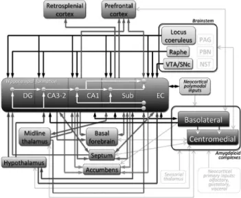
and subiculum inputs.65,68 The hippocampal formation and its main connections are shown in Figures 1 and 2.
In turn, subicular cells that do not project back to the
hippocampal formation send axons through the imbria-fornix
bundles, forwarding hippocampal output to several limbic
sites, including speciic regions of the basal forebrain (e.g.,
vertical limb of diagonal band), septum, nucleus accumbens,
amygdaloid complex (with particularly dense projections to
the basolateral division), mammillary bodies of hypothalamus and adjacent nuclei, midline thalamus (especially nucleus
reuniens), retrosplenial cortex, and prefrontal cortex.63,69,72 The CA1 subield, itself, also sends direct projections to the
vertical limb of diagonal band, septum, nucleus
accum-bens, basolateral amygdaloid complex, and prefrontal cor
-tex,63,70,71,73,74 as well as weaker efferents to retrosplenial cortex and sensorial neocortical areas.74,75 Roughly, all these efferents are accompanied by returning afferents, except for from the nucleus accumbens, retrosplenial cortex, and prefrontal cortex, which do not send projections back to the
hippocampus proper.68,76 In addition to the preferential
hippo-campal output by the CA1/subiculum system, CA3 also sends
direct efferents to the vertical limb of the diagonal band,
septum, and nucleus accumbens, with returning axons from
the septum.70,77,78 Additionally, CA3-2 receives projections – presumably unidirectional – from basolateral amygdala,71
and substance P-containing afferents from the supramam-milary hypothalamic nucleus.79 In contrast with CA subields, the DG is a mere receiver of extrahippocampal afferents, projecting axons solely to CA3.68,80 Beyond the substantial
input from the entorhinal cortex, few subcortical sites target the DG (the septum and some parts of the hypothalamus,
mainly its supramammilary area, are among these sites).81,82
Figure 1 Human hippocampal formation immunostained with the neuronal marker NeuN.
Section from a necropsy subject without history of neurological disorders. The dentate gyrus comprehends the granular layer (GL) and the hilus. CA3-1 are part of Ammon’s horn, and together with the dentate gyrus they constitute the hippocampus proper. PROSUB: prosubiculum; SUB: subiculum; PARASUB: parasubiculum; and EC: entorhinal cortex.
Figure 2 Overview of hippocampal formation connectivity with other limbic structures.
In spite of the mammillary nuclei and adjacencies that are reciprocally connected with the subiculum, none of the
CA subields project to the hypothalamus,83 although CA2
and CA3 do receive hypothalamic projections.79 Mostly, the non-entorhinal afferents directed to the DG and CA subields use the imbria-fornix system as their main route,
along with the majority of hippocampal efferents described above.68 Last but not least, the entire hippocampal formation (Figure 1) receives noradrenergic, serotoninergic, and do-paminergic inputs, respectively from locus coeruleus, raphe nuclei and ventral tegmental area of the brainstem.84,86
Particularly in the DG and CA subfields, dopaminergic
innervation is much more meager than the others,
prefer-entially targeting the entorhinal cortex.68
Because there are unknown efferents from the DG and CA2-3 to the neocortex, it seems that CA1 unidirectional
projections to retrosplenial and prefrontal cortices are the strongest communication between the hippocampus proper and neocortical areas outside the hippocampal formation.63,75 Unlike the hippocampus proper, the entorhinal cortex is re -ciprocally connected with a wider neocortical domain, with a preference for higher-order association areas. Therefore, the
entorhinal cortex acts as the principal link between
the polymodal neocortical inputs and the hippocampus.68,87 Although this neuroanatomical and functional distinction
exists, subcortical connections of the entorhinal cortex are
similar to those of the hippocampus proper, including bidirec-tional pathways with the septum and basolateral amygdala, as well as unidirectional projections to nucleus accumbens, and unidirectional projections from distributed parts of the hypothalamus and midline thalamus, once again with major participation of nucleus reuniens.72,88-91 Among the neocortical
sites reciprocally connected to the entorhinal cortex are the
perirhinal, postrhinal, and prefrontal cortices, including a
weak communication with the retrosplenial cortex.68 As part
of the hippocampal formation, the entorhinal cortex is also subjected to neuromodulatory inluence by the monoami
-nergic ascending systems, including signiicant dopami-nergic
afferents from the ventral tegmental area.90,92 In Figure 2,
the arrows outside the hippocampal formation represent the
extrinsic connections depicted here.
Amygdaloid complex and its efferents and
afferents
Another important part of the mesial temporal lobe is
represented by the amygdaloid complex, a group of func -tionally and structurally diverse nuclei, with intricate interconnections and widely distributed communication with cortical and subcortical limbic sites.93 In particular, the basolateral group projects strong efferents to CA3, CA1, the subiculum, entorhinal cortex and perirhinal cortex, receiving back-projections from all sites except CA3,72,94,95 as
seen in Figure 3. The same basolateral group is responsible for most of the amygdaloid innervation of the prefrontal
cortex (which is also a recipient of CA1/subiculum axons,
as previously mentioned), nucleus accumbens (a target of the whole hippocampal formation), and the striatum.96-98
Speciically, the prefrontal cortex sends returning projec
-tions to the basolateral complex99 (Figure 3). Considering the
emotional roles of the amygdala, as well as the mnemonic/
attentive functions of the hippocampus and the prefrontal
cortex, this basolateral-hippocampal reciprocity, along with
its common limbic targets, comprises a wide network that is well positioned to control cognitive aspects of behavior.100-102
In contrast, the centromedial group of amygdaloid nuclei preferentially sends projections to neuromodulator sites, including brainstem ascending monoaminergic centers (noradrenergic locus coeruleus, dopaminergic substantia nigra and ventral tegmental area, and serotoninergic raphe nuclei), and cholinergic regions of the basal forebrain and septum.103-106 Moreover, this centromedial division of the
amygdala sends efferents to several different autonomic, reproductive, and defensive-related sites, including some brainstem regions (periaqueductal grey, parabrachial nucle-us, and nucleus of the solitary tract), and various portions of the hypothalamus.105,107-110 In particular, the medial nucleus
of the amygdala sends projections to the nucleus reuniens of midline thalamus.104 An important conduit of the amyg-dala ibers that are directed to the brainstem, the basal
forebrain, and the diencephalon is the stria terminalis, whose bed nucleus contributes to additional innervation of these sites.111 Finally, the corticomedial nuclei of the
amyg-dala, which are also referred to as cortical-like nuclei due to
their supericial location and layered structure, are the main
source of efferents to the olfactory system.93 In general, all
of these efferent connections of the centromedial division are reciprocated93 (Figure 3).
The amygdaloid complex, as a whole, receives afferents
from regions that are not preferentially targeted by amygdala projections, such as higher-order areas of somatosensory,
Figure 3 Overview of amygdaloid complex connectivity with other limbic structures.
auditory, and visual cortices, as well as primary areas of the olfactory system, and gustatory and visceral portions
of the insular cortex.112-115 Thalamic nuclei directly related to speciic sensorial functions also contribute with afferents
to the amygdala,116,117 along with the paraventricular and
reuniens nuclei of the midline thalamus 118. Overall, the various afferents terminate onto virtually all the amygdaloid
divisions, despite some intricate patterns of speciicity not detailed here. Nevertheless, the basolateral group acts as the
main input receiver, which processes the information locally, forwarding it to the centromedial nuclei as a preferential output station93 (Figure 3).
Each one of the projection systems mentioned above
have speciic patterns of ipsilateral and/or contralateral
connectivity not detailed in the present article. The majority of connections are mediated by glutamatergic neurotransmis-sion, regardless of termination onto glutamatergic projection
cells or GABAergic interneurons, with speciic exceptions, for
instance, monoaminergic and cholinergic projections arising
from the brainstem and basal forebrain, GABAergic projec
-tions from the septum that coexist with septal cholinergic axons, and GABAergic projections from central amygdala.
There are differential patterns of topographical connectivity
throughout the long axis of the hippocampal formation, which are not explained here. Because comparative neuroanatomy
is not the main purpose of the present review, information on differences between mammalian species was omitted for clarity.
According to Bear,57 psychopathological states in TLE
would derive from intrinsic mechanisms leading to anatomical sensory limbic hyper connectivity. This psychiatric manifes-tation would be the result of a long epileptogenic process throughout the limbic system, and progressive changes in limbic structures secondary to seizure activity would be the underlying physiopathology.57 Such changes would explain
the fact that some patients have incomplete seizure remis-sion after surgical resection of the mesial temporal lobe.1 The extra-temporal commitment would be due to the spread
of seizures by critical subcortical synchronizers, possibly represented by thalamo-hippocampal circuits.1 Speciically,
the midline nuclei of the thalamus are well positioned to act as major synchronizers,118 because some of them are interconnected with both the hippocampal formation and
the amygdaloid complex (Figures 2 and 3). Importantly, the
same thalamic nuclei project to the nucleus accumbens and
are interconnected with the prefrontal cortex, forming a behaviorally lexible circuit whose dysfunction contributes
to the pathophysiology of psychosis and schizophrenia.119
Therefore, the hippocampal and amygdala connectivity
summarized here may be the common context for TLE and
psychotic-like symptoms. Although these network dysfunc-tions are not fully understood, there is increasing evidence from animal models showing how particular pathways of the limbic circuitry may underlie the relationship between psychosis and TLE.
Evidence from animal models
The relationship between psychosis and epilepsy has been
investigated experimentally through electrical kindling in
specific circuits and through single electrically evoked hippocampal afterdischarge protocols,120-123 dopaminergic
sensitization by pharmacological challenge,124,125 and acute
or chronic treatments with non-competitive antagonists of
N-methyl D-aspartate (NMDA) receptors.126,127
Kindling by electrical stimulation in speciic circuits pro -vides evidence for dopaminergic involvement in the putative shared mechanisms between epilepsy and psychosis. For
example, kindling in the ventral tegmental area generates
progressive fearfulness, hiding and loss of social behavior in cats,128 enhancing amphetamine- or methamphetamine-induced locomotor activity in rodents.129,130 Hippocampal
kindling increases the density of dopaminergic receptors, as well as dopamine release in nucleus accumbens.131,132
These dopaminergic changes in mesolimbic circuits after ictal episodes seem to depend on the hippocampal
activ-ity. Hyperlocomotion, PPI deicits and aberrant increases of
fast oscillations elicited by prefrontal or nucleus accumbens kindling are reverted if the hippocampus is previously in-activated.122 Unlike to hippocampal stimulation, prefrontal cortex stimulation is unable to signiicantly depolarize
nucleus accumbens neurons.133,134 Other studies have shown
that kindling in the basolateral amygdala down-regulates dopamine transporters in the striatum, while disrupting PPI.44,135 Altogether, kindling data reinforce the idea that, in experimental psychosis, the hippocampus becomes unable to regulate the source of convergent information low to the
nucleus accumbens, especially those from the prefrontal
cortex, ventral tegmental area and amygdala136 (Figure 4).
Abnormalities in the cortico-frontal-mesolimbic circuits
observed in experimental models may be useful to under -stand how dopaminergic dysfunctions could be related to the high rates of psychosis in TLE patients.137 Postictal psychosis
models based on single electrically evoked hippocampal af-terdischarge also induce psychotic-like behaviors, such as hy-perlocomotion and stereotyped movements.121 This postictal locomotor activity is mediated by D2 dopaminergic receptors
in the nucleus accumbens and requires ventral pallidum activ-ity, suggesting that the nucleus accumbens-ventral pallidum
pathway is critical for the expression of postictal abnormal
behaviors.120 Impairment of sensorimotor gating induced by
afterdischarge is related to increased neural activity in the medial septum and increased hippocampal gamma waves
induced by local afterdischarges through GABAergic, not
cholinergic, septo-hippocampal neurons.123 Experimental
studies on seizure-induced pathological sensitization of the dopaminergic system have also been useful to elucidate the relationship between mesolimbic circuits, psychosis and epilepsy.124 Seizure activity transiently elevates extracel
-lular dopamine levels at various brain sites, including the hippocampus, striatum, nucleus accumbens and prefrontal
cortex132,138,139 (Figure 4). In fact, rats subjected to the
pilo-carpine model of TLE display signiicantly higher spontaneous iring rates of ventral tegmental area neurons compared
to control rats.124 This population activity of dopaminergic
neurons, defined as the proportion of spontaneously
iring neurons, is regulated in intact rats by a ventral subic -ular-nucleus accumbens-ventral pallidal-ventral tegmental area pathway.119 However, the underlying mechanisms by which hippocampal hyperactivity inluences the dopaminergic neurons in TLE are not well recognized. Neonatal seizures
sensitization to methamphetamine in adolescence.125 The increased excitability observed in patients and in TLE models
might be associated with a pronounced neurodegeneration in
layer III of the medial entorhinal cortex.140,141 This pattern of
neuronal loss eventually results in hippocampal and
parahip-pocampal hyperexcitability, which have an important role
in epileptogenesis and enhanced susceptibility to seizures and epileptiform discharges.142,143 Such hyperexcitability could be related to the cognitive deicits and psychiat -ric comorbidities observed in some cases of TLE because entorhinal-hippocampus projections are crucial to temporal association memory in mice.144 Their dysfunction is involved
in the potentiation of drug-induced locomotor hyperactivity in pilocarpine-treated rats.124 In fact, recent data on human
TLE suggest that decreased neuronal density is found in
entorhinal cortex layer III of TLE patients with hippocampal
sclerosis and psychosis.35 Therefore, according to kindling
and hippocampal afterdischarge studies, the increased sensitization to psychostimulants observed in TLE models is likely attributable to an increase in tonic dopaminergic transmission, secondary to an augmented activity within the hippocampal and parahippocampal circuits145 (Figure 4B).
Increased hippocampal and parahippocampal drive is also found in patients with schizophrenia.146
Despite the available data, putative shared mechanisms
between epilepsy and psychosis remain poorly understood.
The non-competitive NMDA antagonists ketamine (KET), phencyclidine (PCP), and dizocilpine (MK-801) produce robust psychotic-like behaviors, PPI deicits, memory impairments
and hippocampal synaptic plasticity disruptions probably
associated with increased excitability in limbic
thalamo-cortical circuits.147,148 Non-competitive NMDA antagonists
generate disinhibition of speciic circuits by blocking the action of NMDA receptors on GABAergic neurons, resulting in decreased iring of GABAergic neurons (mainly parvalbu
-min-positive chandelier cells) and increased excitability of limbic circuits and the prefrontal cortex.149-153 Treatment with non-competitive NMDA antagonists can also decrease parvalbumin expression154 and produce epileptiform activity
in limbic circuits.155 NMDA antagonists can also damage and
kill cortical neurons by increasing cytoplasmic vacuoles in pyramidal neurons of the posterior cingulate and
retrosple-nial cortex of adult rats.156 In the same vein, KET signiicantly
potentiates kainic acid-elicited gamma power in slices of
the mouse prefrontal cortex.157 Moreover, the dopaminergic
system is also involved in the behavioral changes observed
in NMDA-antagonist-based models.158 Impairment of execu
-tive tasks in rodents treated with KET is mediated by D2
receptors.159 In addition, a single dose of PCP promotes an increase of dopamine levels in the prefrontal cortex.160
Jackson et al.152 demonstrated that a single MK-801 injection increases random iring and decreases burst iring activity in the prefrontal cortex. The reduction in burst activity is associ
-ated with a decrease in synaptic eficacy, which in turn could
affect the control of behavioral outputs. Similarly, increases
in the random iring rates of prefrontal neurons are associated with deicits in PPI and working memory.152 Suzuki et al.127 showed that systemic injection with PCP signiicantly changes neuronal iring rates in the prefrontal cortex of rats. No changes were seen after local frontal cortex microinjec
-tion. However, when PCP was microinjected into the ventral Figure 4. Direct and indirect output in psychosis pathways.
A. Non-pathological circuit. B. Hypothetic pathological cir-cuit in temporal lobe epilepsy. Hippocampal hyperactivity may disrupt the fine dopaminergic control of limbic and extralimbic circuits via polysynaptic projections. 1. Epileptogenic process and neuropathological changes may drive hippocampal hyper-excitability. 2. Increased excitability seen in TLE patients and
animal models might be associated with neurodegeneration in the enthorinal cortex. Robust projections between hippocampus and enthorinal cortex reverberate neuronal hyperactivity. 3.
Strong projections between hippocampal formation and amyg-dala may propagate the excitability to the emotional circuit. 4.
Consequently, nucleus accumbens is overactivated by convergent projections. 5. Nucleus accumbens, in turn, strongly inhibits
ventral pallidum. 6. Given that the ventral pallidum provides an
hippocampus, prefrontal neurons showed responses similar to those observed after systemic injection.161 Altogether, a
set of evidence indicates the involvement of the dopaminer-gic system and points to a possible role of hippocampal to
prefrontal cortex projections in the expression of aberrant behaviors induced by non-competitive NMDA antagonists in rats. Considering that the main source of direct hippocampal afferents to the cortex (CA1 subield of the hippocampus) is
especially affected in TLE, and that psychotic-like effects
produced by the administration of NMDA antagonists require a preserved communication between the hippocampus (CA1) and the prefrontal cortex, we speculate that synaptic plastic -ity in this pathway may represent a point of vulnerabil-ity for the development of psychosis in TLE patients.
Conclusions
The investigation on TLE and psychosis has independently pro-vided important pathophysiological hallmarks. The best-known observations include hippocampal sclerosis in TLE, related to a chronic state of recurrent seizures, and heterogeneous cortico-striato-pallido-thalamic disturbances in psychosis, pos-sibly due to interactions between genetic and environmental
inluences on the maturation of these circuits. The relatively
high prevalence of psychotic-like symptoms in TLE patients suggests shared mechanisms and/or substrates between these two conditions. However, few studies have addressed this issue, despite the interesting reports on electrical kindling,
dopaminergic manipulation, and NMDA antagonism reviewed here. Considering mesial temporal lobe connectivity along with the midline thalamus, the prefrontal cortex and the
nucleus accumbens, as well as the ascending
neuromodula-tory inluence on these circuits, future studies involving a
convergence of approaches would be useful. For instance, investigations into the relationship between behavioral symp-toms (e.g., sensorimotor gating), synaptic plasticity (e.g., long-term potentiation/depression in limbic pathways), and pathology in animal models of TLE (e.g., lithium-pilocarpine or electrical kindling in the hippocampus or amygdala) are
scant. Nevertheless, such animal studies would be necessary
to gain a better understanding of clinical cases. In summary, present clinical, neurophysiological and neuropathological
data demonstrate the existence of an intricate mesh of both
antagonistic and also shared mechanisms between TLE and psychosis. As once mentioned by Slater et al. “like effects commonly have similar causes” 26, and the underlying patho-genesis of psychosis in TLE still needs clariication.
Acknowledgments
Supported by Fundação de Amparo à Pesquisa do Estado de
São Paulo (FAPESP), Conselho Nacional de Desenvolvimento Cientíico e Tecnológico (National Counsel of Technological and Scientiic Development - CNPq) and Fundação de Amparo à Pesquisa do Estado do Rio Grande do Norte (FAPERN), Brazil.
Disclosures
Ludmyla Kandratavicius, PhD
Employment:Faculdade de Medicina de Ribeirão Preto, Department of Neurosciences and Behavior, Universidade de São Paulo, Brazil. Cleiton Lopes-Aguiar, MSc
Employment: Faculdade de Medicina de Ribeirão Preto, Department of Neurosciences and Behavior, Universidade de São Paulo, Brazil.
Lézio Soares Bueno-Júnior, PhD
Employment:Faculdade de Medicina de Ribeirão Preto, Department of Neurosciences and Behavior, Universidade de São Paulo, Brazil. Rodrigo Neves Romcy-Pereira, MSc, PhD
Employment:Brain Institute, Universidade Federal do Rio Grande do Norte, Natal, Brazil.
Jaime Eduardo Cecilio Hallak, MD, PhD
Employment:Faculdade de Medicina de Ribeirão Preto, Department of Neurosciences and Behavior, Universidade de São Paulo, Brazil.Other: National Institute of Science and Technology in Translational Medicine (Instituto Nacional de Ciência e Tecnologia - INCT-TM; CNPq).
Joao Pereira Leite, MD, PhD
Employment:Faculdade de Medicina de Ribeirão Preto, Department of Neurosciences and Behavior, Universidade de São Paulo, Brazil. * Modest
** Signiicant
*** Signiicant. Amounts given to the author’s institution or to a colleague for
research in which the author has participation, not directly to the author.
References
1. Bertram EH. Temporal lobe epilepsy: where do the seizures really begin? Epilepsy Behav. 2009;14(Suppl 1):32-7.
2. Swerdlow NR. Integrative circuit models and their implications for the pathophysiologies and treatments of the schizophrenias. Curr Top Behav Neurosci. 2010;4:555-83.
3. Flor-Henry P. Psychosis and temporal lobe epilepsy. A controlled investigation. Epilepsia. 1969;10(3):363-95.
4. Roberts GW, Done DJ, Bruton C, Crow TJ. A “mock up” of schizophrenia: temporal lobe epilepsy and schizophrenia-like psychosis. Biol Psychiatry. 1990;28(2):127-43.
5. Serafetinides EA, Falconer MA. The effects of temporal lobectomy in epileptic patients with psychosis. J Ment Sci. 1962;108:584-93.
6. Stevens JR. Epilepsy, psychosis and schizophrenia. Schizophr Res. 1988;1(1):79-89.
7. Taylor DC. Factors inluencing the occurrence of schizophrenia-like psychosis in patients with temporal lobe epilepsy. Psychol Med. 1975;5(3):249-54.
8. Editorial: Epilepsy, schizophrenia, and limbic system. Lancet. 1974;2(7886):935-6.
9. Papez JW. A proposed mechanism of emotion. J Neuropsychiatry Clin Neurosci. 1937;7(1):103-12.
10. Klüver H, Bucy P. “Psychic blindness” and other symtoms following bilateral temporal lobectomy in rhesus monkeys. American Journal of Physiology. 1937;119:352-3.
11. MacLean P. Psychosomatic disease and the visceral brain; recent developments bearing on the Papez theory of emotion. Psychosom Med. 1949;11(6):338-53.
12. Heimer L, Van Hoesen GW, Trimble M, Zahn DS. The limbic system: a concept in perpetual search for a deinition. In: Heimer L, Van Hoesen GW, Trimble M, Zahn DS, eds. Anatomy of Neuropsychiatry. U.S.A.: Academic Press Elsevier; 2008:1-13. 13. Brain L. Psychosomatic Medicine and the Brain-Mind Relationship.
Lancet. 1964;2(7355):325-8.
14. Cairns H. Disturbances of consciousness with lesions of the brain-stem and diencephalon. Brain. 1952;75(2):109-46. 15. Waxman SG, Geschwind N. The interictal behavior
syndrome of temporal lobe epilepsy. Arch Gen Psychiatry. 1975;32(12):1580-6.
16. Penfield W, Perot P. The Brain’s Record of Auditory and Visual Experience. A Final Summary and Discussion. Brain. 1963;86:595-696.
17. Gloor P, Olivier A, Quesney LF, Andermann F, Horowitz S. The role of the limbic system in experiential phenomena of temporal lobe epilepsy. Ann Neurol. 1982;12(2):129-44.
19. Fujii D, Ahmed I. Psychotic disorder following traumatic brain injury: a conceptual framework. Cogn Neuropsychiatry. 2002;7(1):41-62.
20. Kristensen O, Sindrup EH. Psychomotor epilepsy and psychosis. I. Physical aspects. Acta Neurol Scand. 1978;57(5):361-9. 21. Landolt H. Some correlations between the electroencephalogram
and normal and pathologic mental processes. Epilepsy Behav. 1963;14(3):448-51.
22. Pollock DC. Models for understanding the antagonism between seizures and psychosis. Prog Neuropsychopharmacol Biol Psychiatry. 1987;11(4):483-504.
23. Trimble M. The relationship between epilepsy and schizophrenia: a biochemical hypothesis. Biol Psychiatry. 1977;12(2):299-304. 24. Ogren SO, Pakh B. Effects of dopamine D1 and D2 receptor agonists
and antagonists on seizures induced by chemoconvulsants in mice. Pharmacol Toxicol. 1993;72(4-5):213-20.
25. Turski L, Cavalheiro EA, Bortolotto ZA, Ikonomidou-Turski C, Kleinrok Z, Turski WA. Dopamine-sensitive anticonvulsant site in the rat striatum. J Neurosci. 1988;8(11):4027-37.
26. Slater E, Beard AW, Glithero E. The schizophrenia-like psychoses of epilepsy. Br J Psychiatry. 1963;109:95-150.
27. Krishnamoorthy ES, Trimble MR, Blumer D. The classiication of neuropsychiatric disorders in epilepsy: a proposal by the ILAE Commission on Psychobiology of Epilepsy. Epilepsy Behav. 2007;10(3):349-53.
28. Toone BK. The psychoses of epilepsy. J Neurol Neurosurg Psychiatry. 2000;69(1):1-3.
29. Kanemoto K, Kawasaki J, Kawai I. Postictal psychosis: a comparison with acute interictal and chronic psychoses. Epilepsia. 1996;37(6):551-6.
30. Tarulli A, Devinsky O, Alper K. Progression of postictal to interictal psychosis. Epilepsia. 2001;42(11):1468-71.
31. Braun CM, Dumont M, Duval J, Hamel-Hebert I, Godbout L. Brain modules of hallucination: an analysis of multiple patients with brain lesions. J Psychiatry Neurosci. 2003;28(6):432-49. 32. Torrey EF, Peterson MR. Schizophrenia and the limbic system.
Lancet. 1974;2(7886):942-6.
33. Malamud N. Psychiatric disorder with intracranial tumors of limbic system. Arch Neurol. 1967;17(2):113-23.
34. Taylor DC. Mental state and temporal lobe epilepsy. A correlative account of 100 patients treated surgically. Epilepsia. 1972;13(6):727-65.
35. Kandratavicius L, Hallak JE, Young LT, Assirati JA, Carlotti CG, Jr., Leite JP. Differential aberrant sprouting in temporal lobe epilepsy with psychiatric co-morbidities. Psychiatry Res. 2012;195(3):144-50.
36. Allen P, Laroi F, McGuire PK, Aleman A. The hallucinating brain: a review of structural and functional neuroimaging studies of hallucinations. Neurosci Biobehav Rev. 2008;32(1):175-91. 37. Braff DL. Gating in schizophrenia: from genes to cognition (to
real world function?). Biol Psychiatry. 2011;69(5):395-6. 38. Quednow BB, Ettinger U, Mossner R et al. The schizophrenia
risk allele C of the TCF4 rs9960767 polymorphism disrupts sensorimotor gating in schizophrenia spectrum and healthy volunteers. J Neurosci. 2011;31(18):6684-91.
39. Takahashi H, Iwase M, Yasuda Y et al. Relationship of prepulse inhibition to temperament and character in healthy Japanese subjects. Neurosci Res. 2012;72(2):187-93.
40. Braff DL, Geyer MA, Swerdlow NR. Human studies of prepulse inhibition of startle: normal subjects, patient groups, and pharmacological studies. Psychopharmacology (Berl). 2001;156(2-3):234-58.
41. Pouretemad HR, Thompson PJ, Fenwick PB. Impaired sensorimotor gating in patients with non-epileptic seizures. Epilepsy Res. 1998;31(1):1-12.
42. Syed TU, LaFrance WC, Jr., Kahriman ES et al. Can semiology predict psychogenic nonepileptic seizures? A prospective study. Ann Neurol. 2011;69(6):997-1004.
43. Mokleby K, Blomhoff S, Malt UF, Dahlstrom A, Tauboll E, Gjerstad L. Psychiatric comorbidity and hostility in patients with psychogenic nonepileptic seizures compared with somatoform disorders and healthy controls. Epilepsia. 2002;43(2):193-8. 44. Howland JG, Hannesson DK, Barnes SJ, Phillips AG. Kindling of
basolateral amygdala but not ventral hippocampus or perirhinal cortex disrupts sensorimotor gating in rats. Behav Brain Res. 2007;177(1):30-6.
45. Miller EJ, Saint Marie LR, Breier MR, Swerdlow NR. Pathways from the ventral hippocampus and caudal amygdala to forebrain regions that regulate sensorimotor gating in the rat. Neuroscience. 2010;165(2):601-11.
46. Weinberger DR, Wagner RL, Wyatt RJ. Neuropathological studies of schizophrenia: a selective review. Schizophr Bull. 1983;9(2):193-212.
47. Thom M. Hippocampal sclerosis: progress since Sommer. Brain Pathol. 2009;19(4):565-72.
48. Cavanagh JB, Meyer A. Aetiological aspects of Ammon’s horn sclerosis associated with temporal lobe epilepsy. Br Med J. 1956;2(5006):1403-7.
49. Bonilha L, Rorden C, Appenzeller S, Coan AC, Cendes F, Li LM. Gray matter atrophy associated with duration of temporal lobe epilepsy. Neuroimage. 2006;32(3):1070-9.
50. Roberts GW, Bruton CJ. Notes from the graveyard: neuropathology and schizophrenia. Neuropathol Appl Neurobiol. 1990;16(1):3-16.
51. Sokolov BP, Tcherepanov AA, Haroutunian V, Davis KL. Levels of mRNAs encoding synaptic vesicle and synaptic plasma membrane proteins in the temporal cortex of elderly schizophrenic patients. Biol Psychiatry. 2000;48(3):184-96.
52. Falkai P, Parlapani E, Gruber O, Schmitt A. The Neuropathology of Schizophrenia: Central Role for the Hippocampus? In: Gattaz WF, Busatto G, eds. Advances in Schizophrenia Research 2009. New York: Springer Science; 2010:149-65.
53. Shaw P, Mellers J, Henderson M, Polkey C, David AS, Toone BK. Schizophrenia-like psychosis arising de novo following a temporal lobectomy: timing and risk factors. J Neurol Neurosurg Psychiatry. 2004;75(7):1003-8.
54. Morimoto K, Fahnestock M, Racine RJ. Kindling and status epilepticus models of epilepsy: rewiring the brain. Prog Neurobiol. 2004;73(1):1-60.
55. Flor-Henry P. Lateralized temporal-limbic dysfunction and psychopathology. Ann N Y Acad Sci. 1976;280:777-97.
56. Adamec RE. Does kindling model anything clinically relevant? Biol Psychiatry. 1990;27(3):249-79.
57. Bear DM. Temporal lobe epilepsy--a syndrome of sensory-limbic hyperconnection. Cortex. 1979;15(3):357-84.
58. Duvernoy HM. The Human Hippocampus. Berlin: Springer-Verlag; 1988.
59. Andersen P, Soleng AF, Raastad M. The hippocampal lamella hypothesis revisited. Brain Res. 2000;886(1-2):165-71. 60. Scharfman HE. The CA3 “backprojection” to the dentate gyrus.
Prog Brain Res. 2007;163:627-37.
61. Heinemann U, Schmitz D, Eder C, Gloveli T. Properties of entorhinal cortex projection cells to the hippocampal formation. Ann N Y Acad Sci. 2000;911:112-26.
62. Witter MP, Wouterlood FG, Naber PA, Van Haeften T. Anatomical organization of the parahippocampal-hippocampal network. Ann N Y Acad Sci. 2000;911:1-24.
64. Gigg J. Constraints on hippocampal processing imposed by the connectivity between CA1, subiculum and subicular targets. Behav Brain Res. 2006;174(2):265-71.
65. Naber PA, Lopes da Silva FH, Witter MP. Reciprocal connections between the entorhinal cortex and hippocampal ields CA1 and the subiculum are in register with the projections from CA1 to the subiculum. Hippocampus. 2001;11(2):99-104.
66. Canto CB, Wouterlood FG, Witter MP. What does the anatomical organization of the entorhinal cortex tell us? Neural Plast. 2008;2008:381243.
67. de Curtis M, Pare D. The rhinal cortices: a wall of inhibition between the neocortex and the hippocampus. Prog Neurobiol. 2004;74(2):101-10.
68. Amaral DG, Lavenex P. Hippocampal Neuroanatomy. In: Andersen P, Morris RG, Amaral DG, Bliss TV, O’Keefe J, eds. The Hippocampus Book. New York: Oxford University Press; 2007:37-114.
69. Finch DM, Nowlin NL, Babb TL. Demonstration of axonal projections of neurons in the rat hippocampus and subiculum by intracellular injection of HRP. Brain Res. 1983;271(2):201-16. 70. Friedman DP, Aggleton JP, Saunders RC. Comparison of
hippocampal, amygdala, and perirhinal projections to the nucleus accumbens: combined anterograde and retrograde tracing study in the Macaque brain. J Comp Neurol. 2002;450(4):345-65. 71. Pitkanen A, Pikkarainen M, Nurminen N, Ylinen A. Reciprocal
connections between the amygdala and the hippocampal formation, perirhinal cortex, and postrhinal cortex in rat. A review. Ann N Y Acad Sci. 2000;911:369-91.
72. Saunders RC, Rosene DL, Van Hoesen GW. Comparison of the efferents of the amygdala and the hippocampal formation in the rhesus monkey: II. Reciprocal and non-reciprocal connections. J Comp Neurol. 1988;271(2):185-207.
73. Jay TM, Witter MP. Distribution of hippocampal CA1 and subicular efferents in the prefrontal cortex of the rat studied by means of anterograde transport of Phaseolus vulgaris-leucoagglutinin. J Comp Neurol. 1991;313(4):574-86.
74. van Groen T, Wyss JM. Extrinsic projections from area CA1 of the rat hippocampus: olfactory, cortical, subcortical, and bilateral hippocampal formation projections. J Comp Neurol. 1990;302(3):515-28.
75. Cenquizca LA, Swanson LW. Spatial organization of direct hippocampal ield CA1 axonal projections to the rest of the cerebral cortex. Brain Res Rev. 2007;56(1):1-26.
76. Vertes RP, Hoover WB, Szigeti-Buck K, Leranth C. Nucleus reuniens of the midline thalamus: link between the medial prefrontal cortex and the hippocampus. Brain Res Bull. 2007;71(6):601-9.
77. Gaykema RP, van der Kuil J, Hersh LB, Luiten PG. Patterns of direct projections from the hippocampus to the medial septum-diagonal band complex: anterograde tracing with Phaseolus vulgaris leucoagglutinin combined with immunohistochemistry of choline acetyltransferase. Neuroscience. 1991;43(2-3):349-60.
78. Swanson LW, Cowan WM. An autoradiographic study of the organization of the efferent connections of the hippocampal formation in the rat. J Comp Neurol. 1977;172(1):49-84. 79. Leranth C, Nitsch R. Morphological evidence that hypothalamic
substance P-containing afferents are capable of iltering the signal low in the monkey hippocampal formation. J Neurosci. 1994;14(7):4079-94.
80. Swanson LW, Wyss JM, Cowan WM. An autoradiographic study of the organization of intrahippocampal association pathways in the rat. J Comp Neurol. 1978;181(4):681-715.
81. Vertes RP. PHA-L analysis of projections from the supramammillary nucleus in the rat. J Comp Neurol. 1992;326(4):595-622.
82. Wyss JM, Swanson LW, Cowan WM. Evidence for an input to the molecular layer and the stratum granulosum of the dentate gyrus from the supramammillary region of the hypothalamus. Anat Embryol (Berl). 1979;156(2):165-76.
83. Swanson LW, Cowan WM. Hippocampo-hypothalamic connections: origin in subicular cortex, not ammon’s horn. Science. 1975;189(4199):303-4.
84. Bobillier P, Seguin S, Degueurce A, Lewis BD, Pujol JF. The efferent connections of the nucleus raphe centralis superior in the rat as revealed by radioautography. Brain Res. 1979;166(1):1-8.
85. Loughlin SE, Foote SL, Grzanna R. Efferent projections of nucleus locus coeruleus: morphologic subpopulations have different efferent targets. Neuroscience. 1986;18(2):307-19. 86. Oades RD, Halliday GM. Ventral tegmental (A10) system:
neurobiology. 1. Anatomy and connectivity. Brain Res. 1987;434(2):117-65.
87. Insausti R, Herrero MT, Witter MP. Entorhinal cortex of the rat: cytoarchitectonic subdivisions and the origin and distribution of cortical efferents. Hippocampus. 1997;7(2):146-83. 88. Insausti R, Amaral DG, Cowan WM. The entorhinal cortex
of the monkey: III. Subcortical afferents. J Comp Neurol. 1987;264(3):396-408.
89. Ohtake T, Yamada H. Efferent connections of the nucleus reuniens and the rhomboid nucleus in the rat: an anterograde PHA-L tracing study. Neurosci Res. 1989;6(6):556-68.
90. Room P, Groenewegen HJ. Connections of the parahippocampal cortex in the cat. II. Subcortical afferents. J Comp Neurol. 1986;251(4):451-73.
91. Witter MP, Groenewegen HJ. Connections of the parahippocampal cortex in the cat. IV. Subcortical efferents. J Comp Neurol. 1986;252(1):51-77.
92. Beckstead RM, Domesick VB, Nauta WJ. Efferent connections of the substantia nigra and ventral tegmental area in the rat. Brain Res. 1979;175(2):191-17.
93. Sah P, Faber ES, Lopez De Armentia M, Power J. The amygdaloid complex: anatomy and physiology. Physiol Rev. 2003;83(3):803-34.
94. Canteras NS, Swanson LW. Projections of the ventral subiculum to the amygdala, septum, and hypothalamus: a PHAL anterograde tract-tracing study in the rat. J Comp Neurol. 1992;324(2):180-94.
95. McDonald AJ, Mascagni F. Projections of the lateral entorhinal cortex to the amygdala: a Phaseolus vulgaris leucoagglutinin study in the rat. Neuroscience. 1997;77(2):445-59.
96. Mitrano DA, Pare JF, Smith Y. Ultrastructural relationships between cortical, thalamic, and amygdala glutamatergic inputs and group I metabotropic glutamate receptors in the rat accumbens. J Comp Neurol. 2010;518(8):1315-29.
97. Porrino LJ, Crane AM, Goldman-Rakic PS. Direct and indirect pathways from the amygdala to the frontal lobe in rhesus monkeys. J Comp Neurol. 1981;198(1):121-36.
98. McDonald AJ. Organization of amygdaloid projections to the prefrontal cortex and associated striatum in the rat. Neuroscience. 1991;44(1):1-14.
99. Gabbott PL, Warner TA, Jays PR, Salway P, Busby SJ. Prefrontal cortex in the rat: projections to subcortical autonomic, motor, and limbic centers. J Comp Neurol. 2005;492(2):145-77. 100. Ghashghaei HT, Barbas H. Pathways for emotion: interactions
of prefrontal and anterior temporal pathways in the amygdala of the rhesus monkey. Neuroscience. 2002;115(4):1261-79. 101. Ishikawa A, Nakamura S. Convergence and interaction of
102. Vertes RP. Interactions among the medial prefrontal cortex, hippocampus and midline thalamus in emotional and cognitive processing in the rat. Neuroscience. 2006;142(1):1-20. 103. Caffe AR, van Leeuwen FW, Luiten PG. Vasopressin cells in the
medial amygdala of the rat project to the lateral septum and ventral hippocampus. J Comp Neurol. 1987;261(2):237-52. 104. Price JL, Amaral DG. An autoradiographic study of the
projections of the central nucleus of the monkey amygdala. J Neurosci. 1981;1(11):1242-59.
105. Usunoff KG, Schmitt O, Itzev DE et al. Efferent projections of the anterior and posterodorsal regions of the medial nucleus of the amygdala in the mouse. Cells Tissues Organs. 2009;190(5):256-85.
106. Wallace DM, Magnuson DJ, Gray TS. Organization of amygdaloid projections to brainstem dopaminergic, noradrenergic, and adrenergic cell groups in the rat. Brain Res Bull. 1992;28(3):447-54.
107. Jia HG, Zhang GY, Wan Q. A GABAergic projection from the central nucleus of the amygdala to the parabrachial nucleus: an ultrastructural study of anterograde tracing in combination with post-embedding immunocytochemistry in the rat. Neurosci Lett. 2005;382(1-2):153-7.
108. Petrovich GD, Canteras NS, Swanson LW. Combinatorial amygdalar inputs to hippocampal domains and hypothalamic behavior systems. Brain Res Brain Res Rev. 2001;38(1-2):247-89. 109. Rizvi TA, Ennis M, Behbehani MM, Shipley MT. Connections
between the central nucleus of the amygdala and the midbrain periaqueductal gray: topography and reciprocity. J Comp Neurol. 1991;303(1):121-31.
110. Saha S, Batten TF, Henderson Z. A GABAergic projection from the central nucleus of the amygdala to the nucleus of the solitary tract: a combined anterograde tracing and electron microscopic immunohistochemical study. Neuroscience. 2000;99(4):613-26. 111. Dong HW, Swanson LW. Projections from bed nuclei of the stria
terminalis, dorsomedial nucleus: implications for cerebral hemisphere integration of neuroendocrine, autonomic, and drinking responses. J Comp Neurol. 2006;494(1):75-107. 112. LeDoux JE, Farb CR, Romanski LM. Overlapping projections to
the amygdala and striatum from auditory processing areas of the thalamus and cortex. Neurosci Lett. 1991;134(1):139-44. 113. Scalia F, Winans SS. The differential projections of the olfactory
bulb and accessory olfactory bulb in mammals. J Comp Neurol. 1975;161(1):31-55.
114. Yasui Y, Breder CD, Saper CB, Cechetto DF. Autonomic responses and efferent pathways from the insular cortex in the rat. J Comp Neurol. 1991;303(3):355-74.
115. McDonald AJ. Cortical pathways to the mammalian amygdala. Prog Neurobiol. 1998;55(3):257-332.
116. Doron NN, Ledoux JE. Organization of projections to the lateral amygdala from auditory and visual areas of the thalamus in the rat. J Comp Neurol. 1999;412(3):383-409.
117. Turner BH, Herkenham M. Thalamoamygdaloid projections in the rat: a test of the amygdala’s role in sensory processing. J Comp Neurol. 1991;313(2):295-325.
118. Su HS, Bentivoglio M. Thalamic midline cell populations projecting to the nucleus accumbens, amygdala, and hippocampus in the rat. J Comp Neurol. 1990;297(4):582-93. 119. Floresco SB, Zhang Y, Enomoto T. Neural circuits subserving
behavioral lexibility and their relevance to schizophrenia. Behav Brain Res. 2009;204(2):396-409.
120. Ma J, Brudzynski SM, Leung LW. Involvement of the nucleus accumbens-ventral pallidal pathway in postictal behavior induced by a hippocampal afterdischarge in rats. Brain Res. 1996;739(1-2):26-35.
121. Ma J, Leung LS. Schizophrenia-like behavioral changes after partial hippocampal kindling. Brain Res. 2004;997(1):111-8.
122. Ma J, Leung LS. Kindled seizure in the prefrontal cortex activated behavioral hyperactivity and increase in accumbens gamma oscillations through the hippocampus. Behav Brain Res. 2010;206(1):68-77.
123. Ma J, Leung LW. Medial septum mediates the increase in post-ictal behaviors and hippocampal gamma waves after an electrically induced seizure. Brain Res. 1999;833(1):51-7. 124. Cifelli P, Grace AA. Pilocarpine-induced temporal lobe epilepsy
in the rat is associated with increased dopamine neuron activity. Int J Neuropsychopharmacol. 2011:1-8.
125. Lin TC, Huang LT, Huang YN, Chen GS, Wang JY. Neonatal status epilepticus alters prefrontal-striatal circuitry and enhances methamphetamine-induced behavioral sensitization in adolescence. Epilepsy Behav. 2009;14(2):316-23.
126. Olney JW, Newcomer JW, Farber NB. NMDA receptor hypofunction model of schizophrenia. J Psychiatr Res. 1999;33(6):523-33. 127. Suzuki Y, Jodo E, Takeuchi S, Niwa S, Kayama Y. Acute
administration of phencyclidine induces tonic activation of medial prefrontal cortex neurons in freely moving rats. Neuroscience. 2002;114(3):769-79.
128. Stevens JR, Livermore A, Jr. Kindling of the mesolimbic dopamine system: animal model of psychosis. Neurology. 1978;28(1):36-46.
129. Ehlers CL, Koob GF. Locomotor behavior following kindling in three different brain sites. Brain Res. 1985;326(1):71-9. 130. Watanabe T, Morimoto K, Nakamura M et al. Kindling of the
ventral tegmental area induces supersensitivity in the central dopamine system. Brain Res. 2004;1003(1-2):194-8.
131. Csernansky JG, Kerr S, Pruthi R, Prosser ES. Mesolimbic dopamine receptor increases two weeks following hippocampal kindling. Brain Res. 1988;449(1-2):357-60.
132. Strecker RE, Moneta ME. Electrical stimulation of the kindled hippocampus briely increases extracellular dopamine in the nucleus accumbens. Neurosci Lett. 1994;176(2):173-7. 133. Goto Y, O’Donnell P. Network synchrony in the nucleus
accumbens in vivo. J Neurosci. 2001;21(12):4498-504. 134. O’Donnell P, Grace AA. Synaptic interactions among excitatory
afferents to nucleus accumbens neurons: hippocampal gating of prefrontal cortical input. J Neurosci. 1995;15(5 Pt 1):3622-39. 135. Gordon I, Mintz M, Rosenne E, Rehavi M. Long-term effects of
amygdaloid kindling on striatal dopaminergic terminals. Brain Res Bull. 1995;36(3):235-9.
136. Grace AA. Gating of information low within the limbic system and the pathophysiology of schizophrenia. Brain Res Brain Res Rev. 2000;31(2-3):330-41.
137. Leung LS, Ma J, McLachlan RS. Behaviors induced or disrupted by complex partial seizures. Neurosci Biobehav Rev. 2000;24(7):763-75.
138. Dazzi L, Serra M, Porceddu ML, Sanna A, Chessa MF, Biggio G. Enhancement of basal and pentylenetetrazol (PTZ)-stimulated dopamine release in the brain of freely moving rats by PTZ-induced kindling. Synapse. 1997;26(4):351-8.
139. Meurs A, Clinckers R, Ebinger G, Michotte Y, Smolders I. Seizure activity and changes in hippocampal extracellular glutamate, GABA, dopamine and serotonin. Epilepsy Res. 2008;78(1):50-9. 140. Du F, Eid T, Lothman EW, Kohler C, Schwarcz R. Preferential
neuronal loss in layer III of the medial entorhinal cortex in rat models of temporal lobe epilepsy. J Neurosci. 1995;15(10):6301-13.
141. Du F, Whetsell WO, Jr., Abou-Khalil B, Blumenkopf B, Lothman EW, Schwarcz R. Preferential neuronal loss in layer III of the entorhinal cortex in patients with temporal lobe epilepsy. Epilepsy Res. 1993;16(3):223-33.