I LATIN AMERICAN GUIDELINE FOR
THE DIAGNOSIS AND TREATMENT
of
c
haGas
’ h
eart
d
Isease
This guideline should be cited as:
Andrade JP, Marin-Neto JA, Paola AA, Vilas-Boas F, Oliveira GM, Bacal F, Bocchi EA, Almeida DR,
Fragata Filho AA, Moreira M da C, Xavier SS, Oliveira Junior WA, Dias JC; Sociedade Brasileira de
Cardiologia. I Diretriz Latino Americana para o Diagnóstico e Tratamento da Cardiopatia Chagásica.
Editorial Board
Brazil
Adib D. Jatene (SP) Alexandre A. C. Abizaid (SP) Alfredo José Mansur (SP) Álvaro Avezum (SP) Amanda G. M. R. Sousa (SP) André Labrunie (PR) Andrei Sposito (DF) Angelo A. V. de Paola (SP) Antonio Augusto Barbosa Lopes (SP)
Antonio Carlos C. Carvalho (SP) Antônio Carlos Palandri Chagas (SP) Antonio Carlos Pereira Barretto (SP) Antonio Cláudio L. Nóbrega (RJ) Antonio de Padua Mansur (SP) Ari Timerman (SP)
Armênio Costa Guimarães (BA) Ayrton Klier Péres (DF) Ayrton Pires Brandão (RJ) Barbara M. Ianni (SP) Beatriz Matsubara (SP) Braulio Luna Filho (SP) Brivaldo Markman Filho (PE) Bruce B. Duncan (RS) Bruno Caramelli (SP) Carisi A. Polanczyk (RS) Carlos Alberto Pastore (SP) Carlos Eduardo Negrão (SP) Carlos Eduardo Rochitte (SP) Carlos Eduardo Suaide Silva (SP) Carlos Vicente Serrano Júnior (SP) Celso Amodeo (SP)
Charles Mady (SP)
Claudio Gil Soares de Araujo (RJ) Cleonice Carvalho C. Mota (MG) Dalton Valentim Vassallo (ES) Décio Mion Jr (SP)
Denilson Campos de Albuquerque (RJ) Dikran Armaganijan (SP) Djair Brindeiro Filho (PE) Domingo M. Braile (SP) Edmar Atik (SP) Edson Stefanini (SP) Elias Knobel (SP) Eliudem Galvão Lima (ES) Emilio Hideyuki Moriguchi (RS) Enio Buffolo (SP)
Eulógio E. Martinez Fº (SP) Evandro Tinoco Mesquita (RJ) Expedito E. Ribeiro da Silva (SP) Fábio Sândoli de Brito Jr. (SP) Fábio Vilas-Boas (BA) Fernando A. P. Morcerf (RJ) Fernando Bacal (SP) Flávio D. Fuchs (RS)
Francisco Antonio Helfenstein Fonseca (SP)
Francisco Laurindo (SP) Francisco Manes Albanesi Fº (RJ) Gilmar Reis (MG)
Gilson Soares Feitosa (BA) Ínes Lessa (BA)
Iran Castro (RS) Ivan G. Maia (RJ) Ivo Nesralla (RS)
Jarbas Jakson Dinkhuysen (SP) João Pimenta (SP)
Jorge Ilha Guimarães (RS) Jorge Pinto Ribeiro (RS) José A. Marin-Neto (SP) José Antonio Franchini Ramires (SP) José Augusto Soares Barreto Filho (SE)
José Carlos Nicolau (SP)
José Geraldo de Castro Amino (RJ) José Lázaro de Andrade (SP) José Péricles Esteves (BA) José Teles Mendonça (SE) Leopoldo Soares Piegas (SP) Luís Eduardo Rohde (RS) Luiz A. Machado César (SP) Luiz Alberto Piva e Mattos (SP) Lurildo Saraiva (PE)
Marcelo C. Bertolami (SP) Marcia Melo Barbosa (MG) Marco Antônio Mota Gomes (AL) Marcus V. Bolívar Malachias (MG) Maria Cecilia Solimene (SP) Mario S. S. de Azeredo Coutinho (SC)
Maurício I. Scanavacca (SP) Mauricio Wajngarten (SP) Max Grinberg (SP) Michel Batlouni (SP) Nabil Ghorayeb (SP) Nadine O. Clausell (RS) Nelson Souza e Silva (RJ) Orlando Campos Filho (SP) Otávio Rizzi Coelho (SP) Otoni Moreira Gomes (MG) Paulo A. Lotufo (SP) Paulo Cesar B. V. Jardim (GO) Paulo J. F. Tucci (SP) Paulo J. Moffa (SP) Paulo R. A. Caramori (RS) Paulo R. F. Rossi (PR) Paulo Roberto S. Brofman (PR) Paulo Zielinsky (RS)
Protásio Lemos da Luz (SP) Renato A. K. Kalil (RS) Roberto A. Franken (SP) Roberto Bassan (RJ) Miguel Antônio Moretti
Chief Editor
Luiz Felipe P. Moreira
Associated Editors
Clinical Cardiology
José Augusto Barreto-Filho
Surgical Cardiology
Paulo Roberto B. Evora
Pediatric/Congenital Cardiology
Antonio Augusto Lopes
Arrhythmias/Pacemaker
Mauricio Scanavacca
Non-Invasive Diagnostic Methods
Carlos E. Rochitte
Basic or Experimental Research
Leonardo A. M. Zornoff
Lucia Campos Pellanda
Arterial Hypertension
Paulo Cesar B. V. Jardim
Ergometrics, Exercise and Cardiac Rehabilitation
Dr Ricardo Stein
First Editor (1948-1953)
† Jairo Ramos
Ronaldo da Rocha Loures Bueno (PR)
Sandra da Silva Mattos (PE) Sergio Almeida de Oliveira (SP) Sérgio Emanuel Kaiser (RJ) Sergio G. Rassi (GO) Sérgio Salles Xavier (RJ) Sergio Timerman (SP) Silvia H. G. Lage (SP) Valmir Fontes (SP) Vera D. Aiello (SP) Walkiria S. Avila (SP) William Azem Chalela (SP) Wilson A. Oliveira Jr (PE) Wilson Mathias Jr (SP)
Exterior
Adelino F. Leite-Moreira (Portugal) Alan Maisel (Estados Unidos) Aldo P. Maggioni (Itália) Cândida Fonseca (Portugal) Fausto Pinto (Portugal) Hugo Grancelli (Argentina) James de Lemos (Estados Unidos) João A. Lima (Estados Unidos) John G. F. Cleland (Inglaterra) Maria Pilar Tornos (Espanha) Pedro Brugada (Bélgica) Peter A. McCullough (Estados Unidos)
Vice-President Márcia de Melo Barbosa
Elected President
Jadelson Pinheiro de Andrade Administrative Director Carlos Cleverson Lopes Pereira
Financial Director Andréa Araujo Brandão
Government Liaison Director José Wanderley Neto
Communication Director Miguel Antonio Moretti
Assistance Quality Director José Carlos Raimundo Brito Scientific Director
Ângelo Amato Vincenzo de Paola
Cardiovascular Health Promotion Director - SBC/Funcor
Dikran Armaganijan
Specialized Department Director Djair Brindeiro Filho
Information Technology Director Fernando Augusto Alves da Costa Research Director
Renato A. K. Kalil
Chief Editor of the Brazilian Archives of Cardiology
Luiz Felipe P. Moreira
SBC Journal Editor Ibraim Masciarelli Register Coordinator Luiz Alberto Piva e Mattos
Project Coordinator Fábio Sândoli de Brito
Norms and Guidelines Coordination Iran Castro
Continuing Education Coordination Evandro Tinoco Mesquita
Presidents of State and Regional Brazilian Societies of Cardiology
Ivan Romero Rivera (AL)
Marlucia do Nascimento Nobre (AM) Lucelia Batista N. Cunha Magalhaes (BA) José Sebastião de Abreu (CE)
Luiz Roberto Leite da Silva (DF) Ricardo Ryoshim Kuniyoshi (ES)
Weimar Kunz Sebba Barroso de Souza (GO) José Nicodemo Barbosa (MA)
José Maria Peixoto (MG) Frederico Somaio Neto (MS) Débora Andrea Castiglioni Alves (MT) Kleber Renato Ponzi Pereira (PA) Ana Claudia Andrade Lucena (PB) Carlos Roberto Melo da Silva (PE) Mauricio Batista Paes Landim (PI) Manoel Fernandes Canesin (PR) Roberto Esporcatte (RJ) Itamar Ribeiro de Oliveira (RN) Gilberto Lahorgue Nunes (RS) Ilnei Pereira Filho (SC) Rika Kakuda da Costa (SE) Luiz Antonio Machado Cesar (SP) Ibsen Suetônio Trindade (TO)
Lazaro Fernandes de Miranda (Centro-Oeste) José Xavier de Melo Filho (Norte-Nordeste)
Departaments and Study Groups
SBC/DA - Raul Dias dos Santos Filho
SBC/DECAGE - Roberto Dischinger Miranda
SBC/DCC - Marcelo Westerlund Montera
SBC/DCM - Regina Coeli Marques de Carvalho
SBC/DCP - Ieda Biscegli Jatene SBC/DIC - José Luiz Barros Pena
SBC/DERC - William Azem Chalela
SBC/DFCVR - Frederico Somaio Neto
SBC/DHA - Marcus Vinícius Bolivar Malachias
SOBRAC - Guilherme Fenelon
SBCCV - Gilberto Venossi Barbosa
SBHCI - Mauricio de Rezende Barbosa
SBC/DERC/GECESP - Nabil Ghorayeb SBC/DCC/GAPO- Bruno Caramelli
SBC/DCC/GECETI - Oscar Pereira Dutra
SBC/DCC/GEEL - Carlos Alberto Pastore
SBC/DCC/GEECABE - Alvaro Avezum Júnior
SBC/DEIC - Fernando Bacal SBC/DCC/GEVAL - Flávio Tarasoutchi
SBC/DCP/GECIP - Maria Virginia Tavares Santana
SBC/DIC/GECN - Gabriel Leo Blacher Grossman
Affiliated at the Brazilian Medical Association Indexing: ISI (Thomson Scientific), Cumulated Index Medicus (NLM),
SCOPUS, MEDLINE, EMBASE, LILACS, SciELO, PubMed
The ads showed in this issue are of the sole responsibility of advertisers, as well as the concepts expressed in signed articles are of the sole responsibility of their
authors and do not necessarily reflect the views of SBC.
This material is for exclusive distribution to the medical profession. The Brazilian Archives of Cardiology are not responsible for unauthorized access to its contents and
that is not in agreement with the determination in compliance with the Collegiate Board Resolution (DRC) N. 96/08 of the National Sanitary Surveillance Agency (ANVISA), which updates the technical regulation on Drug Publicity, Advertising, Promotion and Information. According to Article 27 of the insignia, “the advertisement
or publicity of prescription drugs should be restricted solely and exclusively to health professionals qualified to prescribe or dispense such products (...)”.
To ensure universal access, the scientific content of the journal is still available for full and free access to all interested parties at:
www.arquivosonline.com.br.
Adress: Av. Marechal Câmara, 160 - 3º andar - Sala 330 20020-907 • Centro • Rio de Janeiro, RJ • Brazil
Phone.: (21) 3478-2700 E-mail: [email protected] www.arquivosonline.com.br
SciELO: www.scielo.br
Commercial Department Phone: (11) 3411-5500 e-mail: [email protected]
Editorial Production SBC - Internal Design Department
Núcleo Interno de Publicações
Graphic Design and Diagramming SBC - Internal Design Department
1. Introduction and epidemiology ...page 2
1.1. Epidemiological aspects in Brazil ...page 3 1.2. Epidemiology of Chagas disease in Latin America and the rest of the world ...page 3 1.3. Control measures for disease transmission ...page 52. Pathogenesis and pathophysiology of chronic Chagas cardiopathy ...page 5
2.1. Cardiac dysautonomia ...page 5 2.2. Microcirculatory disorders ...page 5 2.3. Immunopathological mechanisms ...page 6 2.4. Inlammation and tissue injury dependent on the parasite presence ...page 6 2.5. Pathophysiology of chronic Chagas cardiopathy ...page 63. Clinical presentation and classiication
...page 6 3.1. Acute phase ...page 7 3.2. Chronic phase ...page 73.2.1. Indeterminate form ...page 7
3.2.2. Cardiac form without ventricular dysfunction ...page 8
3.2.3. Cardiac form with ventricular dysfunction...page 8
4. Clinical diagnosis, differential diagnosis and prognosis of chronic
chagasic cardiopathy...page 8
4.1. Clinical diagnosis of CCC ...page 84.1.1. Serological tests ...page 9
4.1.2. Complementary tests ...page 9 4.1.2.1. Electrocardiography ...page 9 4.1.2.2. Chest radiography ...page 10 4.1.2.3. Echocardiography ...page 10 4.1.2.4. Cardiac magnetic resonance ...page 10 4.1.2.5. Nuclear medicine ...page 10 4.1.2.6. Dynamic electrocardiography (Holter)...page 10 4.1.2.7. Exercise test and cardiopulmonary test ...page 10 4.1.2.8. Electrophysiological study ...page 10 4.1.2.9. Cardiac catheterization ...page 10 4.2. Differential diagnosis of CCC ...page 11 4.3. Prognosis of chronic chagasic cardiopathy ...page 11
5. Etiological treatment of Chagas disease ...page 11
5.1. Parasite participation ...page 115.1.1. Acute phase ...page 11
5.1.2. Chronic phase ...page 12 5.2. Trypanocidal drugs ...page 12
5.2.1. Nifurtimox (nitrofuran)...page 12
5.2.2. Benznidazole (nitroimidazole) ...page 12 5.3. Indication for etiological treatment in CD ...page 13
5.3.2.1. Late chronic phase and indeterminate form in young individuals ...page 13 5.4. Criteria for infection cure ...page 14 5.5. Clinical managements for patients with the indeterminate form ...page 14
6. Treatment of ventricular dysfunction and heart failure ...page 15
6.1. Ventricular dysfunction and heart failure ...page 156.1.1. Renin-angiotensin-aldosterone system blockade ...page 15
6.1.2. Beta-adrenergic blockade ...page 15
6.1.3. Hydralazine and nitrate ...page 16
6.1.4. Digitalis ...page 16
6.1.5. Diuretics ...page 16 6.2. Prevention of thromboembolic events ...page 16 6.3. Treatment of the acute phase cardiopathy ...page 17 6.4. Treatment of decompensated chronic heart failure ...page 17 6.5. Chagasic cardiopathy and co-morbidities ...page 18
6.5.1. Diabetes mellitus ...page 18
6.5.2. Thyroid disorders ...page 18
6.5.3. Systemic arterial hypertension ...page 18
6.5.4. CCC and coronary artery disease ...page 18
7. Treatment of arrhythmias and conduction disorders in chagasic cardiopathy ...page 19
7.1. Mechanisms and substrate of arrhythmias and conduction disorders in chagasic cardiopathy ...page 197.1.1. Mechanisms of rhythm changes in chagasic cardiopathy ...page 19 7.2. Laboratory investigation for diagnostic and therapeutic deinition of arrhythmias in CCC ...page 19
7.2.1. Ambulatory electrocardiography (Holter monitoring) ...page 19
7.2.2. Electrophysiological study ...page 19 7.3. Pharmacological treatment of the arrhythmias of chagasic cardiopathy ...page 20 7.4. Surgical and catheter ablation of ventricular tachycardia in CCC ...page 20
7.4.1. Surgical ablation ...page 20
7.4.2. Catheter ablation of SVT in CCC ...page 21 7.5. Bradyarrhythmia and pacemaker indication in CCC ...page 21 7.6. Indications for CDI ...page 21 7.7. Cardiac resynchronization therapy (CRT) ...page 23
8. Cardiac and cellular transplantation and other surgical therapies in CCC ...page 23
8.1. Etiopathogenic and pathophysiological peculiarities to consider for CT in patients with CCC ...page 248.1.1. Indication and contraindication ...page 24
8.1.2. Organ acceptance criteria ...page 25
8.1.3. Immunosuppression ...page 25
8.1.4. T. cruzi-infection monitoring before and after CT ...page 25
8.1.5. Follow-up chronology of seropositive recipients after CT ...page 25
8.1.6. Methods to assess T. cruzi-infection reactivation ...page 25
8.1.7. Methods to detect T. cruzi in tissues ...page 25
8.3.2.1. Clinical presentation ...page 27 8.3.2.2. Reactivation diagnosis ...page 28 8.3.2.3. Routine etiological treatment before (waiting list) or after transplantation ...page 28 8.3.2.4. Drugs used in the treatment ...page 28
8.3.3. Infection ...page 28
8.3.4. Rejection ...page 28
8.3.5. Neoplasms ...page 29
8.3.6. Graft coronary artery disease ...page 29 8.4. Cellular transplantation and other special therapies ...page 29
8.4.1. Experimental results of cellular transplantation ...page 29
8.4.2. Clinical results ...page 29
8.4.3. Others and new novel surgical therapies in the treatment of HF due to CD...page 29
9. Special subgroups in Chagas disease: coinfection (HIV); immunosuppressive
therapy and non-cardiac transplantation; pregnant women; newborns; children
and adolescents; seropositive individuals and blood banks ...page 29
9.1. HIV coinfection ...page 29 9.2. Noncardiac transplantation and immunosuppressive therapy ...page 30 9.3. Pregnant women ...page 31 9.4. Newborns ...page 31 9.5. Seropositive individuals and blood banks ...page 3110. Recommendations for constituting structured follow-up services for
patients with CCC ...page 32
10.1. Attributions of a structured service ...page 32 10.2. Multiprofessional team ...page 33 10.3. Identiication of comorbidities and creation of reference and counter-reference mechanisms ...page 33 10.4. Education and health ...page 33 10.5. Management model for a structured service ...page 33 10.6. Beneits expected of a structured service for the follow-up of patients with CCC ...page 3311. Preventing transmission and addendum on serological criteria ...page 33
11.1. Introduction ...page 33 11.2. Modes of transmission and factors involved ...page 3311.2.2. Briefly description of the major mechanisms ...page 34 11.2.2.1. Vectorial transmission ...page 34 11.2.2.2. Transfusional transmission ...page 34 11.2.2.3. Congenital transmission ...page 34 11.2.2.4. Accidents in the laboratory ...page 34 11.2.2.5. Oral transmission ...page 34 11.2.2.6. Transmission via organ transplantations ...page 35 11.3. Major programs and strategies to control CD ...page 35
11.3.1. Control of vectorial transmission ...page 35 11.3.1.1. Use of insecticides ...page 35 11.3.1.2. Household improvement ...page 35
11.4.1. Serological diagnosis of chronic T. cruzi infection ...page 36
11.4.2. Practical notes ...page 36
12. Brief retrospective and perspectives ...page 36
r
eaLIzatIon
Sociedade Brasileira de Cardiologia
c
oordInator
of
r
eGuLatIons
and
G
uIdeLInes
of
sBc
Iran Castro
G
eneraL
c
oordInator
Jadelson Pinheiro de Andrade
c
ommIssIon
of
W
ordInG
and
s
ynthesIs
Angelo Amato Vincenzo de Paola, Fábio Vilas-Boas, Gláucia Maria Moraes Oliveira, Jadelson Pinheiro de Andrade and José Antônio Marin Neto
n
atIonaL
o
rGanIzInG
c
ommIttee
Antônio Carlos Palandri Chagas, Fábio Vilas-Boas, Eduardo Augusto Victor Rocha, Gláucia Maria Moraes Oliveira, Jadelson Pinheiro de Andrade, Leandro Ioschpe Zimerman, Luiz Antonio de Almeida Campos, Marcelo Westerlund
Montera, Márcia de Melo Barbosa and Jorge Ilha Guimarães
I
nternacIonaL
o
rGanIzInG
c
ommIttee
Carlos Morillo (Canada), Harry Acquatella (Venezuela), Jorge Mitelman (Argentina),
Juan Bautista Gonzalez Moreno (Uruguay), Luisa Gimenez (Argentina) and Wistremundo Dones (Puerto Rico)
e
dItors
Abílio Augusto Fragata Filho, Angelo Amato Vincenzo de Paola, Dirceu Rodrigues Almeida, Edimar Alcides Bocchi, Fábio Vilas-Boas, Fernando Bacal, João Carlos Pinto Dias, José Antônio Marin Neto, Maria da Consolação Vieira Moreira,
Sérgio Salles Xavier, Wilson Alves de Oliveira Junior
W
ork
G
roups
Group 1 - Introduction and Epidemiology Coordinator: Fernando Bacal
Participants: Armênio Costa Guimarães, Felix Jose Alvarez Ramires and João Manoel Rossi Neto
Group 2 - Pathogenesis and Pathophysiology of Chronic Chagas’ Heart Disease Coordinator: Jose Antonio Marin Neto
Coordinator: Dirceu Rodrigues Almeida
Participants: Alejandro Luquetti Ostermayer, Antonio Carlos Pereira Barretto, Carlos Eduardo Rochitte, Renato Barroso Pereira de Castro
Group 5 - Etiological Treatment of Chagas’ Heart Disease Coordinator: Abilio Augusto Fragata Filho
Participants: Alejandro Luquetti Ostermayer, Maria de Lourdes Higuchi and Salvador Rassi
Group 6 - Treatment of Heart Failure in Chagas’ Heart Disease Coordinator: Maria da Consolação Vieira Moreira
Participants: Barbara Maria Ianni, Carlos Morillo (Canada), Faustino Torrico (Bolivia), Felix José Alvarez Ramires, Luciana Armaganijan (Canada) and Reinaldo Bestetti
Group 7 - Treatment of Arrhythmias and Conduction Disturbances in Chagas’ Heart Disease Coordinator: Angelo Amato Vincenzo de Paola
Participants: Adalberto Menezes Lorga Filho, Diego Vanegas (Colombia), Eduardo Argentino Sosa, Guilherme Drummond Fenelon Costa, Luiz Roberto Leite da Silva, Martino Martinelli Filho and Silas dos Santos Galvão Filho
Group 8 - Heart and Cell Transplantation and Other Surgical Therapies in Chronic Chagas’ Heart Disease Coordinator: Edimar Alcides Bocchi
Participants: João David de Souza Neto, Jose Henrique Andrade Vila, Maria de Lourdes Higuhi, Mirta Diez (Argentina), Reinaldo Bulgarelli Bestetti, Ricardo Ribeiro dos Santos and Victor Sarli Issa
Group 9 - Special Subgroups in Chagas’ Heart Disease: Co-infection (HIV), Immunosuppressive Therapy and Non-heart Transplant, Pregnant; Newborn; Children and adolescents; Seropositive and Blood Banks
Coordinator: Sergio Salles Xavier
Participants: Alejandro Marcel Hasslocher Moreno, Ana Marli C. Sartori, Andréa Silvestre de Sousa, Christina Gallafrio Novaes, Jaime Altcheh and Pedro Emmanuel Alvarenga Americano do Brasil
Group 10 - Recommendations for the Establishment of Structured Monitoring Services to Patients with Chronic Chagas’ Heart Disease
Coordinator: Wilson Alves de Oliveira Junior
Participants: Alejandro Marcel Hasslocher Moreno, Dayse Elizabeth Campos, Divina Seila de Oliveira Marques, Maria da Glória Aureliano de Melo Cavalcanti, Jorge Mitelman (Argentina) and Silvia Marinho Martins
Group 11 - Recommendations for Public Health Strategies, Transmission Control and Eradication of Chagas’ Disease; Identification of Seropositive; Vector Control and Legal Medicine Aspects
Coordinator: João Carlos Pinto Dias
Participants: Alejandro Luquetti Ostermayer, Antônio Carlos Silveira, Hélio de Souza and Roberto Salvatella (Uruguay)
The guideline should be cited as:
Andrade J.P., Marin-Neto J.A., Paola A.A.V., Vilas-Boas F., Oliveira G.M.M., Bacal F., Bocchi E.A, Almeida D.R., Fragata Filho A.A., Moreira M.C.V., Xavier S.S., Oliveira Junior W. A., Dias J.C.P. et al. Sociedade Brasileira de Cardiologia. I Diretriz
Latino Americana para o Diagnóstico e Tratamento da Cardiopatia Chagásica. Arq Bras Cardiol 2011; 97(2 supl.3): 1-48.
Mailing Address:
Sociedade Brasileira de Cardiologia
Class II: Conditions for which there is conflicting evidence and/or divergent opinions about the procedure’s safety and usefulness/effectiveness.
Class IIa: Weight or evidence/opinion in favor of the procedure. Approved by most professionals.
Class IIb: Safety and usefulness/effectiveness less well established, with no predominance of opinions in favor of the procedure.
Class III: Conditions for which there is evidence and/or consensus that the procedure is not useful/effective and, in some cases, that it may be detrimental.
Level of Evidence
Level A: Data derived from multiple significantly sized randomized trials, consistent and/or robust meta-analysis of randomized clinical trials.
Level B: Data derived from less robust meta-analysis, grounded on a single randomized trial or nonrandomized (observational) trials.
Level C: Data derived from consensual opinions of experts.
guideline in question question
If the last three years the author / developer of the Guidelines:
João Carlos Pinto Dias No No No No No No No
João David de Souza Neto No No No No No No No
João Manoel Rossi Neto No No No No No No No
Jorge Mitelman (Argentina) No No No No No No No
Jose Antonio Marin Neto No No No No No No No
Jose Henrique Andrade Vila No No No No No No No
Leandro Ioschpe Zimerman No
Sanoi, Boehringer, Medtronic, St. Jude,
Biotronik No No
Sanoi, Boehringer, Medtronic, St. Jude, Biotronik
Abbott No
Luciana Armaganijan (Canadá) No No No No No No No
Luisa Gimenez (Argentina) No No No No No No No
Luiz Antonio de Almeida Campos No No No No No No No
Luiz Roberto Leite da Silva No Biosense Webster e Jhonson & Jhonson No No No No No
Marcelo Westerlund Montera No No No No No No No
Marcia de Melo Barbosa No No No No No No No
Maria da Consolação
Vieira Moreira No No No No No No No
Maria da Glória Aureliano
Melo Cavalcanti No No No No No No No
Maria de Lourdes Higuchi No No No No No No No
Martino Martinelli No No No No No No No
Mirta Diez (Argentina) No No No No No No No
Pedro Emmanuel Alvarenga
Americano do Brasil No No No No No No No
Reinaldo Bulgarelli Bestetti No No No No No No No
Renato Barroso Pereira
de Castro No No No No Novartis, MSD No No
Ricardo Ribeiro dos Santos No No No No No No No
Roberto Coury Pedrosa No No No No No No No
Roberto Salvatella (Uruguai) No No No No No No No
Salvador Rassi No No Pizer e Novartis No NovartisPizer e Sanoi No
Sergio Perrrone (Argentina) No No
Roche, Myogen, CardioMEMS Inc., Guidant, Medtronic, Pizer,
Abbott, Janssen Cilag, Encysive Pharmaceutical,
Servier, Wyeth - Whitehall,
Janssen Cilag, Bago, Novartis, Raffo, Biotoscana,
Bayer, Asofarma
Victor Sarli Issa No No No No No No No
Wilson Alves de Oliveira Junior No No No No No No No
Presentation
The description of the life cycle of Chagas’ Heart Disease by the Brazilian scientist Carlos Chagas, published in “Memórias do Instituto Osvaldo Cruz”, entitled “Nova tripanosomiaze humana. Estudos sobre a morfolojia e o ciclo evolutivo Schizotrypanum cruzi n. gen. n SP., agente etiológica de nova entidade mórbida no homem” has celebrated its 100-year anniversary in 2009.
Given the relevance and significance of this fact for the Brazilian and international Medicine, the Executive Board of Sociedade Brasileira de Cardiologia (SBC) proposed that this date be celebrated producing a guideline on the diagnosis and treatment of the Chagas’ Heart Disease.
Looking at the overall current scenario for the Chagas’ Heart Disease, a relevant pidemiological issue in several South American and North American countries, including countries in Europe and other continents, SBC chose to invite the South American and Inter-American Societies of Cardiology to prepare together this document and, on account of that, the I Latin American Guideline for the Diagnosis and Treatment of Chagas’ Heart Disease was outlined.
The guideline was prepared with an editorial board made up of Brazilian and Latin American cardiologists. The cardiologists participating in the joint effort have recognized expertise and qualifications in the subject, which is substantiated by a number of papers published in reference scientific journals nationally and internationally. Furthermore, they were supported by a large group of cardiologists, having similar qualifications, and integrating and forming working groups.
SBC intends that the scope of this objective be widened, in concert with the Latin American Cardiology Societies, so as to reflect the international scientific knowledge and thereby gain acceptance, recognition and application by cardiologists of the countries involved.
The Brazilian, South American and Inter-American Societies of Cardiology, in addition to the editors and the entire group of professionals cooperating in the drafting of this guideline expect that its development and disclosure contribute to an enhanced coping and regulating of the behavior towards prevention, diagnosis and treatment of all forms of Chagas’ Heart Disease.
1. Introduction and epidemiology
The 100th anniversary of the discovery of Chagas disease (CD) is celebrated with the publication of the first Latin American Guideline for the Management and Treatment of CD, and with homage paid to Carlos Chagas (Figure 1). With his geniality, he described the disease, from its etiology to its clinical findings, including its major mode of transmission and epidemiology. Differently from what Carlos Chagas did, the clinical-epidemiological presentation of an infectious disease usually motivates the search for its causative agent and mode of transmission, such as happened with Mycobacterium tuberculosis, discovered by Robert Koch in 1882.
At the end of 1907, already a physician at Instituto Oswaldo Cruz, where he entered in 1906, three years after graduating at the Rio de Janeiro Medical School, currently known as Medical School of the Rio de Janeiro Federal University, Carlos Chagas and Belisário Penna were designated by Oswaldo Cruz to control the outbreak of malaria in Lassance, a village in the state of Minas Gerais, close to the margins of the São Francisco river, where malaria hindered the work at the camping site of the Central do Brasil Railroad. Right after arriving at Lassance, the railroad engineer Cantarino Mota made Carlos Chagas aware of an insect that lived in the crevices of the mud walls of the thatched-roof houses. The insects emerge at night, when the inhabitants are sleeping, and tend to feed on blood on people’s faces, being known as “barbers”1. Carlos Chagas initiated then an investigation that showed his brilliant innate talent as a researcher. He found protozoa in the intestine of those hematophagous insects, which he sent to the Instituto Oswaldo Cruz. There those insects infected experimental animals, which became ill, and, in their blood, a new species
of parasite was identified, being named Trypanosoma cruzi (T. cruzi) after Oswaldo Cruz. The human disease was characterized on April 23rd, 1909, when the T. cruzi was identified in the blood of a 2-year-old girl with an acute febrile disease, called Berenice. That was the first case of acute CD described, whose patient survived. In that same year, Carlos Chagas disseminated his discovery, describing the disease cycle in brief notes in the Brasil Médico and in the Archiv fur Schiffs‑und Tropen Hygiene, followed by an extensive article, in Portuguese and German, published in the Memórias do Instituto Oswaldo Cruz, entitled “Nova tripanosomiaze humana. Estudos sobre a morfolojia e o ciclo evolutivo do Schizotrypanum cruzi n. gen., n sp., ajente etiolojico de nova entidade morbida do homem2”.
From the identification of the new disease in 1909 until Carlos Chagas’ death in 1934, he dedicated himself to widen the knowledge on the American trypanosomiasis. The village of Lassance became a permanent site for the study of that and other rural endemic diseases. In 1912, Oswaldo Cruz received federal government funding to map the geographic distribution of the disease, to equip a small hospital in that railroad station, and to begin simultaneously at Manguinhos the construction of a hospital aimed at studying the cases referred from that site. The continuation of the research in Lassance and at the Instituto Oswaldo Cruz allowed Chagas to develop a complete study on the essential features of the new trypanosomiasis. Chagas counted on several researchers from Manguinhos to help him with that task, mainly Gaspar Vianna, Arthur Neiva, Eurico Villela, Magarinos Torres, César Guerreiro, Astrogildo Machado, Evandro Chagas and Emmanuel Dias.
Finishing his cycle of direct contribution on CD, Carlos Chagas has published a review article3 approaching the epidemiology and etiopathogenesis of the disease, with emphasis on its congenital transmission, then described by Gaspar Vianna, one of his collaborators. It is worth noting the description of clinical forms with a rare lethal outcome or remission in the acute phase, as well as the progression to heart failure (HF) and/or sudden death (SD) in the chronic forms. Sudden death, as a primary event in that disease progression, was frequently registered in endemic areas, affecting apparently healthy individuals; HF, however, characterized the clinical form that served as a base for the clinical and pathological recognition of that disease. In addition, atrial, ventricular and atrioventricular arrhythmias, common in that condition, were described in details, the latter as variable bundle-branch block. That review article carefully recorded arterial and venous pulse by use of direct technique, and later by use of electrocardiogram (ECG), a then incipient technique, better mastered by the cardiologist Eurico Villela.
Of the speculations about not granting the Nobel Prize to Carlos Chagas, two aspects are worth noting: first, the lack of massive support of the Brazilian scientific and academic community to the indications; and, second, the fact that the disease was is restricted to Latin American countries, which are not within the colonial interests of European nations and the United States of America. Nevertheless, History has paid homage to the scientific contribution of Carlos Chagas, who should remain as a paradigm of scientist, physician and sanitarian for Brazilians and the rest of the world4.
1.1. Epidemiological aspects in Brazil
Since its description, the attempt to control CD transmission in Brazil has proved to be a challenge. In the 1970’s, there were intense efforts and political and social pressures as organized acts of campaigns to control the disease. However, it was only in 1991 that the development of the Subregional Initiative for CD Control by the Southern Cone Countries boosted that control5. Organized and coordinated campaigns to control the triatomine vector and the transmission via transfusion, with greater strictness in blood banks, have provided a significant reduction in new cases. In June 2006, the World Health Organization (WHO) issued the certificate of elimination of CD transmission via the wild vector Triatoma infestans (T. infestans) to Brazil 6. This, somehow, represents the disease eradication, because isolated outbreaks in different Brazilian states and registration of sporadic acute cases continue to occur. The reduction in transmission is confirmed by comparing 1975-1985 data, approximately 4,500,000 infected individuals, with data from 30 years later, estimated 1,900,000, in 20057. A survey conducted by the Brazilian Ministry of Health, assessing the result of the Southern Cone program for that disease control, evidenced that, from 1975 to 1995, 89% of the potential transmissions were prevented, thus avoiding approximately 2,339,000 new cases and 337,000 deaths. That assessment has also shown the cost-effectiveness of the CD control program, evidencing that, for each US$ 1.00 spent, US$ 17.00 were saved8,9. With better control of the vectorial and transfusional modes of transmission, the oral mode has gained relative importance, as seen in the following outbreaks: 2005, in the states of Santa Catarina and Pará; 2006, in the states of Ceará and Pará; and 2007, in the states of Pará and Amazonas. In the Amazon region, the number of acute cases has increased, from less than 10 in 1968 to almost 100 in 2007. This has mainly occurred as isolated outbreaks usually with oral transmission, or, less frequently, via isolated nondomiciliary vectors, or still via human exposure to vectors in the wild10,11. Because of that increase in the number of cases in the Amazon region, a specific program (AMCHA) was created in 2004 to map and detect the disease transmission12.
A serological inquiry performed with schoolchildren from 7 to 14 years of age, in 1994-1997, revealed a 0.05% seroprevalence13. In 1999, for that same age group, the prevalence of T. cruzi‑infected individuals was 0.04%. In 2005, the screening of Brazilian blood banks showed 100% assessment of the samples regarding a possible T. cruzi infection, with only 0.21% seropositivity14.
Thus, in Brazil, permanent surveillance is required, as well as the continuity of control programs for other modes of transmission, whose results will be assessed in the medium and long run.
1.2. Epidemiology of Chagas disease in Latin America and the rest of the world
Chagas disease is crossing the borders of countries historically known as its major sources.
The exodus of millions of Latin Americans to more developed countries has been decisive to the fact that over 100,000 chronically infected individuals are now living in the United States. In addition, other cases of T. cruzi infection were associated with blood transfusion and transplantations in the United States of America, Canada and several European countries, where screening for CD in donors was not performed until recently15.
In the United States of America, the concern about CD has increased to a point that it is now considered a prevalent disease and an important differential diagnosis in several clinical settings16. A recent analysis with immigrants (data from the PEW Hispanic Center and US Department of Homeland Security) estimates that 300,000 individuals have T. cruzi infection, and between 30,000 and 45,000 have clinical disease. Similarly to classically endemic countries, in the United States of America, most individuals with T. cruzi infection have neither signs nor symptoms of chronic CD, being considered as having the indeterminate form17. Since 2007, comprehensive screening of blood and organ donors have been federally enforced in the United States of America, contributing to increase the visibility of CD18.
In Spain, where most immigrants come from Bolivia, whose CD prevalence is between 20% and 40%, a recent communication has revealed that, in the past two years, the CD prevalence among Bolivian pregnant women at a Spanish hospital was 17.7%, with a vertical transmission rate of 1.4%19.
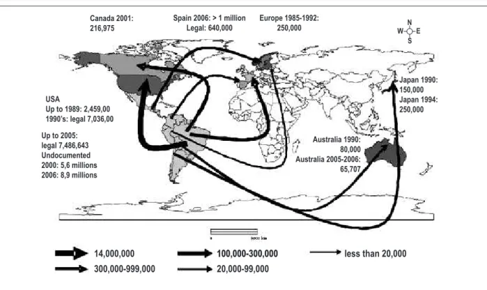
Thus, new epidemiological, socioeconomic and political problems have been recently arisen created, resulting from CD globalization, due to legal and illegal migration from endemic to non-endemic countries, mainly the United States of America, Canada, Spain, France, Switzerland, Italy, Japan, Asian emerging countries and Australia (Figure 2)20. Compounding those problems, the following issues are worth noting: the risk for transfusional and congenital transmission; the need for medical care; difficulties in CD diagnosis due to the lack of experience of physicians in recognizing the disease pathology; and need for additional control at blood banks in countries with little experience in the topic. Such epidemiological aspects differ from those in endemic countries. In the Americas, the CD epidemiological characteristics can be distributed in the following groups of countries according to the transmission cycle and the transfusion and vector control programs (Figure 3)21:
Group I - Argentina, Bolivia, Brazil, Chile, Ecuador, Honduras, Paraguay, Peru, Uruguay and Venezuela have domestic, peridomiciliary and wild transmission cycles, with a high prevalence of human infection and predominance of chronic Chagas cardiopathy (CCC).
Group II - Colombia, Costa Rica and Mexico have domestic and peridomiciliary transmission cycles, and CCC.
Group III - El Salvador, Guatemala, Nicaragua and Panama have domestic, peridomiciliary and wild transmission cycles, with deficient clinical information.
Canada 2001: 216,975
Australia 1990: 80,000 Australia 2005-2006: 65,707
Japan 1990: 150,000 Japan 1994: 250,000
N
S W E Europe 1985-1992:
250,000 Spain 2006: > 1 million
Legal: 640,000
USA
Up to 1989: 2,459,00 1990’s: legal 7,036,00
Up to 2005: legal 7,486,643 Undocumented 2000: 5,6 millions 2006: 8,9 millions
14,000,000 300,000-999,000
100.000-300.000
100,000-300,000 less than 20,000 20,000-99,000
Figure 2 - Globalization of Chagas disease. Potential number of emigrants from countries with T. cruzi infection (*). (*) Adapted from Schmunis G. The globalization of Chagas disease. ISBT Science Series. 2007;2(1):6-11.
USA
Mexico
Guatemala El Salvador
Nicaragua Costa Rica
Panama Colombia Ecuador
Peru
Bolivia Endemic areas
Enzootic areas
Argentina Uruguay
Paraguay Brazil
French Guiana Suriname
Guyana Venezuela Honduras
Belize
Chile
1.3. Control measures for disease transmission
An overview of CD allowing the definition of comprehensive preventive strategies has been provided by a cross-sectional epidemiological survey individually conducted in 15 Latin American countries since the 1980s. The results have shown that the original endemic areas with domiciliary vectorial transmission to humans comprised 18 countries with the highest T. cruzi-infection rates, infested with T. infestans (Southern Cone countries) and Rhodnius prolixus (Andean and Central American countries), which are the best adapted triatomine species to the human household22.
The most effective means to interrupt the transmission of T. cruzi infection have also been indicated: 1) implementation of vector control activities in the household aimed first at reducing, and, then, at eliminating the T. cruzi vectorial transmission; and 2) development and implementation of policy for blood screening for human use to prevent CD transfusional transmission.
In 1975, the program to control CD vectorial transmission was initiated in Brazil. This consisted of insecticide spraying in the houses and around them, aiming at interrupting the domestic and peridomiciliary transmission cycles involving vectors, and animal and human reservoirs. In addition, sanitary educational measures have been adopted and a monitoring system involving the local community members has been established. In Brazil, those programs proved effective to eliminate the T. infestans domiciliary vector, the most important vector from the epidemiological viewpoint23. At the beginning of the program, 711 municipalities had T. infestans‑infested houses. A significant reduction in the number of T. infestans‑infested houses was observed: from 166,000 insects collected in the control program in 1975 to only 6,111 insects collected in 1999. The mean infestation rate dropped to 1 insect to every 10,000 houses investigated, a figure significantly lower than the minimum required for disease transmission21,22. The prevalence of human T. cruzi infection at young age groups can also be considered a control index of disease transmission. As previously reported, for in the age group of 7-14 years, in 1999, a 0.04% positivity rate was observed, representing a 99.8% reduction as compared with 18.5% observed in 198014. In 2007, the results of 94,000 serological tests in samples of 0 to 5 years indicated 0% positivity rate.
2. Pathogenesis and pathophysiology of
chronic Chagas cardiopathy
Chronic Chagas cardiopathy is essentially a dilated cardiomyopathy, in which chronic inflammation, usually mild and continuous, causes progressive tissue destruction and extensive heart fibrosis. Several mechanisms contribute to the pathogenesis of the cardiac lesions and consequent installation of the pathophysiological disorders, according to recent reviews24-28.
2.1. Cardiac dysautonomia
Several independent postmortem studies with chagasic patients and experimental models of T. cruzi infection have
evidenced neuronal depopulation, predominantly of the cardiac parasympathetic system29. Such pathological changes are accompanied by cardiac dysautonomia, confirmed by several researchers with varied methods of functional assessment. They precede the appearance of ventricular dysfunction, being found even in the indeterminate and digestive forms of the disease24,30,31. As a consequence of those functional disorders of the cardiac autonomic regulation, chronic chagasic patients can be deprived of the vagal inhibitory control normally exerted on the sinus node and other cardiac structures. In addition, they can become incapable of rapid chronotropic adjustments in response to physiological stimuli, such as posture changes and physical exercise, mediated by the vagal system24,30. Furthermore, the implication of loss of cardiac parasympathetic control in mechanisms of SD in CCC would be a plausible pathophysiological hypothesis. Although to a lesser extent, structural and functional changes of the cardiac sympathetic system, such as at the ventricular level, also occur in association with myocardial contractility and perfusion disorders32. Circulating antibodies, capable of interfering with receptors of both sympathetic and vagal systems, are likely to affect pathophysiologically the cardiac autonomic behavior and to modulate electrophysiological properties involved in the mechanisms of malignant arrhythmias33. However, the role played by those antibodies in the genesis of myocardial changes is still unclear, and not correlated with ventricular contractility dysfunction34.
Briefly, although morphological and functional changes of the cardiac autonomic system are detected in some chronic chagasic patients, they vary in intensity and do not correlate directly with the degree of ventricular depression. Thus, the “neurogenic theory”, as understood on pioneer studies29, does not convince to explain myocardial destruction in CCC24.
2.2. Microcirculatory disorders
Experimental models of T. cruzi infection have evidenced microvascular changes, such as the formation of microthrombi associated with microcirculatory spasm, endothelial dysfunction and increased platelet activity35. Such microcirculatory disorders might result from the inflammation directly related to T. cruzi or mediated by immune injury36. Those microcirculatory changes are considered to amplify the inflammatory effects and to cause myocardial ischemia24. Several patients with CCC manifest angina-like symptoms, have electrocardiographic changes suggestive of ischemia and various myocardial perfusion defects32,37. Their epicardial coronary arteries are usually angiographically normal, but can react abnormally to vasodilating or vasoconstricting stimuli37,38. It has been postulated that such microcirculatory changes cause hypoperfusion in myocardial areas relatively devoid of coronary branches (zones of marginal perfusion or “watersheds”), being associated with the formation of aneurysms in the apical and posterior basal left ventricular (LV) walls39.
2.3. Immunopathological mechanisms
There is experimental evidence that, after the intense myocarditis of the acute phase of CD, when parasitemia and tissue parasitosis are controlled by immune mechanisms, inflammation subsides and persists focally at low intensity during the indeterminate form of disease40. It has been postulated that the balance and relative pathological stability of that the indeterminate form, in which the immune mechanism should be essentially modulated in the protective direction, are disrupted by still obscure factors, when inflammation, necrosis and fibrosis become more intense, diffuse and progressive24-27,39,41. Several factors can determine the stability or instability of the process: parasite load, parasite strain or tissue tropism, duration of infection, and genetic components of the host. The existence of proper immunoregulatory mechanisms are believed to be crucial in differentiating individuals who control their infection without developing important tissue damage (through limited inflammatory response) from those progressing to severe disease, with intense inflammation, necrosis and reactive fibrosis.
There is unequivocal evidence that autoimmune pathogenic reactions occur in CCC, through molecular mimicry, polyclonal activation and other mechanisms24-27,42.. It is not clear whether the autoimmunity-dependent injury to cardiac structures is decisive for the installation of the characteristic lesions of CCC. Despite the current knowledge limitation, the theory that the immune system reaction to T. cruzi infection is actually a “two-edged sword” and plays as fundamental role in the chronic phase of chagasic myocarditis is supported by an extensive body of experimental and clinical evidence24-27,39-41.
2.4. Inlammation and tissue injury dependent on the parasite presence
In the chronic phase of CD, the classical histologic methods used to emphasize the absence or paucity of parasites in the hearts of T. cruzi‑infected experimental animals or humans29. With more sensitive techniques [immunohistochemistry or polymerase chain reaction (PCR)], the persistence of parasites in myocardial inflammatory foci has been evidenced43,44. In addition, a reduction in the parasite load due to trypanocidal treatment in experimental animals and humans has been observed to attenuate or halt the progression of chronic myocarditis45,46. In contrast, T. cruzi reinfections or its multiplication during immunodepression exacerbate the inflammatory manifestations and CCC course47,48.
Based on that evidence, there is an emerging consensus that the essence of CCC pathogenesis resides on the inflammation directly dependent on the parasite persistence and consequent adverse immune reaction elicited by it24-28,49. This rescues the notion that, even in its chronic phase, the cardiopathy is essentially an infectious inflammatory process, requiring testing whether in that phase the trypanocidal treatment alters the natural history of CCC.
There is substantial evidence that the cytokines produced by T. cruzi-infected patients and animals can modulate the gene and proteomic expression of myocardial cells and other cardiac tissuecomponents50. Polymorphism of genes related to innate immune response and to cytokine production can influence the course of the pathogenetic process of CCC.
2.5. Pathophysiology of chronic Chagas cardiopathy The cardiac injury in CD results from fundamental changes (inflammation, necrosis and fibrosis) caused directly or indirectly by T. cruzi in the specialized conducting tissue, contractile myocardium and intramural nervous system.
The frequent impairment of the sinus node, atrioventricular node and bundle of His due to inflammatory, degenerative and fibrotic changes can cause sinus node dysfunction and various types of varied atrioventricular and intraventricular blocks. The right bundle branch and the left anterior superior fascicle are more vulnerable and frequently affected because they are more individualized structures. Inflammatory foci and areas of fibrosis in the ventricular myocardium, especially in the posterior-lateral and inferior-basal regions, can produce electrophysiological changes, favoring the appearance of reentry. That is the major electrophysiological mechanism of malignant ventricular tachyarrhythmias, which cause SD even in patients with neither HF nor severe LV dysfunction.
Another consequence of the myocardial lesions is the biventricular dysfunction characteristic of CCC. Initially, there is regional impairment, similar to that which occurs in cardiopathy due to coronary obstruction, but, gradually, generalized dilation and hypokinesia develop, conferring the hemodynamic pattern of dilated cardiomyopathy on CCC.
Since the earliest phases, dyssynergia or ventricular aneurysms predispose to thromboembolic complications. In advanced stages, global dilation, venous stasis and atrial fibrillation are additional factors that propitiate the formation of thrombi and consequent pulmonary and systemic embolism, such as in the central nervous system51. That aspect confers on CCC, in addition to the predominant characteristic of causing malignant arrhythmias and refractory HF, the characteristic of essentially causing embolic complicationssm.
3. Clinical presentation and classification
According to its course, CD should be classified into two phases: acute and chronic. The acute phase can be due to primary infection or reactivation of the chronic phase. The chronic phase can have four clinical forms: indeterminate; cardiac; digestive; and mixed (cardiac and digestive impairment in the same patient). The cardiac form can occur with or without global ventricular dysfunction (usually called arrhythmogenic form). The chronic phase can also be classified into stages of cardiac impairment (A, B, C and D), according to international recommendations adapted to the chagasic etiology52.Stage A comprises patients with the indeterminate form, with neither current nor previous HF symptoms and with normal ECG and chest radiography (XR) findings.
Stage C comprises patients with current or previous HF symptoms and ventricular dysfunction [NYHA functional class (FC) I, II, III and IV].
Stage D comprises patients with HF symptoms at rest, refractory to maximized clinical treatment (NYHA FC IV), requiring specialized and intensive interventions.
That classification is simple, operational and consistent, allowing international understanding and comparison with other etiologies (Chart 1).
3.1. Acute phase
After the initial infection, the acute phase of CD lasts 6-8 weeks. The clinical findings are similar to those of other types of myocarditis, with systemic manifestations of fever, disproportional tachycardia, splenomegaly and edema. Inflammation can be observed at the site the parasites penetrate the skin. If the entrance site is the ocular region, conjunctivitis can occur accompanied by unilateral eyelid swelling and preauricular satellite adenopathy (Romaña’s sign). The ECG can reveal sinus tachycardia, low-voltage QRS complexes, prolonged PR and/or QT interval, and alteration of ventricular repolarization. Ventricular arrhythmias, atrial fibrillation and right bundle-branch block can be observed, indicating worse prognosis53. When the disease transmission is congenital, hepatosplenomegaly, jaundice, cutaneous hemorrhage and neurological signs can supervene associate, especially in premature neonates. Other rarer conditions, such as oral contamination and laboratory accident, can lead to the acute form of disease54,55.
The acute phase is most frequently detected in children. If untreated, 5% to 10% of symptomatic patients die during that phase, due to acute HF or meningitis, SD being rare.
The acute phase can also result from the reactivation of a previously established infection in its chronic phase. Immunosuppressive conditions can cause parasite proliferation, necrotic or tumoral lesions in the brain and esophagus, and myocarditis aggravation56. This has been frequently observed in HIV coinfection, particularly with CD4 counts lower than 200/mL, and in organ transplantation57. Acute myocarditis and/or esophagitis have been observed with exacerbation of previous cardiopathy and congestive
HF (CHF). After cardiac transplantation and in the presence of fever, myocarditis and cutaneous lesions, differentiating reactivation from rejection can be difficult58.
3.2. Chronic phase
After the initial acute phase, three clinical conditions can occur: indeterminate form; cardiac form without ventricular dysfunction; and cardiac form with ventricular dysfunction. This new classification should be preferred rather than the one previously used, which defined independent congestive and arrhythmic forms. Ventricular dysfunction, the major prognostic marker, is worth noting. Those clinical forms usually occur after the latency period of several decades that characterizes the indeterminate form59.
Given the current wide availability of echocardiography (ECHO), its performance should be considered as part of the initial assessment of patients with positive serology and whenever there are changes in the clinical or electrocardiographic findings. For patients with electrocardiographic changes, ambulatory ECG (Holter monitoring) should be considered at the initial assessment and later whenever warranted by the symptoms require (Table 1).
3.2.1. Indeterminate form
By definition, patients with the indeterminate form of CD have positive T. cruzi serology and/or parasitological tests, but neither symptoms, nor physical signs; in addition, they have no evidence of organic lesion (cardiac and extracardiac) on ECG and chest XR, and on any other radiological imaging study (esophageal and colon)60-62. However, more strict and sophisticated tests (ECHO, autonomic assessment, exercise testing, Holter monitoring, myocardial scintigraphy, magnetic resonance imaging, cardiac catheterization, and endomyocardial biopsy) can evidence some changes, usually mild and with no prognostic value established in any study61,63-66. Because such patients have chagasic infection, they are believed to be at an increased risk for developing HF and regional contractility abnormalities, and, thus, for progressing to the cardiac form of the disease.
The indeterminate form can last 30-40 years, and 30%-40% of the patients will eventually develop the cardiac form of CD62,67.
Chart 1 – Clinical classiication of left ventricular dysfunction in chagasic cardiopathy
Acute phase
Chronic phase
Indeterminate form Cardiac form with no
ventricular dysfunction Cardiac form with ventricular dysfunction
A B1 B2 C D
Patients with indings
compatible with acute Chagas disease
Patients at risk for developing CHF. They have
positive serology, neither
structural cardiopathy
nor CHF symptoms. No
digestive changes
Patients with structural
cardiopathy, evidenced
by electrocardiographic or echocardiographic
changes, but with normal
global ventricular function and neither current nor previous signs and
symptoms of CHF
Patients with structural cardiopathy characterized
by global ventricular
dysfunction, and neither
current nor previous signs and symptoms of CHF
Patients with ventricular dysfunction and current or previous symptoms of CHF
(NYHA FC I, II, III or IV)
Patients with refractory symptoms of CHF at
rest, despite optimized clinical treatment, requiring
Table 1 – Recommendations and levels of evidence for performing cardiological tests in the initial assessment of chronic chagasic cardiopathy
Recommendation
class Indications Level of evidence
I 12-lead ECG C
Posteroanterior chest XR C
IIa Doppler echocardiography C
Holter monitoring in patients with altered resting ECG C
The other patients will remain asymptomatic during their entire life, maintaining the immune balance between the parasite and the host68. The gradual appearance of electrocardiographic or echocardiographic changes marks the beginning of the chronic cardiac form69.
3.2.2. Cardiac form without ventricular dysfunction
Most commonly, arrhythmic manifestations coexist with congestive findings. Some patients, however, can have a form of chagasic cardiopathy characterized only by arrhythmias and intraventricular and atrioventricular conduction disorders, with normal ventricular function. Although malignant ventricular arrhythmia is more common among patients with concomitant ventricular dysfunction, it can also occur among those with preserved ventricular function, being an important prognostic marker70.
Arrhythmia-related symptoms include palpitations, dizziness, lipothymia and syncope. Syncope in chagasic cardiopathy can be due to both episodes of ventricular tachyarrhythmias and to sinus node dysfunction and atrioventricular blocks (AVB) with asystole71. Sudden death is the major cause of death, has a proteiform multiple mechanism (ventricular tachycardia or fibrillation and asystole) and is associated with multiple scar areas in the myocardium59,70.
Chronotropic incompetence can result from degeneration of the conduction system and autonomic dysfunction, causing symptoms related to intolerance to physical exercise, even in the presence of normal ventricular function30,72.
3.2.3. Cardiac form with ventricular dysfunction
Chronic HF usually installs at least 20 years after the original infection. At that stage, the clinical findings depend on the expression of three frequently coexisting disorders: HF, arrhythmias and thromboembolism. The most frequent clinical presentation is biventricular HF, with predominance of symptoms related to serious higher impairment of the right ventricle function (jugular venous stasis, hepatomegaly, ascitis and lower limb edema), associated with ventricular and atrial arrhythmias and atrioventricular and intraventricular conduction disorders73,74.
Patients usually complain of weakness rather than of dyspnea, which can be partially explained by their lower blood pressure levels as compared with those of other HF etiologies and by the concomitance or preponderance, in some cases,
of right ventricular (RV) dysfunction. Several patients complain of chest pain, usually as like atypical angina, possibly due to microcirculation abnormalities caused by the inflammatory process75. The clinical exam reveals significant cardiomegaly with impulsive and diffuse ictus cordis, murmurs of mitral and tricuspid regurgitation and split of the second cardiac sound.
Dilated ventricles with apical aneurysms, in addition to the high prevalence of atrial fibrillation at more advanced stages, are important sources of mural thrombi, causing systemic and pulmonary thromboembolic phenomena76. Cerebral vascular accidents (CVA) are more common in chagasic HF than in HF of other etiologies, being CD a risk factor for CVA77. The prognosis worsens as congestion progresses and arrhythmias become more difficult to control78-80.
4. Clinical diagnosis, differential diagnosis
and prognosis of chronic chagasic
cardiopathy
4.1. Clinical diagnosis of CCC
4.1.1. Serological tests (Table 2)
Because of the low parasitemia in the chronic phase of CD, parasitological tests are not used, and the serological tests based on the detection of antibodies against T. cruzi should be routinely used to establish the etiology of the cardiopathy81.
The serological diagnosis of T. cruzi infection is confirmed, or excluded, by using at least two serological tests of different principles, which should confirm the existence of anti-T. cruzi antibodies81. Quantifying the concentration of antibodies is desirable. The most commonly used and useful serological tests are the conventional ones: enzyme-linked immunosorbent assay (ELISA), indirect immunofluorescence (IIF) and indirect hemagglutination (IHA). When those three tests are performed, concordance between them is obtained in more than 98% of the sera88,89. Each test has different characteristics regarding sensitivity and specificity: ELISA and IIF have sensitivity greater than 99.5%, but lower specificity (97%-98%), while the IHA tests have lower sensitivity (97%-98%) and higher specificity (99%)90-93. The use of two serological tests prevent false-positive o r f a l s e - n e g a t i v e r e s u l t s , w i t h e t h i c a l a n d l e g a l connotations81,89-94.
4.1.2. Complementary tests (Table 3)
4.1.2.1. Electrocardiography
Electrocardiographic changes are frequently the first indicators of CCC. Initially, the changes are characterized by transient or fixed atrioventricular conduction delays, right bundle-branch conduction delays, ventricular repolarization changes and ventricular ectopies95,96. With disease progression, mainly when global or regional contractility disorders appear, the changes on ECG become outstanding, with relevant prognosticimplications69,97.
In CCC, complete right bundle-branch block associated with left anterior hemiblock is the most frequently found abnormality change (> 50% of the patients)69,95,96. Impairment of the left bundle branch or of the left posterior fascicle is rare. Variable grades of AVB are commonly described in several studies69,95-98. More advanced AVBs result from extensive lesions of the atrioventricular node and bundle of His, and can be the first manifestation of the disease. Dysfunction of the sinus node can cause episodes of sinoatrial block, with bradycardia or ectopic atrial tachycardia. Atrial flutter and fibrillation occur late and usually after the installation of severe ventricular dysfunction, as in other cardiopathies99,100.
Table 2 – Recommendations and levels of evidence for performing serological and parasitological tests in the etiological assessment of patients suspected of having T. cruzi infection
Recommendation
class Indications Level of evidence
I Use of two serological tests of different principles to conirm the etiological diagnosis in the chronic phase of disease (IIF, IHA and ELISA) C
III Use of the Machado Guerreiro reaction C
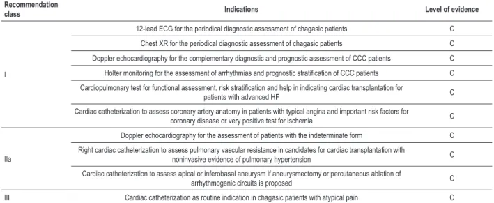
Table 3 – Recommendations and levels of evidence for performing complementary tests for the diagnosis and prognosis of patients with chronic chagasic cardiopathy
Recommendation
class Indications Level of evidence
I
12-lead ECG for the periodical diagnostic assessment of chagasic patients C Chest XR for the periodical diagnostic assessment of chagasic patients C Doppler echocardiography for the complementary diagnostic and prognostic assessment of CCC patients C
Holter monitoring for the assessment of arrhythmias and prognostic stratiication of CCC patients C
Cardiopulmonary test for functional assessment, risk stratiication and help in indicating cardiac transplantation for
patients with advanced HF C
Cardiac catheterization to assess coronary artery anatomy in patients with typical angina and important risk factors for
coronary disease or very positive test for ischemia C
IIa
Doppler echocardiography for the assessment of patients with the indeterminate form C Right cardiac catheterization to assess pulmonary vascular resistance in candidates for cardiac transplantation with
noninvasive evidence of pulmonary hypertension C Cardiac catheterization to assess apical or inferobasal aneurysm if aneurysmectomy or percutaneous ablation of
Polymorphic ventricular extrasystoles are common in the presence of ventricular dysfunction. Complex ventricular arrhythmias, such as non-sustained or sustained ventricular tachycardia (NSVT and SVT, respectively), can even occur in patients with no HF; however, they usually occur in more advanced cases, and their coexistence indicates worse prognosis69,79,85,97.
4.1.2.2. Chest radiography
In advanced phases, severe global cardiomegaly usually contrasts with mild or absent pulmonary congestion. It is worth noting that, due to the frequent and intense RV impairment and tricuspid regurgitation, the enlarged right cavities can stand out on chest XR. Systemic venous congestion, and pleural and pericardial effusion frequently accompany the signs of cardiomegaly.
4.1.2.3. Echocardiography
Echocardiography allows assessing regional and global LV contractility, RV impairment, presence of apical or submitral aneurysms, intracavitary thrombi and diastolic functionchanges74,101. In large case series, even in the indeterminate phase of the disease, ECHO can show segmentary contractility changes in the inferior or apical LV wall in 10%-15% of the patients, and apical aneurysm can be detected in 40%-60% of the patients with CCC74,101,102. Systolic dysfunction has also been detected by using tissue Doppler imaging. The systolic shortening and diastolic stretching velocities, even in patients with normal ECG, can show prolonged isovolumetric contraction times in both ventricles103-106. Thus, a normal ECG reading does not exclude the presence of functional damage to the myocardium103,105-107. Studies with pharmacological stress have reported a reduction in the inotropic and chronotropic response to dobutamine infusion, including biphasic contractile response72. Finally, stress ECHO can induce complex ventricular arrhythmias even in patients at the early phases of cardiopathy108.
It is worth noting that, although some patients with the indeterminate form can have diastolic or systolic function changes, usually mild, on ECHO, several case series have documented the usually excellent prognosis of patients with that form of CD14,62,68,82,109.
The classical echocardiographic aspect of advanced CCC is that of large dilation of the atrial and ventricular cavities, with diffuse biventricular hypokinesia, which is more not so marked than in ischemic cardiomyopathy or in those of other etiologies63,74.In addition, atrioventricular valve regurgitation secondary to the dilation of valvar rings is observed. Despite the predominance of diffuse contractile deficit, ventricular aneurysms detected on ECHO in 47%-67% of the patients are characteristic of CCC, being associated with a higher thromboembolic risk (apical position) and with malignant ventricular arrhythmias (basal inferior or posterior lateral wall)102. Intramural thrombi can be visualized on ECHO also in atria, especially in the presence of atrial fibrillation. All those echocardiographic aspects are relevant to the prognosis of patients with CCC74,101,109.
4.1.2.4. Cardiac magnetic resonance
In CCC, myocardial fibrosis is a constant and intense substrate associated with disease progression and poor prognosis due to the high risk of SD and ventricular arrhythmias39,70. Cardiac magnetic resonance imaging can identify early cardiac involvement, by detecting delayed enhancement areas that indicate fibrosis, thus allowing a more accurate stratification of the stages of disease severity110. The extent of fibrosis correlates directly with the stage of disease and functional class FC, and inversely with the LVEF, contributing to the prognostic stratification of CCC110.
4.1.2.5. Nuclear medicine
Assessing the biventricular function on nuclear angiocardiography with 99mTc is an alternative to ECHO, mainly to assess RV ejection fraction and the conditions of contractile synchronization of both ventricles, which have prognostic value63,86. Myocardial perfusion scintigraphy shows segmental perfusion deficits in as much as 30% of patients with CCC and angina, but with normal coronary angiography, indicating coronary microcirculation changes, and myocardial fibrosis, disorders that correlate with the progressive deterioration of ventricular function75,111.
4.1.2.6. Dynamic electrocardiography (Holter)
Dynamic electrocardiography is indicated to assess the chagasic patient with syncope, which can be due to ventricular bradyarrhythmia or tachyarrhythmia112,113. Both can coexist in the same patient, the most severe being SVT and advanced AVB. In retrospective and prospective series, as well as in a systematic review, NSVT has also been proved to predict worse prognosis79,85,112,113.
4.1.2.7. Exercise test and cardiopulmonary test
Exercise and cardiopulmonary tests, although of limited usefulness to clarify chest pain in chagasic patients, are useful to detect exertion-induced arrhythmias in CCC and to establish their prognosis108,112.The cardiopulmonary test with direct measurement of oxygen consumption (VO2) shows high mortality in one year for patients with VO2 lower than 12 mL/kg/min, being also used as an auxiliary method to indicate cardiac transplantation (CT).
4.1.2.8. Electrophysiological study
The electrophysiological study (EPS) allows the investigation of sinus function and AV conduction, being also indicated to clarify syncope of undetermined origin after noninvasive assessment, in patients with resuscitated SD112,113. It is not indicated for risk assessment in patients with preserved systolic function or with NSVT. It is also indicated to map refractory ventricular tachycardia for possible ablation of arrhythmogenic foci113.
4.1.2.9. Cardiac catheterization