Clinicoradiological Session
Case 6/2007 – A Ten-Year-Old Child with Mild Pulmonary Valve
Stenosis
Edmar Atik
Instituto do Coração do Hospital das Clínicas - FMUSP - São Paulo, SP - Brazil
Mailing address: Edmar Atik •
InCor - Av. Dr. Enéas de Carvalho Aguiar, 44 - 05403-000 - São Paulo, SP - Brazil E-mail: [email protected]
Clinical data
A ten-year-old white male child who had been diagnosed with a heart murmur at six months of age in a routine examination and remained asymptomatic, active and with no physical limitations. On physical examination he was eupneic and with normal pulses. He weighed 47 kg and was 147 cm tall. His blood pressure was 110/70 mm Hg and heart rate was 76 bpm. No cyanosis was observed. The aorta was not palpable in the suprasternal notch. Chest examination showed neither deformities nor cardiac impulses, and a valve and muscular apical impulse + was located at the 4th left intercostal space limited by the breadth of one finger. Cardiac auscultation revealed regular rate and rhythm, and a split second sound with both components of equal intensity. A +/++ systolic ejection murmur was heard in the 2nd, 1st, and 3rd left intercostal spaces, accompanied by a soft systolic thrill. The liver was not palpable.
The electrocardiogram showed sinus rhythm with mild right ventricular overload, R-wave amplitude of 8 mm in V1 and mild ventricular conduction delay. P-axis: +45°, QRS-axis:+100°, T-axis:+40°.
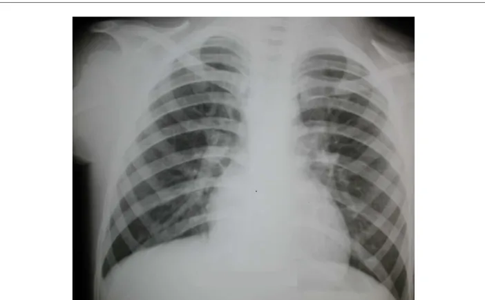
Radiographic examination
Radiographic images showing a straight mid-arch and normal pulmonary vasculature (Figure 1).
Diagnostic impression
This image may correspond to that of a normal heart or even an acyanotic congenital heart disease with little repercussion.
Differential diagnosis
Both the obstructive heart diseases and those with left-to-right shunting , with little repercussion, share similar features. It is difficult to differentiate between pulmonary valve stenosis and atrial septal defect when both conditions are mild. Therefore, other clinical data should be considered.
Diagnostic confirmation
The systolic murmur in the pulmonary area accompanied by systolic thrill suggests the diagnosis of pulmonary valve stenosis. The normal splitting of the second sound demonstrates the little repercussion of the defect, as well as the electrocardiographic (Figure 2) and radiographic findings. The echocardiogram corroborated this hypothesis, with pressure gradient of 34 mm Hg between the right ventricle and pulmonary trunk. The pulmonary annulus was18 mm in diameter, the pulmonary valve was thick and trileaflet, and the pulmonary trunk was slightly dilated. Other measurements were as follows: left ventricular diastolic diameter, 40 mm; aorta, 25 mm; left atrium, 26 mm; right ventricle, 19 mm; septum, 7 mm; and myocardial fiber shortening, 30%. The interatrial septum was intact.
Management
The boy should be treated expectantly, given the mild degree of the defect, with pressure gradient less than 40 mm Hg, since he was six months old.
Key words
Infant; heart defects, congenital; pulmonary valve stenosis; heart murmurs.
Clinico Radiological Session
Edmar Atik
Arq Bras Cardiol 2007; 89(5) : 309-310
Fig. 1 - Chest radiography showing cardiac silhouette and pulmonary vascularity close to normal, which may be consistent with any acyanotic heart disease with little repercussion.
Fig. 2 - Electrocardiogram showing evidence of mild right ventricular overload.