2 7 6
Dias et al
Aneurysmal dilation of the reimplant segment of the visceral vessels
Arq Bras Cardiol 2003; 81: 276-8.
Instituto do Coração do Hospital das Clínicas da FMUSP, São Paulo (Brazil) and Methodist Hospital, Houston, TX (USA)
Mailing address: Ricardo Ribeiro Dias – Av. Cotovia, 80/141 - 04517-000 – São Paulo, SP – Brazil - E-mail: [email protected]
Received - 12/2/02 Accepted - 1/14/03
Arq Bras Cardiol, volume 81 (nº 3), 276-8, 2003
Ricardo R. Dias, Joseph S. Coselli, N oedir A. G. Stolf, Altamiro R. Dias, Charles Mady, Sérgio A. O liveira
São Paulo, SP - Houston, TX - Brazil
Aneurysmal Dilation of the Reimplant Segment of the Visceral
Vessels After Thoracoabdominal Aneurysm Correction
Case Report
We present a case of aneurysmal dilation of the aortic residual segment, involving abdominal vessels in correc-tive surgeries for thoracoabdominal aortic aneurysm, through the identification of risk groups for recurrent dilation, aiming at using a specific operative technique with a branched graft, to prevent aneurysm relapse.
Advances in surgery for thoracoabdominal aneurysm correction occurred after the systematization proposed by Crawford et al in 1978 1. Once diagnosed, the patient must be
carefully and invasively investigated to determine an objective approach, because the natural history of thoraco-abdominal aortic aneurysm, without surgical intervention, is well known and causes increased morbidity and mortality, especially due to the comorbidities associated with the spread of vascular disease.
Griepp et al 2 presented a follow-up of 156 patients with
descending aortic aneurysms and thoracoabdominal abdo-minal aneurysm without surgery, and they pointed out that 22.4% evolved to rupture in 1 year. Of the 44 deaths in this cohort, 79.5% were due to aortic rupture, 9 in 10 dissections, and 26 in 34 degenerative aneurysm (P=0.004). Mean dia-meter, when rupture occurred also was significantly diffe-rent between the groups; in dissections it was 5.4 cm, and in degenerative aneurysms it was 5.8 cm (P=0.05).
Pressler and McNamara3 described 72% mortality in a
mean 3-year follow-up in asymptomatic patients with thoracic aortic aneurysm under clinical treatment. Forty per cent of the mortality was secondary to aortic rupture, and 32% was secondary to cardiovascular disease.
The severity and the incidence of thoracoabdominal aortic aneurysm are increasing, especially because of the ageing of the population, surpassing 5 cases per 100,000
people/year 4. However, current surgical results are
encou-raging, providing a great therapeutic possibility to this group of patients. Coselli et al 5 obtained a hospital mortality
rate of 7.1%, and paraplegia of 4.6% in 1220 patients under-going surgery.
According to the surgical technique used to correct thoracoabdominal aortic aneurysm, in case of a remaining diseased aortic segment, this region may dilate again and have complications, especially in young patients with con-nective tissue disease (Marfan’s syndrome and Ehlers-Danlos syndrome).
Case Report
A 77-year-old-male patient underwent surgery to correct thoracoabdominal aortic aneurysm (Crawford extent III 6) in 1991, and myocardial revascularization in 1996. During
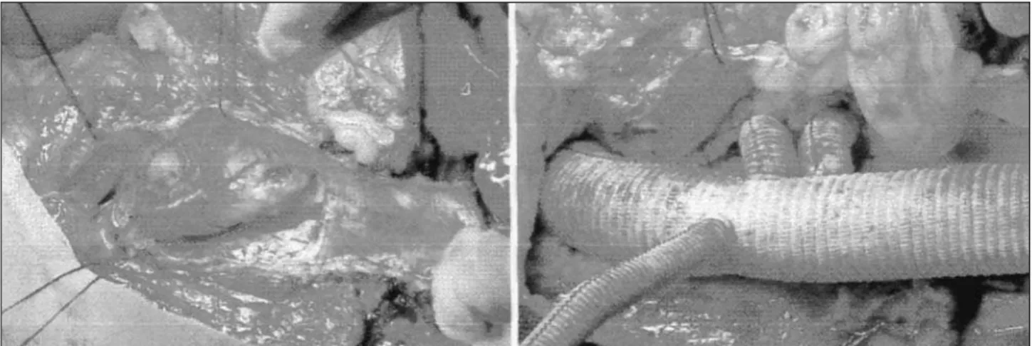
follow-up, we observed abdominal aortic dilation that involved the celiac trunk, the superior mesenteric artery, right and left renal arteries, anastomosed in Dacron graft 11 years ago to correct thoracoabdominal aortic aneurysm (fig. 1). This aortic segment had grown 0.4 cm/year in the previous 2 years, and at the time of surgical indication, it was 5.9 cm in the major transversal diameter. The patient reported scattered episodes of lumbar pain not related to effort. He also had mild left ventricular dysfunction with an ejection fraction of 50%, systemic blood hypertension, chronic obstructive pulmona-ry disease, hypercholesterolemia, peripheral vasculopathy, obstructive carotid disease (left carotid with 50% obstruction in the carotic bulb), atrophic left kidney, and borderline renal function (serum creatinine 1.6 mg/dL).
Arq Bras Cardiol 2003; 81: 276-8.
Dias et al Aneurysmal dilation of the reimplant segment of the visceral vessels
2 7 7
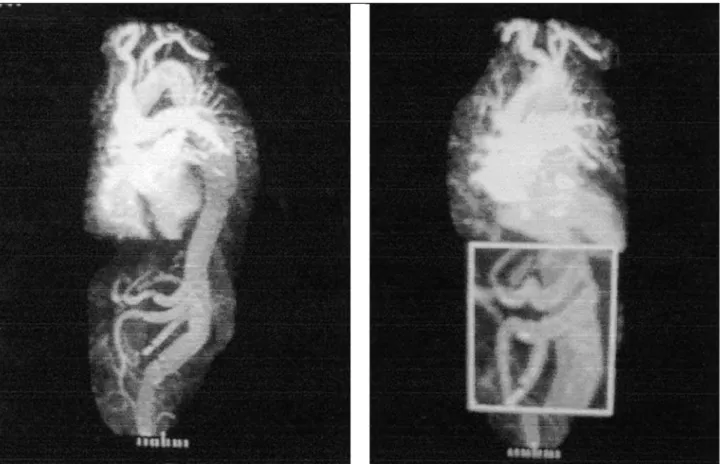
celiac trunk were 35 minutes, 41 minutes, and 59 minutes, respectively. Cerebrospinal fluid drainage and renal perfu-sion were performed with intermittent cold crystalloid solu-tion, for spinal cord and renal protecsolu-tion, during the period of ischemia. The patient evolved with worsening of renal function (creatinine peak of 2.6 mg/dL on the 8th postope-rative day) without the need for dialysis. He also had pul-monary hypersecretion requiring orotracheal reintubation during the 4th postoperative day (extubated again in the 8th postoperative day). Amylase and hepatic enzyme did not alter. The patient was discharged in the 26th postopera-tive day after an adequate control examination (fig. 3).
Discussion
The greater the experience of the surgical staff, the lower the operative risk of thoracoabdominal aortic aneu-rysms, despite being associated with important morbidities to its correction, especially, due to the associated organs (bone marrow, liver, pancreas, kidneys, and other abdominal viscera) by the access and surgical thoracolaparotomy.
Predictor factors of elective operative mortality are the following: advanced age, renal failure, symptomatic aneu-rysms, and thoracoabdominal aneurysms with Crawford ex-tent II 6. Reoperation and chronic dissections do not interfere
with mortality 7.
The incidence of paraplegia has been reduced by maintaining distal aortic perfusion during surgery, cere-brospinal fluid drainage, moderated hypothermia, and reim-plantation of intercostal vessels (T8-L1), especially in the longer aneurysms (Crawford type II) 8,9.
In this case, we used the most frequently applied technique for insertion of abdominal vessels through direct suture of the aortic segment (containing the celiac trunk and the superior mesenteric and renal arteries) in the Dacron graft. This kind of approach simplifies and reduces the pro-cedure’s duration, thereby reducing ischemic time, which is decisive in the evolvement of the patient. However, young patients with connective tissue disease or those whose re-sidual aortic segment is extensive (due to the distance bet-ween vessels), are more prone to have dilation of this region of diseased aorta during their follow-up.
Fig. 1 – Scanned images demonstrating aneurysmal dilation of the residual segment of the aorta used for insertion of the abdominal vessels in Dacron graft. A) transversal plane; B) sagittal plane.
2 7 8
Dias et al
Aneurysmal dilation of the reimplant segment of the visceral vessels
Arq Bras Cardiol 2003; 81: 276-8.
Dardick et al 10 reported that of 107 patients operated on
due to thoracoabdominal aneurysms, 8 patients (7.5%) evolved with aneurysmal expansion of this region. Three of them were women with connective tissue disorders and a mean age of 36 years, and 5 were men with degenerative aneurysm and a mean age of 73 years. Mean follow-up time was 6.5 years. With the purpose of avoiding this complication, we
Fig. 3 – Total aortic magnetic resonance imaging performed on the 18th postoperative day. A) Panoramic view of the cardiovascular system with a view of the graft with its pervious branches. B) Close-up of the anastomoses performed.
suggest that the risk population or those whose vessels are distant use branched vascular prostheses inserted during reoperation, because although making the procedure more complex and increasing ischemia time, they offer a more radi-cal treatment with the possibility of complete resection of the involved aorta, avoiding the relapse of the aneurysm in the residual aorta.
References
1. Crawford ES, Snyder DM, Cho GC, Roehm JO. Progress in treatment of tho-racoabdominal and abdominal aortic aneurysms involving celiac, superior me-senteric, and renal arteries. Ann Surg 1978; 188:404-10.
2. Griepp RB, Ergin A, Galla JD, et al. Natural history of descending thoracic and thoracoabdominal aneurysms. Ann Thorac Surg 1999; 67:1927-30. 3. Pressler V, McNamara JJ. Thoracic aortic aneurysm: natural history and treatment.
J Thorac Cardiovasc Surg 1980; 79: 489-98.
4. Fann JI. Descending thoracic and thoracoabdominal aortic aneurysms. Coron Art Dis 2002; 13: 93-102.
5. Coselli JS, LeMaire SA, Miller CC, et al. Mortality and paraplegia after thoracoabdominal aortic aneurysm repair: a risk factor analysis. Ann Thorac Surg 2000; 69:409-14. 6. Crawford ES, Crawford JL, Safi HJ, et al. Thoracoabdominal aortic aneurysms:
preoperative and intraoperative factors determining immediate and long-term results of operation in 605 patients. J Vasc Surg 1986; 3: 389-404.
7. Coselli JS, Figueiredo LFP, LeMaire SA. Impact of previous thoracic aneurysm repair on thoracoabdominal aortic aneurysm management. Ann Thorac Surg 1997; 64:639-50.
8. Coselli JS, LeMaire SA. Left heart bypass reduces paraplegia rates after thoraco-abdominal aortic aneurysm repair. Ann Thorac Surg 1999; 67:1931-4. 9. Coselli JS, LeMaire SA, Schmittling ZC, Koksoy C. Cerebrospinal fluid drainage
in thoracoabdominal aortic surgery. Semin Vasc Surg 2000; 13: 308-14. 10. Dardik A, Perler BA, Roseborough GS, Williams GM. Aneurysmal expansion of