Rev Bras Cardiol Invasiva. 2013;21(3):299-300
© 2013 Sociedade Brasileira de Hemodinâmica e Cardiologia Intervencionista. Published by Elsevier Editora Ltda. All rights reserved.
Endovascular Correction of Pararenal Abdominal
Aortic Aneurysm using the Chimney Technique
Frederico Augusto de Carvalho Linhares Filho
1, Antonio Massamitsu Kambara
2, Nilo Mitsuru Izukawa
3,
Samuel Martins Moreira
1M
ale patient, 75 years old, with a history of cramp-like, intermittent abdominal pain, was submitted to CT angiography due to evidence of pararenal abdominal aortic aneurysm. As comorbidity, the patient had chronic obstructive pulmonary disease requiring home oxygen therapy; for this reason, the endovascular treatment was chosen.1 Vascular surgeon. Enhancing surgeon at the Center of Endovascular Inter-ventions of Instituto Dante Pazzanese de Cardiologia. São Paulo, SP, Brazil. 2 Interventionist radiologist. Head of the Radiology Section of Instituto Dante Pazzanese de Cardiologia. São Paulo, SP, Brazil.
3 Vascular surgeon. Head of the Vascular Surgery Section of Instituto Dante Pazzanese de Cardiologia. São Paulo, SP, Brazil.
4 Vascular surgeon. Assistant at the Center of Endovascular Interventions of Instituto Dante Pazzanese de Cardiologia. São Paulo, SP, Brazil.
Correspondence to: Frederico Augusto de Carvalho Linhares Filho. Av. Dr. Dante Pazzanese, 500, prédio III, 1o andar, Hemodinâmica − Ibirapuera − São Paulo, SP, Brazil − CEP 04012-180
E-mail: [email protected]
Received on: 06/07/2013 • Accepted on: 08/11/2013
The abdominal aneurysm correction was performed using the chimney technique, which consists of the implantation of a coated stent in parallel to the aortic endoprosthesis and allows for continuous perfusion of the renal arteries. This technique permits the endovas-cular correction of pararenal or juxtarenal abdominal aortic aneurysms.
Figure 1 – Angiography showing coated stent placement and aortic endoprosthesis.
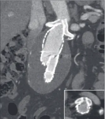
Figure 2 – CT angiography of the abdominal aorta with 3-D volu-metric reconstruction of the treated aortoiliac segment, performed postoperatively (30 days).
Linhares-Filho et al. Chimney Technique
Rev Bras Cardiol Invasiva. 2013;21(3):299-300
300
The procedure was performed through dissection of the bilateral common femoral artery and puncture of the bilateral brachial artery, with the introduction of a long 8F sheath in the abdominal aorta. The renal arteries were catheterized through brachial access and 0.035 extra-stiff guidewires were positioned; then, the 5 × 50 mm Viabahn coated stents (WL Gore & Associates – Flagstaff, USA) were released. The Excluder® aorta endoprosthesis (WL Gore &
As-sociates, Flagstaff, USA) was positioned through the femoral artery above the anatomical origin of the renal arteries and below the proximal portion of the coated stents (Figure 1).
A CT angiography performed in the postoperative period showed correct coated-stent positioning in the renal arteries, with maintenance of renal perfusion and positioning of the proximal portion of the endoprosthesis above the anatomical origin of the renal arteries, with no evidence of leaks. The blood flow was bilaterally directed to the common iliac arteries, with pre-existing occlusion observed in the right internal iliac artery at its origin (Figures 2 and 3).
CONFLICTS OF INTEREST
The authors declare to have no conflicts of interest.