CASE REPORT
Endotension: rupture of abdominal aortic aneurysm
Endotensão: ruptura de aneurisma de aorta abdominal
Alexandre Campos Moraes Amato1, Flávio Amim Abraham2, Henrique Dini Kraide2,
Leandro Teixeira Rocha2, Ricardo Virginio dos Santos1
Introduction
With the development of endovascular surgery, a new treatment option for high-risk patients who require con-ventional aneurysm surgery, but are not suited for standard endovascular procedures, is being considered. he new technique (called ‘chimney’) is the percutaneous translumi-nal placement of a stent through the aortic visceral and re-nal branches1. A prosthesis is placed to exclude the
thoraco-abdominal aneurysm. his method is mainly used when the aneurysm afects the renal and visceral vessels, and open surgery is considered too risky and not feasible.
One of the complications of an endovascular aneurysm repair (EVAR) is the endoleak, which maintains the low within the excluded aneurysm sac and pressurises it. hese leaks are divided into ive kinds, according to their mecha-nisms and locations. he type-V endoleak, also called en-dotension, is deined by the persistent or recurrent pressuri-sation of the aneurysm sac through the parietal thrombi. What diferentiates an endoleak from endotension is that, in the latter, pressure is transmitted without the presence of blood low within the aneurysm2-4.
he prevalence of endoleaks ater an EVAR is high-ly variable. In the literature, it has been reported to be
Abstract
Aortic endovascular exclusion technique called ‘chimney’ consists of placing stents through abdominal aortic visceral branches and a prosthesis that excludes the thoraco-abdominal aneurysm. Stents and an aortic endoprosthesis are placed in the renal arteries. his method is primarily used when open surgery is too risky. he mechanism that provides aneurysm sac increase without the visible presence of endoleaks has not been fully elucidated. he expansion of the aneurysm sac, due to endotension, is diicult to diagnose, even with the use of advanced imaging tests. Its diagnosis is made by exclusion. We present a case of a late complication in a high-risk patient after a ‘chimney’ endovascular procedure. Following the surgery, the patient presented a ruptured aneurysm sac without a visible endoleak. A second intervention was not feasible due to the high risk of occluding all of the branches, and complicated by previous ‘chimney’. Endotension is a possible cause of aneurysm rupture and death.
Keywords: aortic rupture; endoleak; postoperative complications.
Resumo
A técnica de exclusão endovascular conhecida como ‘chaminé’ consiste na colocação de stent em ramos viscerais e de endoprótese excluindo o aneurisma toracoabdominal. São colocados stents revestidos nas artérias renais e uma endoprótese aórtica, que o método utilizado quando a cirurgia aberta tem risco muito alto. O mecanismo que causa a expansão aneurismática sem a presença detectável de vazamento pelos métodos de imagem não está completamente esclarecido. A expansão do saco aneurismático por endotensão é de difícil diagnóstico, mesmo com o uso de técnicas de imagem avançadas, como tomograia computadorizada e eco-Doppler, sendo o diagnóstico por exclusão. Apresenta-se um caso de complicação tardia após o tratamento endovascular pela técnica da ‘chaminé’. Após a cirurgia, o paciente apresentou ruptura sem endoleak visível. Outro procedimento endovascular foi impossibilitado pela técnica da ‘chaminé’, que diiculta novos procedimentos e há alto risco de oclusão dos ramos. Endotensão é causa de ruptura e óbito.
Palavras-chave: ruptura aórtica; endoleak; complicações pós-operatórias.
Study carried out at the Vascular Surgery Discipline at the Santo Amaro University (Unisa) – Santo Amaro (SP), Brazil.
1 Professor of the Vascular Surgery Discipline at Unisa – São Paulo (SP), Brazil. 2 Medical students attending the sixth year at Unisa – São Paulo (SP), Brazil. Financial support: none.
Endotension: rupture of abdominal aortic aneurysm - Amato ACM et al. J Vasc Bras 2012, Vol. 11, Nº 2 163
between 2.4 and 45.5%5. he clinical importance of
en-doleaks is directly related to an increased risk of aneurysm rupture3, although endotension-causing ruptures have
rarely been reported3,6.
Case report
he patient was a 66-year-old male, who was hyper-tensive, obese, with chronic obstructive pulmonary disease and dyslipidaemic. A computed tomography scan was con-ducted in October, 2006, and a type-IV thoraco-abdominal aortic aneurysm was diagnosed. Since then, the aneurysm has showed continuous growth in subsequent bi-annual ex-aminations without visible endoleak.
In July, 2008, a computed tomography angiography (CTA) revealed that the aneurysm dilatation in the distal abdominal aorta was juxtarenal and extended up to the bi-furcation. he centre of the predominating concentric pari-etal thrombus had a maximum proximal diameter of 5.2 x 4.1 cm. he CTA presented a lower calibre in the let renal artery, with an ipsilateral renal reduction (Figure 1).
he patient underwent an endovascular aneurysm ex-clusion (Powerlink bifurcated stent, Endologix, Inc., Irvine, CA, the USA) with an endovascular revascularization of the renal arteries, celiac trunk, and superior mesenteric artery by the chimney technique (Figure 2).
Two years later, the patient presented with increasing lower abdominal pain, which radiated to the lumbar region and had evolved over three weeks. he patient was conscious and eupneic and showed no signs of shock. he abdomen was slightly distended, produced bowel sounds, and was some-what painful to palpation. he posterior tibial pulse was bi-laterally present, and the let pedal pulse was absent.
he patient was admitted into the Intensive Care Unit (ICU), where he was held under tight blood-pressure con-trol, and a CTA was performed, as an endoleak was sus-pected from previous procedure. Aneurysm growth was observed (Figure 3).
he aneurysm had a maximum diameter of approxi-mately 13x12 cm. he dilation and tortuosity of the entire abdominal aorta relected the largest diameter in its infra-renal portion. In the distal region on the same side and near the bifurcation of the iliac artery, irregular areas (suggestive of ulcers) were observed. A ruptured aneurysm wall without a contrast leak was observed on the let anterior side.
Conservative medical management and observa-tion were performed, as the patient was deemed to have a high anaesthetic risk given his age and comorbidities. Angiography was requested to conirm the absence of en-doleaks2. herefore, the patient was kept in the ICU with
a normo/hypotensive blood pressure and rigorous clini-cal support. He worsened cliniclini-cally with no signs of initial
Figure 1. Angio-tomography of the aortic aneurysm.
Endotension: rupture of abdominal aortic aneurysm - Amato ACM et al. J Vasc Bras 2012, Vol. 11, Nº 2
164
hypovolemic shock. However, he died three days ater his hospital admission, due to sudden hypovolemic shock. Angiography was not performed for it required moving the patient to another facility2.
Discussion
he chimney technique essentially consists of deploy-ing a covered stent parallel to the main aortic stent-grat. It protrudes similarly to a chimney and preserves the low of the vital side branches, which are covered by the aortic stent grat. he chimney technique enables the use of a stan-dard of-the-shelf endoprosthesis to treat a lesion, with an inadequate ixation zone and it provides an alternative to a fenestrated stent grat in urgent cases and in aneurysms with challenging neck morphologies. his technique also enables the rebuilding of the side branches of the aorta that might have been involuntarily covered during the en-dovascular repair1,7.
Several theories have been proposed in an attempt to elucidate the mechanisms of expansion of the aneurysm sac, without the presence of a detectable endoleak. hese include increased porosity of the prosthesis5,direct
trans-mission of pressure from the stent to the lumen of the an-eurysm sac4,8, and a low low endoleak which is
undetect-able by the imaging methods2,9. Mennander et al. found ive
patients (3.1%) with endotension within 160 patients who underwent endovascular aneurysm repair, and endoleaks were not detected. hree sufered aneurysm sac ruptures during the study period, but none of them presented clini-cal evidence of major bleeding9.
he best explanation for these cases may be the associa-tion of more than one of the aforemenassocia-tioned theories. Of these, the exudate is the most accepted one that may explain the cases of endotension. However, in this case, it cannot be exclusively attributed to the exudate, since there are no studies demonstrating the efectiveness of the chimney seal between the endoprosthesis (Figure 2).
he continuous sweating through the endoprosthe-sis may result in the rupture of the sac without any evi-dent hemodynamic changes. But also, the sac rupture may cause endoprosthesis migration due to sudden morpho-logic changes of the aneurismal sac and lack of satisfac-tory anchoring of the stent-grat. Patients with aneurysm sac enlargements ater EVAR may be diicult to diagnose. herefore, the irst step is to check for the presence of en-doleaks. Multi-slice CT, high-resolution Doppler echocar-diography, and angiography were the most widely used imaging modalities at the beginning of the investigation.
Figure 2. 3D Angio-computed tomography reconstruction and scheme of the chimney procedure, demonstrating the patent and lost branches, and the main endoprosthesis. he axial view demonstrates the chimney seal between grafts.
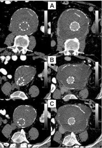
Figure 3. Angio-computed tomography demonstrating the rup-tured wall of the aortic aneurysm without a contrast leak. Left images are noncontrasted from before rupture, and right images contrasted after rupture. A: calcium inside aneurysm sac, not to be mistaken with endoleak; B: aneurism growth after the proce-dure and point of rupture; C: ruptured aneurysm without bleeding or contrast leak.
A
B
Endotension: rupture of abdominal aortic aneurysm - Amato ACM et al. J Vasc Bras 2012, Vol. 11, Nº 2 165
Endotension has also been measured by indirect methods using arterial punctures6,10.
In the present case, the endoprosthesis expanded poly-tetraluoroethylene (ePTFE) grat material had low porosity. We cannot exclude the possibility that the increased pres-sure in the aneurysm sac was developed due to the passage of exudates through the prosthesis material or the sealing diiculty associated with the chimney technique.
Kougias et al. have recently reported a less-invasive technique, which uses stent-based reinforcement with the deployment of new grat components to re-line the en-dograt and to minimize the porosity-related endotension. his technique is applicable to patients with a porous en-doprosthesis6. However, it does not apply to this case
be-cause stent reinforcement would increase the main radial grat strength and possibly occlude the parallel visceral branches.
Conclusions
he development of aneurysm sac expansion due to endotension is diicult to diagnose, even with the use of advanced imaging tests, such as multi-slice CT, Doppler echocardiography, and angiography, as they may not be suiciently sensitive to detect small endoleaks. By deini-tion in endotension, there must be no leaks. his techni-cal limitation of imaging may falsely categorize a patient as having endotension when the actual reason for the expan-sion is a very small, but persistent, endoleak. he chimney technique may be associated with an apparently technically successful procedure because images do not show any en-doleaks between stents. here may be small enen-doleaks that are responsible for future sac expansion and rupture.
Endotension diagnosis is currently made by exclusion and therefore should encourage the development of new techniques, which would allow the detection of small pres-sure changes in the aneurysm sac. Aneurysm sac prespres-sure measured by an implantable remote sensor may help to elu-cidate endotension causes11.
he chimney technique is not a standard procedure and should only be performed in few selected cases, be-cause it can compromise the ability to perform further pro-cedures to treat possible complications. he sealing of the chimney technique is not guaranteed, as our current imag-ing techniques are not suiciently sensitive to detect small endoleaks2. Even without any detectable endoleaks, and the
aneurysm sac may grow due to endotension or undetectable endoleaks, this aneurysm sac may rupture and cause death. In the event of an aneurysm sac rupture, ater a chimney
procedure, no endovascular procedure is safe, and open surgery that was once avoided now has a higher risk and is characterized by even greater technical diiculties3,5,8-10.
References
1. Ohrlander T, Sonesson B, Ivancev K, et al. he chimney graft: A technique for preserving or rescuing aortic branch vessels in stent-graft sealing zones. J Endovasc her. 2008;15:427-32.
2. Chagas Neto FAD, Barreto ARF, Reis HFD, et al. A importância do diagnóstico por imagem na classiicação dos endoleaks como complicação do tratamento endovascular de aneurismas aórticos. Radiol Bras. 2010;43:289-94.
3. Görich J, Rilinger N, Sokiranski R, et al. Leakages after endovascular repair of aortic aneurysms: classiication based on indings at CT, angiography, and radiography. Radiology. 1999;213:767-72.
4. White GH. What are the causes of endotension? J Endovasc her. 2001;8:454-6.
5. Williams GM. he management of massive ultrailtration dis-tending the aneurysm sac after abdominal aortic aneurysm re-pair with a polytetraluoroethylene aortobiiliac graft. J Vasc Surg. 1998;28:551-5.
6. Kougias P, Lin PH, Dardik A, et al. Successful treatment of endoten-sion and aneurysm sac enlargement with endovascular stent graft reinforcement. J Vasc Surg. 2007;46:124-7.
7. Schlösser FJ, Gusberg RJ, Dardik A, et al. Aneurysm rupture after EVAR: can the ultimate failure be predicted? Eur J Vasc Endovasc Surg. 2009;37:15-22.
8. Meier GH, Parker FM, Godziachvili V, et al. Endotension after en-dovascular aneurysm repair: he ancure experience. J Vasc Surg. 2001;34:421-6.
9. Mennander A, Pimenof G, Heikkinen M, et al. Nonoperative ap-proach to endotension. J Vasc Surg. 2005;42:194-9.
10. Risberg B, Delle M, Lönn L, et al. Management of aneurysm sac hygroma. J Endovasc her. 2004;11:191-5.
11. Hoppe H, Segall JA, Liem TK et al. Aortic aneurysm sac pressure measurements after endovascular repair using an implantable remote sensor: Initial experience and short-term follow-up. Eur Radiol. 2008;18:957-65.
Correspondence
Alexandre Campos Moraes Amato Avenida Juriti, 144 CEP 05612-010 – São Paulo (SP), Brazil E-mail: [email protected]
Author’s contributions