Instituto de Cardiologia do Rio Grande do Sul/Fundação Universitária de Cardiologia – Porto Alegre
Mailing address: Jose Luiz da Costa Vieira – Rua Coraci Prates da Veiga, 73 – 94410-290 – Viamão, RS, Brazil
English version by Stela Maris C. Gandour
Objective – To report the effects of 2 regimens of hor-mone replacement therapy during the postmenopausal pe-riod on the profile of the major lipoprotein subfractions (HDL, LDL, and VLDL).
Methods – We carried out a cohort study in 38 post-menopausal patients who were starting their hormone re-placement therapy due to gynecological indications, for a period of 12 weeks. Analysis of lipoprotein subclasses was performed through nuclear magnetic resonance spectros-copy.
Results – Hormone replacement therapy cause an increase in the proportion of larger subfractions of VLDL and HDL (p=0.008 and 0.03, respectively) and in the proportion of larger particles of VLDL due to a 36% increase in the levels of larger particles (p=0.004), concomitantly with a 15% re-duction in the levels of smaller particles (p=0.04). In regard to HDL, the increase occurred only a 17% increase in the levels of larger particles (p=0.002). No significant change occurred in the distribution pattern of LDL subfractions.
Conclusion – The proportion of larger subfractions of VLDL and HDL increases after hormone replacement the-rapy. The increase in the proportion of larger particles of VLDL occurs due to an increase in the levels of the larger subclasses concomitantly with a reduction in the smaller particles. However, an increase in the proportion of larger particles of HDL occurs only due to an increase in the le-vels of the larger subfractions.
Key words: lipoproteins, hormone replacement therapy,
coronary heart disease
Arq Bras Cardiol, volume 76 (nº 3), 183-88, 2001
José Luiz da Costa Vieira, Marcos Emanuel W. Gomes, Áurea Beltrão de Almeida, Emílio H. Moriguchi
Porto Alegre, RS - Brazil
Changes in the Profile of Lipoprotein Subfractions Associated
with Hormone Replacement Therapy
Despite the evidence, at the present time challenged by the results of the first 2 randomized clinical trials of se-condary prevention in ischemic heart disease 1,2, that
estro-gen may protect against coronary heart disease, the me-thod by which this protective effect can be mediated has never been properly explained. Among the proposals, the estrogen impact on the lipid profile has always been high-lighted, with a reduction in LDL-cholesterol and an increase in HDL-cholesterol. However, some studies have shown that only 25% to 50% of risk reduction could be attributed to this change 3-5. In the last decade, addition of progestin
has been routinely recommended in postmenopausal hor-mone replacement to reduce the risk of endometrial carcino-ma induced by estrogen 6. In observational studies 7-9, this
practice seems not to have attenuated the cardioprotective effect of postmenopausal estrogen therapy.
In recent years, has gained relevance that each group of lipoproteins – high density lipoprotein (HDL), low density lipoprotein (LDL), and very low density lipoprotein (VLDL) – is formed by very heterogeneous and different particles in their association with coronary heart disease 10-12. Based on
this, increasing importance has been attributed to the influ-ence of the distribution pattern of subfractions of each of these major lipoproteins in coronary risk. Therefore, indivi-duals with similar serum levels of LDL-cholesterol and HDL-cholesterol may also have very different levels of risk for co-ronary heart disease because of differences in the distri-bution of subclasses. Currently, evidence exists that for a certain level of LDL-cholesterol, patients with a profile of li-poprotein subclasses in which small and dense LDLs (pat-tern B) predominate have a substantially higher risk of deve-loping coronary heart disease and acute myocardial infarc-tion than those patients in whom large and floating LDLs (pattern A) predominate 10-14. Likewise, subclasses of HDL
seve-rity of the disease, but the 2 smallest subclasses (HDL3b and HDL3c) show a positive correlation 15,16.
Only some studies considered the possibility that the cardioprotective mechanism of postmenopausal hormone replacement therapy could also be mediated by changes imposed on the profile of these subfractions 17-19.
We report a cohort study aiming to assess the effects of an estrogen-progestin replacement regimen and another of estrogen replacement, classically used in the period following menopause, on the distribution of the subfrac-tions of the major lipoproteins – HDL, LDL, and VLDL. These subfractions were determined by nuclear magnetic resonance to test the hypothesis that hormone replacement therapy may lead to a less atherogenic distribution profile of the lipoprotein subfractions.
Methods
We carried out a cohort study on postmenopausal patients from the Outpatient Care Clinics of Climacterium of the Gynecological Service of the Irmandade da Santa Casa de Misericórdia de Porto Alegre, who had gynecological indication for initiating hormone replacement therapy. Postmenopausal patients of any age referred to the ambulatory clinics due to symptoms of climacterium or for treatment or prevention of osteoporosis were accrued. After complete gynecological assessment, including mammography and endometrial evaluation, these patients were referred for hormone replacement therapy during the period from July ‘97 to December ‘97. Two regimens of hormone replacement were used out of those usually employed in the outpatient clinics. The regimens comprised equine conjugated estrogens at the dosage of 0.625mg associated with 2.5mg of medroxyprogesterone (Premelle®), administered daily in a continuous way for those patients with an intact uterus, and equine conjugated estrogens at the dosage of 0.625mg daily (Premarin 0.625®) used isolated for hysterectomized patients. Patients on isolated or combined estrogen therapy who effectively used the medication for more than 75% of the days were enrolled in the same group (group of hormone replacement therapy) because, after the statistical analysis, no difference was found between the 2 forms of therapy in regard to reper-cussions on the profile of distribution of the subclasses of lipoproteins. The patients who did not use the medication for at least 75% of the days were excluded from the treat-ment group. Those patients with less than 50% adherence or who did not use the medication for at least 4 weeks when returning at the 12th week became part of the control group
(group C). The patients with an intermediary level of adherence to treatment were considered as lost and excluded from the study. Of the 44 postmenopausal patients who were effectively enrolled, 7 were included in group C because of the already cited reasons. Two patients did not return and 4 used the medication during 50% to 75% of the days; therefore, they were considered as lost. The group of hormone replacement therapy comprised 31 patients, 26
with combined estrogen-progestin therapy, and 5 with iso-lated estrogen therapy. Group C comprised 7 patients.
Right after arriving at the laboratory, the frasks with withdrawn blood were centrifuged for 15 minutes at 1,600 G for plasma separation. The plasma was then frozen in a spe-cial freezer at the temperature of -75°C. Measurements of the subfractions of lipoproteins were performed at Wake Forest University School of Medicine, Winston-Salem, NC, USA, in a Brucker WM-250 spectrometer. The basis of the ana-lysis through nuclear magnetic resonance is that, in plasma, each particle of lipoprotein, within a diameter band, irradia-tes a distinct signal of nuclear magnetic resonance, which is proportional to the total concentration of the lipid mass. Quantification is obtained through a 3-step process compri-sing measurement of the plasma lipid spectrum through nu-clear magnetic resonance, followed by splitting, and compu-terized calculation using special software. The entire pro-cess lasts approximately 1 minute, and an excellent degree of accuracy and precision was demonstrated in validation tests. A strict concordance exists between distribution of the LDL and HDL subclasses determined by electropho-resis and nuclear magnetic resonance 20-23. In addition to
chylomicrons, this method quantifies 15 different subclas-ses of lipoproteins as follows: 6 subclassubclas-ses of VLDL, 1 of IDL, 3 of LDL, and 5 of HDL, exploring natural differences of proton nuclear magnetic resonance spectra exhibited by li-poprotein particles of different sizes, without requiring rea-gents, and using minimum manipulation.
The significance level was ≤0.05 in the 2-tailed test. Analysis of efficacy was performed for the patients who ef-fectively used the medication. The basal differences betwe-en the groups, in regard to demographic and biochemical variables, were analyzed with the Student # test for inde-pendent samples when continuous variables were conside-red, and with the chi-square test for categorical variables. For each major lipoprotein (VLDL, LDL, and HDL), the sub-fractions were grouped into large and small for analysis of their distribution. In regard to VLDLs, the V6, V5, and V4 subfractions (corresponding to the largest VLDLs) consti-tuted the large VLDL subgroup, and V3, V2, and V1 (corres-ponding to the smallest VLDLs) constituted the small VLDL subgroup. In regard to LDLs, the L3 and L2 subfractions (corresponding to large, floating, and intermediate LDLs) constituted the large LDL group, and L1 (corresponding to small and dense LDLs) constituted the small LDL group. Of the 5 HDL subfractions, H5 and H4 (corresponding to HDL2b and HDL2a) constituted the large HDLs, and H3, H2, and H1 (corresponding to HDL3a, HDL3b, and HDL3c) constituted the small HDL group. Values within each group of subfrac-tions were transformed in percentage of the total sum of lipoprotein subfractions for analysis of their distribution. For analyzing the results of the variations of the profile of subfractions from the week 0 to week 12, we used the Stu-dent t test for samples paired for each group.
Results
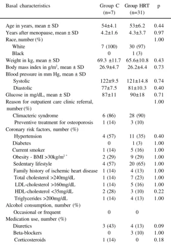
weight, body mass index, blood pressure, reason for searching the outpatient care clinic of climacterium, alcohol ingestion, coronary risk factors, and use of medication are shown in table I and do not differ in the 2 groups.
During the 12-week follow-up, no significant change occurred in the mean weight of the patients in both groups. Two patients in the group using hormone replacement the-rapy and 1 patient in the control group started regular phy-sical exercise practice (p=0.46).
Basal and final distribution profiles of lipoprotein sub-fractions in the different groups studied, according to grou-ping for analysis, are shown in table II. The corresponding variation of this distribution is depicted in figure 1.
A significant increase in the proportion of the larger VLDL subfractions (large VLDLs comprising V6, V5, and V4) occurred in regard to the smaller subfractions (small VLDLs comprising V3, V2, and V1) in the hormone replacement the-rapy group (p<0.008). In this group, the proportion of larger particles of this lipoprotein increased from 47% to 57% in the 12 weeks of treatment, and the proportion of smaller par-ticles decreased correspondingly from 53% to 43%. Group C showed no variation in the proportion of particles.
In regard to LDL subfractions, no significant variation in the proportion of large (corresponding to L3 and L2) and small (corresponding to L1) LDLs occurred in both groups during the 12 weeks of treatment.
In regard to HDLs, a significant increase in the propor-tion of large HDLs (corresponding to H5 and H4, or HDL2b and HDL2a) occurred as compared with small HDLs (corres-ponding to H3, H2, and H1, or HDL3a, HDL3b, and HDL3c) in the group of hormone replacement therapy (p<0.025). This resulted from an increase in the larger subfractions from 46% to 50% and correspondingly reduction in the smaller subfractions from 54% to 50%. Group C showed no variation in the proportion of particles.
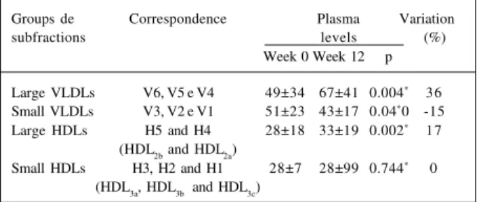
Analyzing the changes occurring in plasma levels of VLDL subclasses to verify how alterations in proportion oc-curred after hormone replacement therapy, we observed a 36% increase in the larger VLDL subfractions (p<0.004) concomitantly with a 15% reduction in the smaller VLDL subfractions (p<0.036). In regard to HDL, a 17% increase in the larger subfractions (p<0.002) occurred with no change in the smaller subfractions (Table III and fig. 2).
Discussion
Even though the clinical relevance of the heteroge-neity of the lipoprotein subfractions has been difficult to assess because of the extremely laborious nature of the methods for determining the subfractions of lipoproteins, differences in association with coronary heart disease suggest that data on lipoprotein subfractions may improve the evaluation of coronary risk. This situation is similar to
VLDLs VLDLs LDLs LDLs HDLs HDLs
large small large small large small
Fig. 1 - Variação média porcentual da distribuição das subfrações de lipoproteínas entre a 12ª semana e a basal nos dois grupos.
Table I – Basal characteristics of patients in the different groups
Basal characteristics Group C Group HRT p (n=7) (n=31)
Age in years, mean ± SD 54±4.1 53±6.2 0.44 Years after menopause, mean ± SD 4.2±1.6 4.3±3.7 0.97
Race, number (%) 1.00
White 7 (100) 30 (97)
Black 0 1 (3)
Weight in kg, mean ± SD 69.3 ±11.7 65.6±10.8 0.43 Body mass index in g/m2, mean ± SD 26.9±4.7 26.2±4.4 0.73
Blood pressure in mm Hg, mean ± SD
Systolic 122±9.5 121±14.8 0.74 Diastolic 77±7.5 81±10.3 0.40 Glucose in mg/dL, mean ± SD 87±11 90±18 0.71 Reason for outpatient care clinic referral, 1.00 number (%)
Climacteric syndrome 6 (86) 28 (90) Preventive treatment for osteoporosis 1 (14) 3 (10) Coronary risk factors, number (%)
Hypertension 4 (57) 11 (35) 0.40
Diabetes 0 1 (3) 1.00
Current smoker 1 (14) 5 (16) 1.00 Obesity - BMI >30kg/m2 * 2 (29) 9 (29) 1.00
Sedentary lifestyle 4 (57) 20 (65) 1.00 Family history of ischemic heart disease 1 (14) 4 (13) 1.00 Total cholesterol >240mg/dL 1 (14) 7 (23) 1.00 LDL-cholesterol >160mg/dL 1 (14) 5 (16) 1.00 HDL-cholesterol <35mg/dL 2 (28) 3 (10) 0.22 Triglycerides >200mg/dL 1 (14) 4 (13) 1.00 Alcohol consumption, number (%)
Occasional or frequent 0 0 Medication use, number (%)
Diuretics 3 (43) 4 (13) 0.09
Beta-blockers 0 3 (10) 1.00
Corticosteroids 1 (14) 0 0.18
SD- standard deviation; * - criteria for obesity according to Wood et al 37.
Table II – Distribution of lipoprotein subfractions (% of total ± standard deviation) in the two groups
Groups of subfractions Group C Group HRT
(n=7) (n=31)
Week 0 Week 12 p Week 0 Week 12 p
Large VLDLs 51±15 50±6 0.83 47±18 57±15 0.008*
Small VLDLs 49±15 50±6 0.83 53±18 43±15 0.008*
Large LDLs 67±21 65±22 0.69 80±17 79±17 0.814*
Small LDLs 33±21 35±22 0.69 20±17 21±17 0.814*
Large HDLs 37±23 37±22 0.75 46±19 50±31 0.03*5
Small HDLs 63±23 63±22 0.75 54±22 50±21 0.03*5
*p≤0.05; Week 0 - basal measurement; Week 12 - measurement after 12
that observed in the ‘50s and ‘60s, when data on cholesterol levels of lipoproteins could better predict the coronary risk than the total cholesterol serum levels isolated.
Most methods usually used for separating and quantifying subfractions of lipoproteins include techniques such as ultracentrifugation, electrophoresis, or chromato-graphy, which are very laborious and slow and, therefore, become unfeasible for clinical research on a large scale or for assessment of coronary risk in the general population. A recently validated method that employs nuclear magnetic resonance spectroscopy is a more precise, rapid, and less expensive alternative as compared with the already existing methods for assessing and handling coronary risk based on measurements of subfractions 21. This technique explores
the natural spectral difference that exists between the subfractions of each lipoprotein, depending only on the size of the particles. The technique avoids the steps of physical separation, the use of reagents, and employs a totally automated process, thus providing a high rate of efficiency and precision in the evaluation. This allows the simultaneous calculation of the concentrations of each of the 16 lipoprotein subclasses, 1 being chylomicron, 6 of VLDL, 1 of IDL, 3 of LDL, and 5 of HDL 24.
Despite the great methodological difference between nuclear magnetic resonance and the other methods used to quantify lipoproteins, some studies have shown a proximity between the sizes of LDL and HDL particles measured by nuclear magnetic resonance and by electrophoresis with gel gradient 20,25. As no appropriate and high-resolution
technique was available for measuring VLDL subfractions, only a few studies could separate VLDL subfractions that effectively represented distinct subclasses 21,26,27.
There-fore, comparison of nuclear magnetic resonance with other methods is impaired in this regard.
In our study, we observed an increase in the proportio-n of larger VLDLs as compared with that of smaller VLDLs due to an increase in the level of larger particles and a con-comitant reduction in smaller particles. Another study 28
using the same method has already shown a reduction in the smaller VLDL subfractions after hormone replacement therapy. In the literature, only a few studies report the effect of hormone replacement therapy on the profile of VLDL sub-fractions, because of the difficulties in separating these subfractions by the conventional techniques used prior to nuclear magnetic resonance. Estrogens seem to increase hepatic synthesis of VLDL-triglycerides forming particu-larly large VLDLs, rich in triglycerides, acting directly upon the hepatocyte 29. Most VLDLs are withdrawn from
circula-tion directly by the liver without being metabolized to small VLDL or LDL. In this way, the elevation in triglycerides in-duced by estrogens is supposed to be less harmful than that present in other clinical situations 30. The increase in the
proportion of larger VLDL subfractions was similar in pa-tients using isolated or combined estrogen therapy, which could be expected because progestins do not block the me-tabolic effects of estrogen in the hepatocyte. Whether large VLDLs actually produce small and dense LDLs 22 in case of
no inhibition of the hepatic lipase by estrogen, with the increase in VLDLs caused by hormone replacement therapy, a greater amount of small and dense, more atherogenic, LDLs could be produced, which in reality does not occur 30.
No change was observed in the distribution of LDL subfractions in the present study. Even though an induc-tion to a predominance of larger and less atherogenic (pattern A) particles could be expected because of the supposed cardioprotective effect, a few studies have found an opposite trend 17,31. This action of reducing the larger
and less dense LDLs in detriment to the smaller and denser particles could be due to the fact that the former are more easily captured by hepatic receptors of LDL, whose action is stimulated by estrogens 32.
The increase in the proportion of larger subfractions of HDL found agrees with data in the literature 28,33. An increase
in the proportion of larger particles (H5 and H4, corresponding to HDL2b and HDL2a) as compared with that of smaller particles (H3, H2, and H1, corresponding to HDL3a, HDL3b, and HDL3c) occurred due to an increase in the larger subfractions with no changes in the levels of the smaller fractions, which is in accordance with data in the literature 29,30,34,35. The
catabo-lism of HDL2 is mediated by hepatic lipase, a lipolytic en-zyme present in the endothelial cells lining the hepatic si-nusoids and highly specific for HDL2b, the larger subfrac-tion of HDL 36. The effects of sexual steroids on the levels of
HDL, particularly HDL2, are believed to be mediated by changes in the activity of hepatic lipase 33, because the
in-crease in HDL2 with the use of estrogen is associated with a Fig. 2 - Variação média porcentual dos níveis médios das subfrações de lipoproteínas
entre a 12ª semana e a basal no grupo TRH.
Table III – Plasma levels of the groups of VLDL and HDL ubfractions (mg/dL ± standard deviation) and their variation (%) in the group HRT
Groups de Correspondence Plasma Variation
subfractions levels (%)
Week 0 Week 12 p
Large VLDLs V6, V5 e V4 49±34 67±41 0.004* 36
Small VLDLs V3, V2 e V1 51±23 43±17 0.04*0 -15
Large HDLs H5 and H4 28±18 33±19 0.002* 17
(HDL2b and HDL2a)
Small HDLs H3, H2 and H1 28±7 28±99 0.744* 0
(HDL3a, HDL3b and HDL3c)
*p≤0.05; Week 0 – basal measurement; Week 12 – measurement after 12 weeks;
reduction in the activity of this enzyme. On the other hand, addition of a progestin has an inverse effect, which is more marked as the androgenic effect increases (the effect of me-droxyprogesterone is low), and it may even reverse the in-crease in HDL2 and the reduction in the activity of the he-patic lipase 34.
Clinical significance of the changes imposed by hor-mone replacement therapy in the profile of lipoprotein sub-fractions should be better evaluated in further larger cli-nical trials.
In the present study, in addition to the possibility of potential contamination of the cohort model by confoun-ding factors, the control group was composed of patients not adhering to the treatment. As all patients starting hor-mone replacement therapy in the outpatient care clinics had precise medical indications for it (climacteric symptoms or osteoporosis), from the ethical point of view they were not amenable to being directly allocated to the control group.
Even though all basal variables observed were not signifi-cantly different in both groups, a tendency to a higher pre-valence of hypertension occurred in the control group, as did HDL below 35mg/dL and the use of diuretics. This and the known fact that the adherence factor in many studies may be associated with healthier habits may have interfered with the final result of the study.
Aknowledgement
We thank the Laboratório Weinmann from Porto Ale-gre for the biochemical measurements performed and the shipping of samples to the USA; the Department of Bioche-mistry and Department of Comparative Medicine of Wake Forest University School of Medicine, Winston-Salem, NC, for the analysis of lipoproteins with nuclear magnetic reso-nance; and the Laboratório Wyeth, for providing the medi-cation used in the study.
References
1. Hulley S, Grady D, Bush T, et al. Randomized trial of estrogen plus progestin for secondary prevention of coronary heart disease in postmenopausal women. JAMA 1998; 280: 605-13.
2. Herrington D. The Estrogen Replacement and Atherosclerosis in older women (ERA) Trial, Late-Breaking Clinical Trial Results. American College of Cardiology 49th Annual Scientific Session, Anaheim, 2000.
3. Grodstein F, Stampfer M. The epidemiology of coronary heart disease and estrogen replacement in postmenopausal women. Prog Cardiovasc Dis 1995; 38: 199-210.
4. Manson JE. Postmenopausal hormone therapy and atherosclerotic disease. Am Heart J 1994; 128: 1337-43.
5. Barrett-Connor E. Heart disease in women. Pharmacologic Fertil Steril 1994; 62(suppl.2): 127S-32S.
6. Thorneycroft IH. Practical aspects of hormone replacement therapy. Prog Cardiovasc Dis 1995; 38: 243-54.
7. Grodstein F, Stampfer MJ, Manson JE, et al. Postmenopausal estrogen and progestin use and the risk of cardiovascular disease. N Engl J Med 1996; 335: 453-61. 8. Grodstein F, Stampfer MJ, Colditz GA, et al. Postmenopausal hormone therapy
and mortality. N Engl J Med 1997; 336: 1769-75.
9. Gotto AM Jr. Postmenopausal hormone-replacement therapy, plasma lipoprotein[a], and risk for coronary heart disease. J Lab Clin Med 1994; 123: 800-3.
10. Austin M, Breslow J, Hennekens C, Buring J, Willett W, Krauss R. Low-density lipoprotein subclass patterns and risk of myocardial infarction. JAMA 1988; 260: 1917-21.
11. Superko HR, Haskell WL, Krauss RM. Association of lipoprotein subclass distribution with use of selective and non-selective beta-blocker medications in patients with coronary heart disease. Atherosclerosis 1993; 101: 1-8. 12. Austin M, Mykkanen L, Kuusisto J, et al. Prospective study of small LDLs as a
risk factor for non-insulin dependent diabetes mellitus in elderly men and women. Circulation 1995; 92: 1770-8.
13. Krauss RM, Blanche PJ. Detection and quantification of LDL subfractions. Curr Opin Lipidol 1992; 3: 377-83.
14. Austin A, Hokanson J, Brunzell J. Characterization of low-density lipoprotein subclasses: methodologic approaches and clinical relevance. Curr Opin Lipidol 1994; 5: 395-403.
15. Tornvall P, Karpe F, Proudler A, et al. High-density lipoprotein: relations to metabolic parameters and severity of coronary artery disease. Metabolism 1996; 45: 1375-82.
16. Johansson J, Walldius G, Carlson LA. Close correlation between high-density lipoprotein and triglycerides in normotriglyceridaemia. J Intern Med 1992; 232: 43-51.
17. Seed M, Crook D. Post-menopausal hormone replacement therapy, coronary heart disease and plasma lipoproteins. Curr Opin Lipidol 1994; 5: 48-58. 18. Paganini-Hill A. Estrogen replacement therapy and stroke. Prog Cardiovasc Dis
1995; 38: 223-42.
19. Bagatell CJ, Bremner WJ. Androgen and progestagen effects on plasma lipids. Prog Cardiovasc Dis 1995; 38: 255-71.
20. Otvos JD, Jeyarajah EJ, Bennett D, Krauss RM. Development of a proton nuclear magnetic resonance spectroscopic method for determining plasma lipoprotein concentrations and subspecies distributions from a single, rapid measurement. Clin Chem 1992; 38: 1632-8.
21. Otvos J, Jeyarajah E, Bennet D. A spectroscopic approach to lipoprotein subclass analisys. J Clin Ligand Assay 1996; 19: 184-9.
22. Freedman DS, Otvos JD, Jeyarajah EJ, Barboriak JJ, Anderson AJ, Walker JA. Relation of lipoprotein subclasses as measured by proton nuclear resonance spectroscopy to coronary heart disease. Arterioscler Thromb Vasc Biol 1998; 18: 1046-53. 23. Otvos J. Measurement of triglyceride-rich lipoprotein by NMR spectroscopy.
Clin Cardiol 1999; 22: 21-7.
24. Parsons SK, Skapek SX, Neufeld EJ, et al. Asparaginase-associated lipid abnormalities in children with acute lymphoblastic leukemia. Blood 1997; 89: 1886-95. 25. Ala-Korpela M, Korhonen A, Keisala J, et al. 1H NMR-based absolute
quantitation of human lipoproteins and their lipid contents directly from plasma. J Lipid Res 1994; 35: 2292-304.
26. Otvos JD. Measurement of lipoprotein subclass profiles by NMR spectroscopy. In: Rifai N, Warnick R, Dominiczak M, eds. Handbook of Lipoprotein Testing. Washington DC: AACC Press, 1997: 497-508.
27. Zhao SP, Bastiaanse EM, Hau MF, et al. Separation of VLDL subfractions by density gradient ultracentrifugation. J Lab Clin Med 1995; 125: 641-9. 28. Tangney CC, Mosca L, Otvos JO, et al. Hormone replacement therapy in
post-menopausal women increases HDL particle size. Circulation 1998; Supplement (abstracts from the 71st Scientific Sessions. November, 1998): 32.
29. Nabulsi AA, Folsom AR, White A, et al. Association of hormone-replacement therapy with various cardiovascular risk factors in postmenopausal women. N Engl J Med 1993; 328: 1069-75.
30. Walsh B, Schiff I, Rosner B, Greenberg L, Ravnikar V, Sacks F. Effects of postmenopausal estrogen replacement on the concentrations and metabolism of plasma lipoproteins. N Engl J Med 1991; 325: 1196-204.
31. Lahdenperä S, Puolakka J, Pyörälä T, Luotola H, Taskinen MR. Effects of postmenopausal estrogen/progestin replacement therapy on LDL particles; comparison of transdermal and oral treatment regimens. Atherosclerosis 1996; 122: 153-62.
33. Thuren TY, Rudel LL, Brinton EA, et al. Effect of estrogen on hepatic lipase and HDL particle size in post-menopausal women. from the 71st Scientific Sessions.
November, 1998): Circulation 1998; (Suppl.): 2377.
34. Tikkanen M, Nikkila E, Kuusi T, Sipinen S. High density lipoprotein-2 and hepatic lipase: reciprocal changes produced by estrogen and norgestrel. J Clin Endocrinol Metab 1982; 54: 1113-7.
35. Sotelo MM, Jonhson SR. The effects of hormone replacement therapy on coronary heart disease. 1997; 26: 313-326.
36. Moriguchi EH, Fusegawa Y, Tamachi H, Goto Y. Age-related modi-fications in high density lipoprotein particle size and chemical composition - effects of reduction in hepatic lipase activity with age. Asian Med J 1990; 33: 641-50.
Paranapiacaba - São Paulo Mucio Tavares de Oliveira Jr
Seção de fotografias artísticas Cícero Piva de Albuquerque