Instituto de Cardiologia do Rio Grande do Sul - Fundação Universitária de Cardiologia Mailing address: Gabriel Lorier – IC/FUC - Av. Princesa Isabel, 395 - 90620-001 - Porto Alegre, RS – E-mail: [email protected]
Objective - To analyze late clinical evolution after surgical treatment of children, with reparative and reconstructive techniques without annular support.
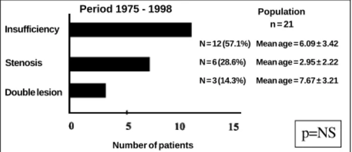
Methods - We evaluated 21 patients operated upon between 1975 and 1998. Age 4.67±3.44 years; 47.6% girls; mitral insufficiency 57.1% (12 cases), stenosis 28.6% (6 ca-ses), and double lesion 14.3% (3 cases). The perfusion 43.10±9.50min, and ischemia time were 29.40±10.50min. The average clinical follow-up in mitral insufficiency was 41.52±53.61 months. In the stenosis group (4 patients) was 46.39±32.02 months, and in the double lesion group (3 pa-tients), 39.41±37.5 months. The echocardiographic follow-up was in mitral insufficiency 37.17±39.51 months, stenosis 42.61±30.59 months, and in the double lesion 39.41±37.51 months.
Results - Operative mortality was 9.5% (2 cases). No late deaths occurred. In the group with mitral insufficiency, 10 (83.3%) patients were asymptomatic (p=0.04). The majorit y with mild reflux (p=0.002). In the follow-up of the stenosis group, all were in functional class I (NYHA); and the mean transvalve gradient varied between 8 and 12mmHg, average of 10.7mmHg. In the double lesion group, 1 patient was reoperated at 43 months. No endo-carditis or thromboembolism were reported.
Conclusion - Mitral stenosis repair has worse late re-sults, related to the valve abnormalities and associated le-sions. The correction of mitral insufficiency without annu-lar support showed good long-term results.
Key-words: congenital cardiopathy, valvuloplasty, sur-gery, stenosis, mitral insufficiency
Arq Bras Cardiol, volume 76 (nº 3), 215-20, 2001
Gabriel Lorier, Renato A.K. Kalil, Gustavo R. Hoppen, Christiano Barcellos, Nicolas Teleo, Abud Homsi Netto, Javier Gonzalez, Paulo R. L. Prates, Paulo R. Prates, João R. M. Sant’Anna,
Ivo A. Nesralla
Porto Alegre, RS - Brazil
Unsupported Valvuloplasty in Children with Congenital
Mitral Valve Anomalies. Late Clinical Results
Congenital malformations of the mitral valve are com-plex lesions resulting from several morphological abnorma-lities that, generally, involve more than 1 valve component 1-3 and take place in a population with high prevalence of associated cardiac anomalies 4-6.
Isolated congenital lesions, such as stenosis or mitral insufficiency, are rare 1,7,8, representing 1% of the population of congenital cardiopathy patients 5.
Congenital mitral insufficiency is extremely uncommon 8, being found in childhood associated with other cardiac de-fects, disorders of conjunctive tissue, and in acquired inflam-matory conditions, such as myocarditis, endocarditis, rheu-matic fever, Kawasaki’s disease, and other colagenosis with vascular impairment 8. In congenital mitral stenosis, obs-truction to the flow results from morphological anomalies at different levels1,2,5 and is more frequent than in cardiac in-sufficiency 2.
The purpose of the present study was analysis of late clinical evolution after the surgical treatment of congenital mitral anomalies, with or without associated malformations in children up to 12 years of age treated with reparative and reconstructive techniques without annular support, and to carry out a literature review.
Methods
was reduction of leaflet mobility in 100% of patients (Table I). Associated intracardiac malformations were found in 61.9% of the patients, in the stenosis group, 83.3%; in the insufficiency group 41.6%; and in the double lesion group, all patients; the individual description of the different associated intracardiac malformations are presented in Table I. More severe malforma-tions were found in 28.6% of the patients, all with valvular ste-nosis. Parachute valve was found in 3 cases, all with stenosis; 2 of them were associated with Shone syndrome. Isolated an-nular dilation without other malformations was present in 2 ca-ses (Table I).
Table I - Valvular morphology, surgical technique, and long-term results of the population under study
Patient Morphology Associated Surgical Surgical Surgical Reope- Functional ECO malformations Technique morbidity mortality ration class
n / age Hospital Late Hospital Late Pre-OP P o s t - O P * P R E POST**
MITRAL INSUFFICIENCY
1 –6 y A C PAVSD Anterior cleft suture N o N o N o N o N o I I severe R. moderate R.
2 – 4 y A D TI Wooler + De Vega D P N o N o N o N o I I “ light R.
3 - 7 y AD + PC PAVSD Wooler + cleft N o N o N o N o N o III I “ light R. at
suture release
4 - 2 y AD e at of reflux H L L Wooler Atelectasis N o N o N o N o III I “ Absenc
5 - 9 y P C PAVSD Posterior cleft N o N o N o N o N o II I “ light R.
suture
6 - 6 y AD + PAL N o Wooler + chord N o N o N o N o 4 years PS IV I “ “
shortening (new
valvu-loplasty)
7 - 6 y A C N o Anterior cleft suture N o N o N o N o N o I I “ “
8- 5 y AD + AC PAVSD Wooler + anterior N o N o N o N o N o I I “
light/mo-cleft suture derate R.
9 - 1 y AD + PAL + AIPM N o Wooler + chord N o N o N o N o N o IV I “ moderate R. shortening
10 – 13 y A D N o Wooler + chord Atelectasis N o N o N o N o II I “ light R. shortening
11 – 8 y AD + PAL N o Wooler + chord N o N o N o N o N o I I “ “
shortening
12 – 2 y AD + AC N o Wöoler + anterior N o N o N o N o N o I I “ light R.
cleft suture at release
MITRAL STENOSIS
1 - 3 y P PAC + IVC + Papillotomy N o N o N o N o N o III I severe stenosis l i g h t
SUB. Ao STE double lesion
2 - 16 y CF N o Comissurotomy N o N o N o N o 8 years PS I I “ Light stenosis
+ Papillotomy (valvuloplasty)
3 – 12 d HMV Ao STE + FE Comissurotomy N o N o Death - - III - “
-4 – 18 mo. CF Ao. STE Comissurotomy N o N o N o N o N o III I “ Light double lesion
5 - 3 y P Ao STE + Papillotomy RRI N o N o N o N o II I “ “
SUB.Ao STE
6 - 7 mo. P FE Comissurotomy N o N o Death N o 9 days PS IV - “
-DOUBLE LESION
1 – 6 a DLM + CF N o Comissurotomy N o N o N o N o N O II I “ Moderate
double lesion 2 – 10 a DLM + AC Ao STE Papillotomy N o N o N o N o N O III I “ l i g h t
+ FEPM + S. Noonam (at release)
3 - 4 a DLM + FEPM Light TI Papillotomy S W I N o N o N o 4 year PS III II “ No gradient (prosthesis)
AC: anterior cleft; PAVSD: partial atrioventricular septum defect; ASYMPT: asymptomatic; AD: annular dilation; PH: pleural hemorrhage; PC: posterior cleft; HLL: hypoplasic left lung; PAL: prolapse of the anterior leaf; PS: postsurgery; AIPM: anomalous implant of the papillary muscle; P: parachute; PAC: patent arterial canal; IVC: interventricular communication; SUB.Ao STE: subaortic stenosis; CF: commissural fusion; HMV: hypoplasic mitral valve; Ao STE: aortic stenosis; FE: fibroelastosis; RRI: repetitive respiratory infection; DLM: decreased leaf mobility; FEPM: fibroelastosis in papillary muscles; IT: tricuspid insufficiency; SWI: surgical wound infection. No episodes of endocarditis or lung thromboembolism occurred. None of the patients presented had previous mitral valve corrective surgery. (*) In the insufficiency group p=0.004. (**) In the insufficiency group p=0.002.
Fig. 1 - Patients’ distribution by groups with congenital mitral valve malforma-tions. Patients with complete defects of the atrioventricular septum were excluded from the sample.
p=NS
Period 1975 - 1998
Double lesion Insufficiency
Stenosis
Population n = 21
Number of patients
N = 12 (57.1%) Mean age = 6.09 ± 3.42
N = 6 (28.6%) Mean age = 2.95 ± 2.22
All patients were operated on with median sternotomy and with conventional extracorporeal circulation, using a disposable bubble or membrane oxygenator and moderate hypothermia between 28° and 30°C. Hyperkalemic crys-talloid cardioplegia, with cooling of the pericardial cavity with saline solution at 4°C was used for myocardial pro-tection. The approach to the mitral valve was performed by left longitudinal atriotomy. In 2 patients (33.3%), isolated mitral commissurotomy was carried out. In the insufficiency group, annuloplasty was performed with the Wooler’s tech-nique in 9 (75%) patients, and the associated procedures in 75% of the cases were 4 chord shortenings, 3 closings of the anterior leaflet cleft, and 2 closings of the posterior leaflet cleft (Table I). The associated cardiac lesions were all corrected after the mitral valvuloplasty.
The mean extracorporeal circulation and aortic clam-ping times were 43.1±9.5min and 29.4±10.5min.
The postsurgical follow-up was performed by perio-dic clinical, radiological, and echocardiographic evaluation. The follow-up period varied from 7 days to 15.7 years (mean of 3.8±4.1 years).
Results
The operative mortality in mitral stenosis was 8.9% (2 deaths). One patient, 12 days of age, had hypoplastic mitral valve associated with left ventricular fibroelastosis (Table I).
Overall morbidity was 28.5% (of the total population of patients), being higher in the mitral insufficiency group. Two patients had atelectasis after chest tube withdrawal and pleural effusion due to cardiac failure. One patient in the stenosis group had repetitive respiratory infection. One pa-tient in the double lesion group had infection of the surgical wound.
No episodes of endocarditis or thromboembolism during the follow-up were reported (Table I). The incidence of reoperation was 13% (3 cases). The associated anoma-lies, as well as the surgical techniques employed, are shown in Table I.
In the insufficiency group, the mean follow-up was 41.52±53.61 months. In the last clinical evaluation, 10 (83%) patients were asymptomatic without medication. One patient was in NYHA functional class I, and 1 patient was in functional class II, both receiving medication (Table I). One patient was operated on again 48 months after surgery, when a new valvuloplasty was performed. The mean echocardiographic follow-up was of 37.17±39.51 months, most of the patients had a light level of valvular regurgitation p=0.002 (Table I).
In the stenosis group (6 cases), 2 deaths and 1 reopera-tion that evolved to death occurred. Four patients were in functional class I (2 were receiving medication). The echocardiographic follow-up was performed in 4 of these patients (42.61±30.59 months) and showed an average transvalve gradient between 8 and 12mmHg; the mean was 10.7mmHg.
In the double lesion group (3 cases), 1 patient was out-of-control, in whom a slight reflux occurred on the echocar-diogram at the time of release from the hospital, and in 1 case, the valve was replaced by a bioprosthesis 43 months after the first surgery. The third patient of the group was in functional class II, and the echocardiography showed stenosis and light insufficiency at 75 months of the surgery. At the echocardiographic evaluation of the whole sample, with a mean follow-up of 39.89 months, most patients had light lesions (p=0.002) (Table I). In the insufficiency group, 83.3% had absence or light reflux.
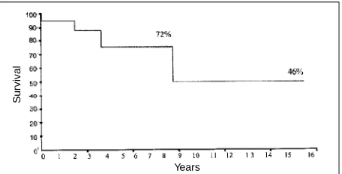
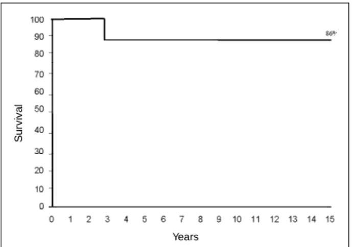
The actuarial survival curve in the insufficiency group at 5, 10, and 15 years was of 90% (Fig. 2). The actuarial survi-val probability free of reoperation in the whole sample (n=21) was 72% at five years and of 46% at 10 and 15 years (Fig. 3). In the insufficiency group, the actuarial survival probability free of reoperation was 86% at 5, 10 and 15 years (Fig. 4).
Discussion
The clinical presentation, as well as the surgical indication, of stenosis or insufficiency depends on the severity of the mitral lesion and the association with intracardiac defects 10. The surgery in our series was indicated for refractory cardiac failure or important pulmo-nary hypertension, or both. Preferably, the surgery should not be performed before 6 months of life, according to Carpentier 3. During childhood, before 3 months of age, with inadequately matured collagen, the valvular tissue is particularly friable, making the manipulation difficult 3. In
Fig. 3 - Actuarial survival probability curve free of reoperation of the whole population studied.
S
u
rv
iv
a
l
Years
Fig. 2 - Actuarial survival probability curve in the group of with congenital mitral insufficiency.
S
u
rv
iv
a
l
our sample, the mean age of the patients was 5.7 ± 3.3 years in the congenital mitral insufficiency group and 4.0±5.9 years in the congenital mitral stenosis group, without statistical significance (Fig. 1). In Carpentier’s series 3, the age of the congenital mitral insufficiency group was 6.1±3.2 years and in the congenital mitral stenosis group 5.1±3.2 years. In the report by Uva and colleagues 11, from Marie-Lannelongue Hospital, the congenital mitral insufficiency group was 7.4±2.7 months old, and the congenital mitral stenosis group was 5.8±3.9 months old. The factors that explain the lower age of the congenital mitral stenosis patients at the time of surgery are 1) the stenotic lesion is less tolerated than the insufficiency; 2) the association with higher and more severe intracardiac anomalies; 3) the abnormalities of the subvalvular apparatus are more frequent in these patients.
Congenital mitral stenosis occurs in 0.6% of the auto-psies and in 0.2% and 0.4% of the clinical series 1,12. In the Ruckman and Van Praagh’s 13 series of 49 autopsies with congenital mitral stenosis, typical mitral stenosis was found in 49% of the cases, with coarctation of the aorta as the most common associated lesion. The size of the left ventricle was normal in 96% of these patients. Congenital hypoplasia of the mitral valve was the second cause of congenital mitral stenosis (41%), and it was always associated with left ven-tricular hypoplasia. Supravalvular ring was found in 12% of the cases and the parachute mitral valve in 8%.
Traditionally, the lesions that make the effective and long-lasting repair more difficult are those that have al-terations in the subvalvular system with abnormal papillary muscles, including the parachute valve, the hammock valve, and agenesis of the papillary muscles. These lesions determine stenosis with greater frequency 10 and are asso-ciated with the high frequency of complex malformations. Barbero-Marcial and co-workers 14 achieved good short-and long-term results in 7 patients with mitral parachute valve with stenosis 14. According to the review presented in Table II, the different publications during the last decade show important progress regarding the results, where it was possible to perform the valvuloplasty in 91.4% of the cases
of mitral stenosis with parachute and hammock valve in 50% of the cases.
In the Moore et al. 12 series, the typical mitral hypopla-sia with symmetric papillary muscles was the first cause of congenital mitral stenosis (52% of the cases), followed by the supravalvular ring (in 20%), double orifice (in 11%), mi-tral hypoplasia with asymmetry of the papillary muscles (in 8%), and parachute-shaped mitral valve (in 8%). Contrary to this series, Embrey and Behrendt 5 state that the ring is ra-rely so small that stenosis results, unless left ventricular hy-poplasia is present. According to these authors, chordal malformation is the most common cause of stenosis 5.
In a series of 50 patients, Fuzzelier et al. 15 reported commissura fusion as the most common cause of congenital mitral stenosis with normal papillary muscles (in 17 pa-tients). With abnormal papillary muscles, the most common was the hammock mitral valve in 11 patients. In our series, 50% of the cases of congenital mitral stenosis had abnormal papillary muscles; as the most prevailing cause, the parachute mitral valve in 3 patients, 2 of them associated with Shone syndrome, with possible recovery (Table I); 38% typical mitral stenosis and 12.5% hypoplasic mitral valve (Table I). Left ventricular hypoplasia was present in 50% of the cases of congenital mitral stenosis, associated in 50% with left ventricular fibroelastosis.
According to McGiffin 2, congenital mitral insuffi-ciency is less frequent than congenital mitral stenosis. In our series, we observed 12 cases of insufficiency and 6 ca-ses of stenosis. The most frequent cause in congenital mitral stenosis is annular dilation 2,16; this finding was confirmed in our sample. Seventy-five percent of the patients in the congenital mitral insufficiency group had this malformation, and in 25% this was the only malformation.
Congenital or acquired abnormalities of the mitral valve in children can be surgically managed with mechanical or biological prosthesis or valvuloplasty 4,17. Valvular replacement is accompanied by a high mortality rate and by anticoagulation problems and the impossibility of annular growth, leading inevitably to re-operation 2,4,17-22.
The use of a rigid or flexible ring as a prerequisite for efficacy and durability of annular remodeling 23 is currently being questioned in the adult 24,25 and a tendency to reduce its use has been observed 26,27.
In children and adolescents, the prosthetic rings must be avoided for they do not allow a normal annular growth 5, in addition to being a risk factor for distortions of the
ventri-Fig. 4 - Actuarial survival probability free of reoperation in the group of congenital mitral insufficiency.
S
u
rv
iv
a
l
Years
Table II - Results of valvuloplasty in stenosis with abnormal papillary muscles
Hammock valve Parachute Repair Replace Repair Replace
Uva (Planché)11 1 1 2 2
Barbero-Marcial14 9
Mc Carthy4 5
Stellin26 4 1
Fuzellier15 7 7 9
cular cavity and for contributing to the obstruction of the left ventricular outlet 18,28. Without adding the fact that the annular segment of mitral-aortic continuity is capable of contracting and relaxing during the cardiac cycle in the left ventricular outlet 25,26,28, because it is the only annular portion that does not dilate, which leads to the conclusion that at the descri-bed level no prosthetic structure should exist.
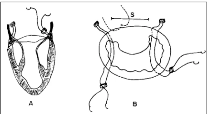
The long-term stability of mitral valve repair within the concept of remodeling annuloplasty does not imply the use of a rigid or flexible ring to decrease the antero-posterior ring diameter. Since 1975, we have believed that the stability in Wooler’s technique 9 of unsupported annuloplasty is con-tained in the anchorage of the right and left fibrous trigones (Fig. 5), keeping the normal variable anatomical relationshi-ps with the aortic valve and the left ventricular outlet, and the annular segment of mitral-aortic continuity with the capacity of contraction and relaxation during the cardiac cycle 28.
At the time of performing Wooler’s technique, it is of essential importance to long-term repair stability and to avoid failure, that the points undergo through the fibrous trigones, reminding their position related with the anterior
cuspid 29. This technique has a late result comparable to more complex support technqiues 30-34, but with a lower inci-dence of technical repair failure. The reported inciinci-dence of repair failure due to technical problems with the prosthetic ring in the adult is varied (Aharon 10 - 2.9%; Cosgrove 34 -3.2%; Deloche 35 - 4.3%; Cosgrove 24 - 3.3%).
Regarding intraoperative deaths, patients with con-genital mitral stenosis who were in NYHA functional class IV had in our series 100% mortality and in Kirklin’s series 50% mortality 36,37; both patients had endocardial fibroelas-tosis. The 12-day-old patient had left ventricular hypo-plasia, and the 7-month-old had Shone syndrome. Recent studies carried out by Ni and colleagues 38 suggest a viral infection as the cause of endocardial fibroelastosis, sup-porting the hypothesis that this disease is a sequela of viral myocarditis, particularly due to the mumps virus.
The mitral corrections were performed by longitudinal atriotomy of the left atrium’s right wall, without using other approaches 14,39-43.
The comparative results among the studies found in the literature about congenital mitral insufficiency, presen-ted in chart I, corroborate the good long-term results of the unsupported annuloplasty in this population of patients, also found in our series. However, the population of patients with congenital mitral stenosis has a variety of mitral valve malformations, frequent associations with cardiac malfor-mations, and variable age, making it difficult to obtain generalized conclusions, according to chart II.
In conclusion, mitral valvuloplasty in isolated conge-nital lesions or in association with other cardiac malfor-mations has good long-term results. Failure to repair stenosis and double lesions is due to the complexity of malformations detected.
In the case of congenital mitral insufficiency, the unsupported mitral valvuloplasty with Wooler’s technique had a low surgical risk and good long-term results. The use of prosthetic rings in these patients is considered unnecessary.
Fig. 5 - Representation of the Wooler’s type placement of annuloplasty points. A) Stitches of braided polyester string anchored in Teflon felt are crossed through the mitral leaves insertion ring; B) stitches aiming at reducing the mural leaf, without compromising the width (S) of the septal leaf. The projection of the aortic valve is represented by the dotted line.
Chart I - Congenital mitral insufficiency (CMI). Surgical Results in Different Publications
Mean age N Hospital Late Actuarial Free of re- Type of
(years) mortality (%) mortality (%) survival (%) operation (%) annuloplasty
Okita, 1988 39 5.5 66 1.5 6.0 93.1-7a 89-10a Key-Reed
Kirklin, 1993 37 - 18 18 - - - Reed
Kalil, 1996 30 12.4 13 7.7 0 89-5a 91-5a Wooler
Uva, 1991 11 1 10 0 0 100-7a 61.2-7a Wooler
Carpentie, 1994 3 6.1 105 5 4 62-15a 62-15a Rigid ring 50%
This series 6.09 12 0 0 100-15a 86-15a Wooler
Chart II - Congenital mitral stenosis (CMS). Surgical Results in Different Publications
Mean age N Hospital Late Actuarial Free of
(years) mortality (%) mortality (%) survival (%) reoperation (%)
Kirklin, 1993 37 4.5 19 21 - -
-Uva, 1991 11 0.5 10 0 10 94.1-7y 54.8-7y
Carpentier, 1994 3 5.1 50 26 3 47-10y 47.10y
Fuzillier, 1997 15 4.5 58 25.8 5 67-15y
1. Anderson RH, Tynan M, Shinebourne EA, Macartney FJ. Pediatric Cardiology. Edinburgh: Churchill-Livingstone, 1984: 1023.
2. McGiffin DC. Surgery of the mitral valve in children. In: Wells FC, Shapiro LM. Mitral Valve Disease. Oxford: Butterworth-Heinemann, 1996; 16: 149-59. 3. Carpentier A. Congenital malformations of the mitral valve. In: Stark J, de Leval M.
Surgical for Congenital Heart Defects. Philadelphia: WB Saunders, 1994: 599-614. 4. McCarthy JF, Neligan MC, Wood AE. Ten years’ experience of an aggressive reparative approach to congenital mitral valve anomalies. J Cardio-Thorac Surgery 1996; 10: 534-9.
5. Embrey RP, Behrendt DM. Congenital abnormalities of the mitral valve. In: Bave AE, Geha AS, Laks H, Hammond GL, Naunheim KS. Glenn’s Thoracic and Cardiovascular Surgery. 6th ed. Vol II. Stanford: Appleton & Lange, 1996. 6. Aharon AS, Laks H, Drinkwater DC, et al. Early and late results of mitral valve
repair in children. J Thorac Cardiovas Surg 1995; 107: 1262-71.
7. Medeiros Sobrinho JH, Fernandes V, Cunha S. Anomalias da valva mitral. In. ___. Cardiopatias Congênitas. São Paulo: Sarvier, 1990; 22: 386.
8. Baylen BG, Eriley JM. Diseases of mitral valve. In: Adams FH, George C. Heart Disease in Infants, Children and Adolescents. 5th ed. Baltimore: Williams & Wilkins, 1995; 32: 516-26.
9. Wooler GH, Nixon PG, Grimsaw VA, Watson DA. Experience with the repair of the mitral valve in mitral incompetence. Thorax 1962; 17: 49. Apud Kalil RAK. Valo-rização da valvoplastia para correção de insuficiência mitral (Tese de Doutorado). Porto Alegre. Universidade Federal do Rio Grande do Sul - UFRGS, 1987. 10. Aharon AS, Laks H, Milgater E. Congenital malformations of the mitral valve - In:
Sabiston DC, Spencer FC. Surgery of the Chest. 6th ed. Vol. II. Philadelphia: WB Saunders Co., 1995.
11. Uva MS, Galletti L, Gayet FL, et al. Surgery for congenital mitral valve disease in the first year of life. J Thorac Cardiovasc Surg 1995; 109: 164-76.
12. Moore P, Adatia I, Spevak PJ, et al. Severe congenital mitral stenosis in infants. Circulation 1994; 89: 2099-106.
13. Ruckman RN, van Praagh R. Anatomic types of congenital mitral stenosis; report of 49 autopsy cases with consideration of diagnosis and surgical implications. Am J Cardiol 1978; 42: 592-601.
14. Barbero-Marcial M, Riso A, Albuquerque A, et al. A ventriculotomia apical esquerda para tratamento cirúrgico da estenose mitral congênita. Rev Bras Cir Cardiovasc 1991; 6: 167-73.
15. Fuzellier J-F, Chauvaud S, Houel R, Berrebi A, Mihaileanu S, Carpentier AF. Surgery for congenital mitral valve stenosis in pediatric age group. Prognostic factors and long-term results. 70th AMA Meeting. Orlando, 1997: 1030. 16. Anderson R, Becker EA. El Corazón, Estructura Normal y Patológica.
Barcelo-na: Doyma Libros SA, 1994: 6-30.
17. Castañeda AR, Jonas RA, Mayer JE, Hanley FL. Cardiac Surgery of the Neonate and Infant. Philadelphia: WB Saunders Co., 1994: 388.
18. Pomerantzeff PMA. Plástica da valva mitral. Revista do InCor 1997; 2: 38-42. 19. Kutsche L, Oyer P, Shumway N, Baum D. An important complication of hancock
mitral valve replacement in children. Circulation 1979; 48: II-148.
20. Gerola LR, Pomerantzeff PMA, Pêgo-Fernandes PM, et al. Cirurgia valvar em cri-anças e jovens: resultados de 131 casos. Rev Bras Cir Cardiovas 1990; 5: 187-94. 21. Taybi H, Capitanio MA. Tracheobronchial calcification: an observation in three children after mital valve replacement and warfarin sodium therapy. Radiology 1990; 17: 728-30.
References
22. Carpentier A. Plastic and reconstructive mitral valve. In: Kalmanson D. The Mitral Valve a Pluridisciplinary Approach. London: Sciences Group Inc., 1976; (I) 45: 529.
23. David TE, Armstrong S, Sun Z, Daniel L. Late results of mitral valve repair for mi-tral regurgitation due to degenerative disease. Ann Thorac Surg 1993; 56: 7-14. 24. Cosgrove III DM, Arcidi JM, Rodriguez L, et al. Initial experience with the cosgrove-edwards annuloplasty system. Ann Thorac Surg 1995; 60: 466-504. 25. Carpentier AF, Lessana A, Relland JYM, et al. The “Physio-Ring”: an advanced
concept in mitral valve annuloplasty. Ann Thorac Surg 1995; 60: 1177-86. 26. Stellin G, Bortolotti U, Mazzucco A, et al. Repair of congenitally malformed
mitral valve in children. J Thorac Cardiovasc Surg 1988; 95: 480-5. 27. Muehrcke DD, Cosgrove DM. Mitral valvuloplasty. In: Edmunds Jr H. Cardiac
Surgery in the Adult. Philadelphia: McGraw-Hill, 1997; 33: 991-1024. 28. Jatene FB, Monteiro R, Jatene MB, Magalhães MHG, Fukushima JT, Jatene AD.
Estudo do anel mitral e trígonos fibrosos com diferentes variáveis. Rev Bras Cir Cardiovasc 1991; 6: 190-4.
29. Kalil RAK, Lucchese FA, Prates PR, et al. Anuloplastia sem suporte para trata-mento da insuficiência mitral reumática. Rev Bras Cir Cardiovas 1992; 7: 186-93. 30. Kalil RAK, Lucchese FA, Prates PR, et al. Late outcome of unsupported annulo-plasty for rhematic mitral regurgitation. J Am Coll Cardiol 1993; 22: 1915-20. 31. Kalil RAK. Valorização da valvoplastia para correção de insuficiência mitral (Tese
de Doutorado). Universidade Federal do Rio Grande do Sul. Porto Alegre, 1987. 32. Bordignon S. Anuloplastia mitral sem suporte em crianças e adolescentes: resultado clínico tardio (Dissertação de Mestrado). Fundação Universitária de Cardiologia - IC-FUC. Porto Alegre, 1995. 75p.
33. Bordignon S, Kalil RAK, Sant’Anna JRM, et al. Resultado clínico tardio da anuloplastia mitral sem suporte em crianças e adolescentes. Rev Bras Cir Cardiovasc 1996; 11: 263-9.
34. Cosgrove DM, Chavez AM, Lytle BW, et al. Results of mitral valve recons-truction. Circulation 1986; 74: I-82-I-7.
35. Deloche A, Jebara VA, Relland JYM, et al. Valve repair with carpentier techniques. J Thorac Cardiovas Surg 1990; 99: 990-1002.
36. Kirklin JW, Barret-Boyes BG. Congenital Mitral Valve Disease. Cardiac Surgery. 2nd ed. vol 2. London: Churchill Livingstone, 1993.
37. Kirklin JW, Barret-Boyes BG. Endomyocardial Fibroelastosis. Cardiac Surgery. 2nd. ed. Vol. 2. London: Churchil Livingstone, 1993.
38. Ni J, Bowles NE, Kim Y-H, et al. Viral infection of the myocardium in endocardial fibroleastosis. Circulation 1997; 95: 133-9.
39. Okita Y, Miki S, Kusuhara K, et al. Early and late results of reconstructive operation for congenital mitral regurgitation in pediatric age group. J Thorac Cardiovasc Surg 1988; 96: 294-8.
40. Hisatomi K, Isomura T, Sato T, et al. Mitral valve repair for mitral regurgitation with ventricular septal defectin children. Ann Thorac Surg 1996; 62: 1773-7. 41. Matsuwaka R, Sakakibara T, Mitsuno M, Yagura A, Yoshikawa M, Ishikura F.
Valve repair for mitral regurgitation associated with isolated double-orifice mitral valve. J Thorac Cardiovasc Surg 1996; 112: 1666-7.
42. Hurst JW, Anderson RH, Becher AE, Wilcox BR. Atlas del Corazon. Mexico, DF: Interamericana, 1989: 1.3.