Instituto do Coração do Hospital das Clínicas - FMUSP
Mailing address: Marcelo Park – Estrada de Itapecerica, 1187/52 – Bl. 1 – 05835-002 São Paulo, SP, Brazil – E-mail: [email protected]
English version by Stela Maris C. Gandour
Objective – To compare the effects of 3 types of noninva-sive respiratory support systems in the treatment of acute pulmonary edema: oxygen therapy (O2), continuous positive airway pressure, and bilevel positive pressure ventilation.
Methods – We studied prospectively 26 patients with acute pulmonary edema, who were randomized into 1 of 3 ty-pes of respiratory support groups. Age was 69±7 years. Ten pa-tients were treated with oxygen, 9 with continuous positive airway pressure, and 7 with noninvasive bilevel positive pres-sure ventilation. All patients received medicamentous therapy according to the Advanced Cardiac Life Support protocol. Our primary aim was to assess the need for orotracheal intubation. We also assessed the following: heart and respiration rates, blood pressure, PaO2, PaCO2, and pH at begining, and at 10 and 60 minutes after starting the protocol.
Results – At 10 minutes, the patients in the bilevel po-sitive pressure ventilation group had the highest PaO2 and the lowest respiration rates; the patients in the O2 group had the highest PaCO2 and the lowest pH (p<0.05). Four patients in the O2 group, 3 patients in the continuous posi-tive pressure group, and none in the bilevel posiposi-tive pres-sure ventilation group were intubated (p<0.05).
Conclusion – Noninvasive bilevel positive pressure ventilation was effective in the treatment of acute cardio-genic pulmonary edema, accelerated the recovery of vital signs and blood gas data, and avoided intubation.
Key words: acute pulmonary edema, noninvasive venti-lation, respiratory failure
Arq Bras Cardiol, volume 76 (nº 3), 226-30, 2001
Marcelo Park, Geraldo Lorenzi-Filho, Maria Inês Feltrim, Paulo Ricardo Nazário Viecili, Márcia Cristina Sangean, Márcia Volpe, Paulo Ferreira Leite, Alfredo José Mansur
São Paulo, SP - Brazil
Oxygen Therapy, Continuous Positive Airway Pressure, or
Noninvasive Bilevel Positive Pressure Ventilation in the
Treatment of Acute Cardiogenic Pulmonary Edema
Application of positive pressure ventilation with a face mask has been suggested in association with the conventional medicamentous treatment as an effective therapeutical modality in acute cardiogenic pulmonary edema. It provides more rapid recovery of vital signs and blood gas parameters when compared with the conventio-nal treatment with oxygen by face mask 1,2. A few studies have also shown a reduction in the need for tracheal intuba-tion and mechanical ventilaintuba-tion 1,3-5. The mechanisms invol-ved in improving the respiratory discomfort of patients with acute pulmonary edema by using positive pressure are multiple, and we can cite the following: improvement of hy-poxemia, a reduction in the left ventricular preload and afterload, and an increase in pulmonary compliance due to recruiting of previously collapsed alveolar units 6-10.
Two noninvasive methods for applying positive res-piratory pressure exist as follows: by mask with continuous positive pressure in the airways (continuous positive airway pressure) or by ventilation with 2 levels of pressure (bilevel positive pressure ventilation). In the case of continuous po-sitive airway pressure, the predetermined value of pressure remains constant during the entire respiratory cycle, and the respiratory work is completely performed by the patient. During bilevel pressure ventilation, the pressure is higher during inspiration and decreases during expiration. It is a mo dality that supports inspiration and, therefore, directly reduces the patient’s respiratory work.
determinant of poor evolution, and, when accompanied by pulmonary edema, it has even higher morbidity and mor-tality. Therefore, no absolute definition exists in regard to the use of noninvasive ventilation in acute ischemic heart disease.
Patients with acute pulmonary edema have increased respiratory work. We hypothesized that bilevel positive pressure ventilation is a better ventilatory modality than continuous positive airway pressure, because it adds the beneficial effects of expiratory positive pressure to a reduction in the respiratory work provided by inspiratory support. In this study, we prospectively compared the need for orotracheal intubation and observed the clinical response of patients with acute pulmonary edema, who were randomized into 1 of 3 groups, each using a different form of respiratory support, as follows: oxygen therapy, continuous positive airway pressure, or bilevel positive pressure ventilation. All forms of treatment were applied in a noninvasive way, using a face mask. Medicamentous treatment was standardized in the 3 groups according to the Advanced Cardiac Life Support protocol 12.
Methods
We studied 26 patients, 16 females and 10 males, whose ages ranged from 51 to 87 years (mean of 69±7 years). They sought emergency treatment because of severe respiratory failure due to acute cardiogenic pulmonary edema during the period from May to October ’97. The patients were consecutively randomized as follows: 10 of these patients were treated with oxygen therapy, 9 with continuous positive airway pressure, and 7 with bilevel positive pressure ventilation. The inclusion criteria were as follows: dyspnea of acute onset or worsening, respiration rate >25 inspirations per minute, and pulmonary findings compatible with pulmonary congestion, which was radio-graphically confirmed later. Patients with the following findings were excluded from the study: systolic blood pressure <90mmHg, cardiac arrhythmias requiring electric cardioversion, decrease of the consciousness level, bradypnea, lack of cooperation or agitation, repetitive vomiting despite the use of antiemetics, upper digestive hemorrhage, facial deformities, or any other decompensated respiratory disease.
After formal consent provided by the patient or guardian, randomization to 1 of the 3 ventilation modalities was performed. The patients were kept in the sitting position, monitored with continuous electrocardiography, their blood pressure was measured noninvasively, and pulse oxymetry was performed with a 56S Hewlett Pa-ckardTM monitor. If the patient had systolic blood pressure >100mmHg, 5mg of isosorbide dinitrate was sublingually administered. A venous access was acquired by a puncture in the upper limb with a flexible catheter. Concomitantly, vital signs, such as heart and respiration rates, blood pressure, and noninvasive oxygen saturation, were recorded, and analysis of arterial blood gases collected in the
environ-mental air was carried out. After collecting these data, the randomized ventilatory modality was applied.
Vital signs and arterial blood gases were reassessed at 10 and 60 minutes after randomization. Medicamentous therapy was the same for all patients, regardless of the modality of ventilation.
The criterion for intubation was clinical and was not determined by any member participating in the protocol but by the physician responsible for the patient.
The patients were randomized into 3 groups receiving different modalities of respiratory support, as follows: 1) oxygen group (group I) – oxygen was provided by an open semirigid facial mask (OxigelTM) at a continuous flow of 15 L/ min; 2) continuous positive airway pressure group (group II) – a closed face mask was used with a high continuous flow of compressed air provided by a flow generator of the Venturi type fed with 15 L/min of humidified O2 in parallel and proximally to the circuit. At the end, a valve of conti-nuous positive pressure in the airways was installed, and it was regulated with a coil (Vital SignsTM), initially with 5cm of H2O, which was gradually increased by 2.5 cm H2O every 5 minutes, up to a maximum of 12.5cm H2O, if O2 saturation was lower than 90% or if the presence of bronchospasm was observed; 3) bilevel positive pressure ventilation group (group III) – ventilation was applied by nasal mask with the BiPAP ST/D 30® Respironics® system, in the spontaneous modality with expiratory pressure of 3cm H2O and inspira-tory pressure of 8cm H2O fed with humidified O2 at the rate of 15 L/min in parallel and proximally to the circuit. If the patient persisted with respiratory discomfort, the inspiratory pressure was elevated by 2cm H2O every 5 minutes; if O2 saturation was lower than 90%, or if bronchospasm occurred, inspiratory and expiratory pressures were equally elevated by 2cm H2O, with the difference between these pressures kept constant, until improvement of O2 saturation (higher than or equal to 90%) or of bronchospasm.
All 3 ventilatory support modalities were applied according to a pre-established pattern, varying according to the needs of each patient. The reduced pressures used were based on prior reports of success in cases of severe respiratory failure due to acute pulmonary edema 19 and also based on the interest of researchers in better understanding the clinical effects of low pressures in these circumstances. Oxygen flow was maintained constant at 15 L/min in the 3 groups during the first 10 minutes. When the patients were able to maintain O2 saturation above 90% and a comfortable respiratory pattern with a respiration rate below 30 breaths per minute, we started to withdraw ventilatory support slowly and gradually every 10-20 minutes. Initially, we reduced oxygen flow by 2 L/min at each step up to a minimum value of 5 L/min. In the continuous positive pressure mask modality, pressures were gradually reduced by 2.5 cm H2O until a minimum value of 5cm H2O was obtained. In the bilevel positive pressure ventilation mo-dality, we started to reduce the inspiratory pressure by 2 cm H2O until the difference between inspiratory and expiratory
expiratory pressures were simultaneously reduced by 2cm H2O until the initial values of 8cm H2O and 3cm H2O, respectively, were reached. Once the minimum values of continuous positive pressure or inspiratory and expiratory pressure were reached, the mask was removed and oxygen support was provided with a catheter.
We compared the need for tracheal intubation between the groups and also vital signs and blood gas data at 3 particular times (at randomization, and 10 and 60 minutes after starting the protocol). We used two-way variance analysis (treatment and time) and the Friedman test, adopting a significance level of 0.05.
Results
Ten patients (5 females and 5 males) were randomized for the use of an oxygen mask, 9 patients (6 females and 3 males) for the use of a mask with continuous positive pressure, and 7 patients (5 females and 2 males) for the use of bilevel positive pressure ventilation. The causes of acute pulmonary edema in the 3 groups are shown in table I.
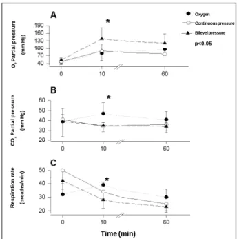
The period of time during which the respiratory support was used did not significantly differ among nonintubated patients, and the oxygen group did not have its time measured. Respiration rates (breaths per minute) at the time of randomization were 32±11 in group I, 50±10 in group II, and 42±6 in group III. At 10 minutes, the respiration rates were 39±2 in group I, 34±5 in group II, and 28±6 in group III. At 60 minutes, the respiration rates were 30±6 in group I, 25±5 in group II, and 23±4 in group III. The difference between group III and the 2 other groups was significant at 10 minutes (fig. 1).
Heart rates (bpm) at randomization were 120±36 in group I, 101±13 in group II, and 75±15 in group III. At 10 minutes, heart rates were 112±19 in group I, 118±22 in group II, and 106±29 in group III. At 60 minutes, heart rates were 100±15 in group I, 89±16 in group II, and 84±16 in group III. This difference was not significant.
Systolic blood pressures (mm Hg) at randomization were 173±48 in group I, 169±40 in group II, and 139±44 in
group III. At 10 minutes, systolic blood pressures were 163±35 in group I, 149±30 in group II, and 139±30 in group III. At 60 minutes, systolic blood pressures were 123±27 in group I, 122±8 in group II, and 124±17 in group III. This difference was not significant.
Diastolic blood pressures (mmHg) at randomization were 102±23 in group I, 100±36 in group II, and 114±23 in group III. At 10 minutes, diastolic blood pressures were 94±16 in group I, 95±25 in group II, and 91±25 in group III. At 60 minutes, diastolic blood pressures were 82±8 in group I, 70±9 in group II, and 82±17 in group III. A difference oc-curred at 60 minutes, when the results of the continuous positive pressure group were significantly lower compared with those of the other 2 groups. This may have happened casually due to the small sample.
Partial oxygen pressures (mmHg) at randomization we-re 46±11 in group I, 47±12 in group II, and 53±9 in group III. At 10 minutes, partial oxygen pressures were 80±29 in group I, 89±29 in group II, and 138±43 in group III. At 60 minutes, partial oxygen pressures were 95±24 in group I, 77±17 in group II, and 121±37 in group III. The difference was statistically significant at 10 minutes, when the partial oxy-gen pressure in group III was higher than that in the other 2 groups, which did not differ among themselves (fig. 1).
Partial carbon dioxide pressures (mmHg) at randomi-zation were 39±7 in group I, 41±11 in group II, and 39±15 in group III. At 10 minutes, partial carbon dioxide pressures were 47±11 in group I, 34±4 in group II, and 35±6 in group III. At 60 minutes, partial carbon dioxide pressures were 41±8 in group I, 36±2 in group II, and 34±6 in group III. At 10 minutes, the results in group I were significantly higher than those in groups II and III (fig. 1).
Table I – Distribution of patients according to the ventilatory modality, duration of mask use, and causes of acute pulmonary edema
Group Oxygen Continuous pressure Bilevel pressure
N 10 9 7
Duration of * 170 ± 90 ’ 155 ± 38 ‘ mask use
Etiology
2 acute myocardial 1 acute myocardial 1 acute myocardial infarctions infarction infarction 5 hypertensive 5 hypertensive 4 hypertensive
emergencies emergencies emergencies 1 acute ischemic 2 acute ischemic 2 acute ischemic
heart disease heart diseases heart diseases 1 undetermined
1 infectious endocarditis
* time not measured
Fig. 1 – Partial oxygen pressure was higher and respiration rate was lower in the bilevel positive pressure ventilation group as compared with the other groups at 10 minutes, when the group treated with oxygen therapy had a higher value for PaCO2 as compared with the other groups (p<0.05).
O2
P
a
rt
ia
l
p
re
s
s
u
re
(m
m
H
g
)
C
O2
P
a
rt
ia
l
p
re
s
s
u
re
(m
m
H
g
)
R
e
s
p
ir
a
ti
o
n
r
a
te
(b
re
a
th
s
/m
in
)
Time (min)
Oxygen
Continuous pressure
Bilevel pressure
At randomization, pH was 7.33±0.05 in group I, 7.30±0.1 in group II, and 7.35±0.12 in group III. At 10 minu-tes, pH was 7.23±0.08 in group I, 7.38±0.05 in group II, and 7.36±0.07 in group III. At 60 minutes, pH was 7.35±0.04 in group I, 7.41±0.02 in group II, and 7.38±0.06 in group III. At 10 minutes, acidosis occurred in group I, but not in the other 2 groups. This fact was also associated with an increa-sed PaC O
2 in group I at 10 minutes; therefore, we conclude that the origin of this alteration may be predominantly respiratory.
In regard to the need for tracheal intubation and deaths, 4 patients were intubated in the oxygen group, 3 patients in the continuous positive pressure group, and none in the bilevel positive pressure ventilation group. One death occurred in the continuous positive pressure group 3 days after the acute event, and it was caused by a new acute myocardial infarction.
Discussion
Our results indicate that the patients with acute pul-monary edema of cardiogenic origin benefited from the noninvasive ventilation with 2 pressure levels (noninvasive bilevel positive pressure ventilation). This benefit was evident not only by assessment of the analyzed data but also by the good acceptance by and cooperation of the pa-tients, in addition to the report of improvement of dyspnea. Even though the use of continuous positive pressure has also had good acceptance by the patients, this was not as immediate as that for the patients treated with the noninva-sive bilevel positive pressure ventilation. Even though the patients received standardized medical assistance in the 3 groups, only the patients randomized to the bilevel positive pressure ventilation modality needed less tracheal intuba-tion as compared with those of the remaining groups. In addition, the respiration rate, the PaO2, the PaCO2, and the pH of the patients undergoing bilevel positive pressure ventilation improved more rapidly. On the other hand, patients treated with continuous positive airway pressure showed an intermediate result as compared with the other groups, because they had a more rapid improvement in vital signs and in blood gases only when compared with the group on oxygen. Our results are consistent with our initial hypothesis. The bilevel positive pressure ventilation modality combines the beneficial effects of intrathoracic positive pressure, provided by the continuous positive airway pressure modality, and the ventilatory assistance, provided by the additional inspiratory pressure.
In our study, we used relatively low pressure levels (mean continuous positive pressure of 7.5cm H2O, mean expiratory positive pressure of 4cm H2O, and mean inspira-tory pressure of 12cm H2O). We aimed to assess whether lower pressures would have a lower effect in preload, which, theoretically, would expose the patient to a lower risk of hypotension. Most studies available in the literature used continuous positive airway pressure values of 10cm H2O 1,7. Ideal levels of continuous positive pressure to be used in
the treatment of acute pulmonary edema have not yet been established. A recent study suggests that a final expiratory pressure of 10cm H2O in severely ill patients with significant ventricular dysfunction improves heart work without im-pairing the cardiac index 7. This level of pressure determines a low transmural pressure in the ventricular wall and an ef-ficient recruiting of collapsed alveolar units. This recruiting improves the pulmonary shunt, reduces hypoxemia, and increases the residual functional capacity and pulmonary compliance. In addition, the use of positive pressure in-creases the caliber of the airways, leading to a decrease in their resistance 9,11,12. All these effects result in a reduction in the ventilatory and cardiac work in this phase, where the respiratory stress may cause muscle fatigue and death by relative hypoventilation 13. However, these beneficial ef-fects of the positive pressure in the airways may be can-celled by the noxious action on heart preload, which may cause hypotension. In our study, the patients undergoing continuous positive airway pressure required a number of intubations similar to that required by patients in the oxy-gen group. This may be explained because of the relatively low number of patients and also the low values of positive pressure used.
Bilevel positive pressure ventilation as a modality of treatment for acute pulmonary edema has been studied little. Only one study 14 exists in the literature comparing, in a prospective and randomized way, the effects of con-tinuous positive airway pressure with bilevel positive pressure ventilation in the treatment of acute pulmonary edema. This study shows that patients who underwent bi-level positive pressure ventilation had more marked im-provement as compared with patients who underwent con-tinuous positive airway pressure. Unfortunately the study was interrupted because of an unexpected result; the group treated with bilevel positive pressure ventilation had a sig-nificantly greater number of patients with acute myocardial infarction. The causes of this result, which led to the early interruption of the study, are unknown. The authors raised the hypothesis that the group treated with bilevel positive pressure ventilation had a significant drop in blood pressure. They speculate that they may have used very high pressure values in the airways (inspiratory pressure of 15cm H2O and expiratory pressure of 5cm H2O), which may have led to a significant reduction in cardiac preload, hypotension, and consequent worsening of cardiac is-chemia. In our study, we used an inspiratory pressure of 8cm H2O and an expiratory pressure of 3cm H2O, ie, mean pres-sures equivalent to those of the group treated with conti-nuous positive airway pressure.
also because our case series was small. Studies on modali-ties of ventilatory support using noninvasive pressure and their methodology should continue with larger case series
References
1. Bersten AD, Holt AW, Vedig AE, Skowronski GA, Baggoley CJ. Treatment of severe pulmonary edema with continuous positive airway pressure delivered by face mask. N Engl J Med 1991; 325: 1825-30.
2. Sachetti AD, Harris RH, Paston C. Bi – level positive airway pressure support system use in acute congestive failure: Preliminary case series. Acad Emerg Med 1995 ; 2: 714-8.
3. Lin M, Chiang HT. The efficacy of early continuous airway pressure therapy in patients with acute cardiogenic pulmonary edema. J Formosan Med Assoc 1991; 90: 736-43.
4. Lin M, Yang YF, Chiang HT, Chang MS, Chiang BN, Cheitlin MD. Reapraisal of continuous positive airway pressure therapy in acute cardiogenic pulmonary edema – Short term results and long term follow – up. Chest 1995; 107: 1379-86. 5. Pang D, Kenan SP, Cook DJ, Sibbald WJ. The effect of positive airway support on mortality and the need of intubation in cardiogenic pulmonary edema – a systematic review. Chest 1998; 114: 1185-91.
6. Buda A, Pinsky MR, Ingels NB, Daughters GT, Stinson EB, Alderman EL. Effect of intrathoracic pressure on left ventricular performance. N Eng J Med 1979; 301: 453-9.
7. Lenique F, Habis M, Lofaso F, Dubois-Randé JL, Harf A, Brochard L. Ventilatory and hemodynamic effects of continuous positive airway pressure in left heart failure. Am J Resp Crit Care Med 1997; 155: 500-5.
8. Montner PK, Greene ER, Murata GH, Stark DM, Timms M, Chick TW. Hemo-dynamic effects of nasal and face mask continuous positive airway pressure. Am J Respir Crit Care Med 1994; 149: 1614-8.
9. Sharp JT, Grifffith GT, Bunnell IL, Greene DG. Ventilatory mechanics in pulmonary edema in man. J Clin Invest 1958; 37: 111-7.
and higher pressures to better elucidate their potential benefits in the treatment of patients with acute pulmonary edema of cardiogenic origin in emergency services.
10. Advanced Cardiac Life Support. 2th ed. Dallas. American Heart Association. Hypotension/Shock/Pulmonary Edema 1997; 1.40 - 1.47.
11. Jones JG, Lemen R, Graf PD. Changes in airway calibre following pulmonary venous congestion. Br J Anaesth 1978; 50: 743-51.
12. Blomqvist H, Wickerts CJ, Berg B, Frostell C, Jolin A, Hedenstierna G. Does PEEP facilitate the resolution of extravascular lung water after experimental hydrostatic pulmonary edema? Eur Resp J 1991; 4: 1053-59.
13. Aubier M, Trippenbach T, Roussos C. Respiratory muscle fadigue during cardiogenic shock. J Appl Physiol 1981; 51: 499-508.
14. Mehta S, Jay GD, Woolard RH, Hipona RA, Conolly EM, Cimini DM, Drinkwine JH, Hill NS. Randomized, prospective trial of bilevel versus continuous positive airway pressure in acute pulmonary edema. Crit Care Med 1997; 25: 620-8. 15. Barbas CSV, Bueno MAS, Amato MBP, Hoelz C, Rodrigues-Junior M. Interação
cadiopulmonar durante a ventilação mecânica. Rev Soc Cardiol Estado de São Paulo 1998; 3: 28-41.
16. Rasanen J, Heikkila J, Downs J, Nikki P, Vaissanen I, Viitanen A. Continuous positive airway pressure by face mask in acute cardiogenic pulmonary edema. Am J Cardiol 1985; 55: 296-300.
17. Philip–Joet FF, Paganelli FF, Dutau HL, Saadjian AY. Hemodynamic effects of bilevel nasal positive airway pressure ventilation in patients with heart failure. Respiration 1999; 66: 136-43.
18. Hoffmann B, Welte T. The use of noninvasive pressure support ventilation for severe respiratory insufficiency due to pulmonary oedema. Intensive Care Medicine 1999; 25: 15-20.