Effect of Penicillin G Every Three Weeks on Oral Microflora by
Penicillin Resistant Viridans Streptococci
André Andrade de Aguiar
1,2, Roney Orismar Sampaio
1,2, Jorge Luiz de Mello Sampaio
3, Guilherme Sobreira
Spina
1,2, Ricardo Simões Neves
1,4, Luiz Felipe Pinho Moreira
1,5, Max Grinberg
1,2Universidade de São Paulo1; Departamento de Doença Valvular – Instituto do Coração (InCor)2; Fleury Medicina Diagnóstica3; Departmento de Odontologia – Instituto do Coração (InCor)4; Departmento de Cirurgia Torácica – Instituto do Coração (InCor)5, São Paulo, SP – Brazil
Abstract
Background: Benzathine penicillin G every 3 weeks is the standard protocol for secondary prophylaxis for recurrent
rheumatic fever.
Objective: Assess the effect of Benzathine penicillin G on Streptococcus sanguinis and in patients
with cardiac valvular disease due to rheumatic fever receiving secondary prophylaxis.
Methods: Oral streptococci were evaluated before (baseline) and 7 days (day 7) after Benzathine penicillin G in 100
patients receiving routine secondary rheumatic fever prophylaxis. Saliva samples were evaluated for colony count and presence of S. sanguinis and S. oralis. Chewing-stimulated saliva samples were serially diluted and plated onto both nonselective and selective 5% sheep blood agar containing penicillin G. The species were identified using conventional biochemical tests. Minimal inhibitory concentrations were determined with the Etest.
Results: No statistical differences were found in the presence of S. sanguinis comparing baseline and day 7 (p =
0.62). However, the existing number of positive cultures of S. oralis on day 7 after Benzathine penicillin G presented a significant increase compared to baseline (p = 0.04). No statistical difference was found between baseline and day 7 concerning the number of S. sanguinis or S. oralis CFU/mL and median minimal inhibitory concentrations.
Conclusion: This study showed that Benzathine penicillin G every 3 weeks did not change the colonization by S.
sanguinis, but increased colonization of S. oralis on day 7 of administration. Therefore, susceptibility of Streptococcus sanguinis and Streptococcus oralis to penicillin G was not modified during the penicillin G routine secondary rheumatic fever prophylaxis. (Arq Bras Cardiol 2012;98(5):452-458)
Keywords: Penicilin G benzathine/therapeutic use; rheumatic fever; heart valve diseases; mouth; Viridans streptococcus; Streptococcus oralis.
Mailing Address: André Andrade de Aguiar •
Rua Botucatu, 572 - conjunto 31 - Vila Clementino – 04023-061 - São Paulo, SP – Brazil
E-mail: drandreaguiar@yahoo.com.br
Manuscript received July 05, 2011; revised manuscript received July 18, 2011; accepted December 01, 2011.
RF with BPG. Also, none of the studies have assessed this issue related to S. sanguinis and S. oralis, which are predominant species recovered in IE7,8.
BPG prophylaxis protects valvular heart disease patients of new RF recurrences. However, no study has evaluated the oral flora with an expressive casuistic and specificity for these pathogens.
Therefore, the aim of this study is to evaluate if there is an association between BPG prophylaxis and the colonization of oral cavity by penicillin resistant Viridans Streptococci.
Methods
Cohort
A hundred patients were selected and evaluated in 2 periods:
BPG baseline and day 7 – One hundred RF patients previously receiving a secondary prophylaxis regimen with benzathine penicillin G 1,200,000 IU for at least 6 months before study admission. Patients with rheumatic activity or patients with diagnosis of infection were excluded.
Introduction
Rheumatic fever (RF) is the leading cause of valvular heart disease in developing countries. RF causes significant morbidity and mortality, causing 90% of cardiac surgeries in children and over 30% of cardiac surgeries in adults. Secondary prophylaxis of 1,200,000 U benzathine penicillin G (BPG) every 3 weeks is the standard regimen for the prevention of recurrent rheumatic fever in developing countries. Valvular sequelae is the most dreadful consequence of acute rheumatic fever, and such cardiac lesions can also predispose a patient to infective endocarditis (IE), a morbid disease that worsens the prognosis of these patients1-4.
In spite of the widely recommended secondary prophylaxis of RF with BPG every 3 weeks2,5,6, very few studies have assessed
All patients were assessed clinically, and they underwent oral cavity inspection to avoid admission of patients with acute systemic or oral infection.
This study was approved by the institutional ethics committee, and all study patients gave informed consent.
Sample collection, transport, and culture
Samples were collected at baseline (100 samples) and day 7 (100 samples) after BPG prophylactic dosage. Saliva samples were obtained from patients who chewed paraffin tablets9 and
were collected into a sterile disposable plastic vial. Approximately 1 mL was transferred to a vial containing 5.7 mL of Göteborg viability medium, anaerobically prepared and sterilized (VMGA II S)10. Samples in transport medium were maintained at 2ºC to
8ºC before plating. Saliva samples were serially diluted tenfold in phosphate-buffered solution pH 7.2 before plating onto Columbia nalidixic agar plus 5% sheep blood (CNASB), CNASB plus penicillin G (0.25 µg/mL)11, and amphotericin B (0.5 µg/
mL)12-15. Media were incubated at 35ºC in 5% CO
2 for 72 h and
then analyzed for colony count for each colonial morphotypes.
Species identiication
Each colony morphotype was subcultivated onto tryptic soy agar plus 5% sheep blood (SBA) and tested for catalase and Gram stain. Catalase negative Gram-positive cocci were subsequently evaluated for the following characteristics/substrate utilization: hemolysis, arginine, urea, L-pyrrolidonyl-β-naphthylamide and esculin hydrolysis, β-N-acetyl glycosidase, α-D-glycosidase, β -N-acetyl galactosidase, optochin susceptibility, Voges-Proskauer test, and acid production from inulin, mannitol, raffinose, and melibiose, as recommended16.
Susceptibility testing
Minimal inhibitory concentration (MIC) for penicillin G was determined using the Etest as recommended by the manufacturer (AB Biodisk, Solna, Sweden). Results were interpreted as recommended by the Clinical and Laboratory Standards Institute (CLSI)11.
Statistical analysis
The Logistic Regression model was used to check the group’s association with the occurrence of S. sanguinis and S. oralis with age and sex corrections.
The qualitative analysis was performed using the Mc Nemar nonparametric test.
Quantitative analysis was performed with the use do the Wilcoxon nonparametric test.
Statistical significance of 5% was applied for all tests.
Results
The group studied was composed of 100 patients, 38 males, aged 10 to 53 years old (26.5 ± 8 years).
In the group, 54 patients had mitral regurgitation, 27 had aortic regurgitation, 25 had mitral stenosis, and 14 had aortic stenosis. Eight patients had biological mitral prosthesis, 3 had biological
aortic prosthesis, 2 had mechanical mitral prosthesis, and 1 had mechanical aortic prosthesis.
There were no differences comparing the number of positive cultures existed for S. sanguinis at baseline and day 7 after BPG (p = 0.62, Table 1). However, the existing number of positive cultures for S. oralis at day 7 after BPG presented a significant increase compared to baseline (p = 0.04, Table 1).
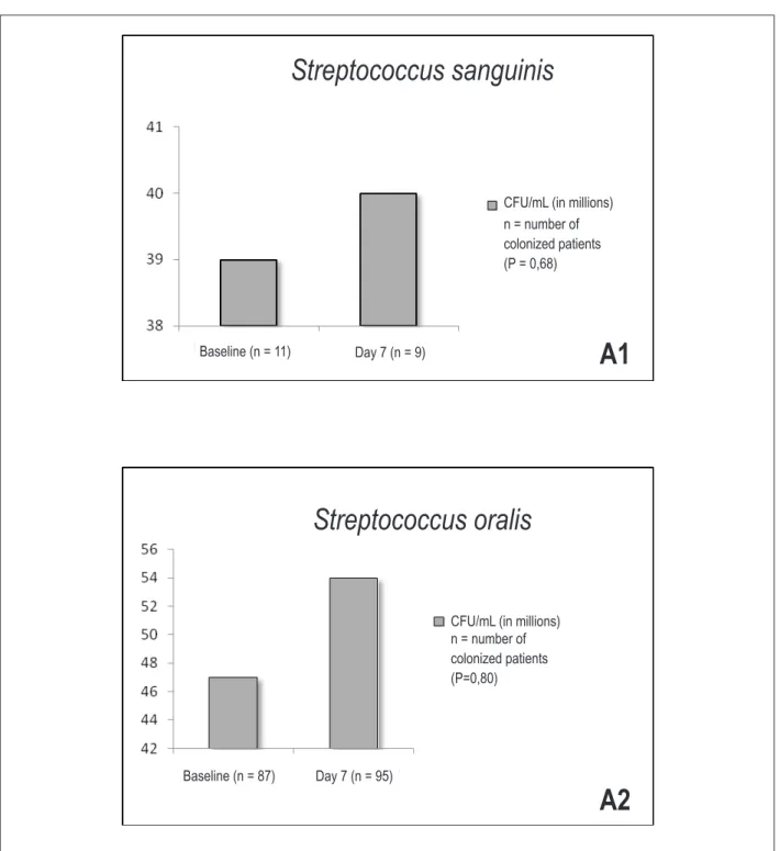
The assessments of the CFU/mL number in saliva of the patients at baseline and day 7 after BPG were subdivided into S. sanguinis and S. oralis.
The CFU/mL values in saliva for S.sanguinis and S. oralis did not differ between baseline and day 7 after BPG (p = 0.68 and p= 0.80 respectively; Figure 1).
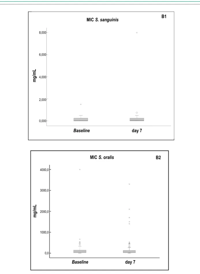
The minimal inhibitory concentrations for penicillin G values were subdivided into S. sanguinis and S. oralis. No statistical difference was found between baseline and day 7 after BPG concerning the MIC of S. sanguinis and S. oralis (p = NS; Figure 2).
Table 2 shows data as MIC50 and MIC90, which represents the minimal inhibitory concentrations for penicillin G to inhibit 50% and 90%, respectively, of the susceptible Streptococcus sanguinis and Streptococcus oralis to penicillin G.
Discussion
This group of patients with valvular heart disease is predisposed to infective endocarditis. Infective endocarditis results from bacteremia, often related to oral infectious focuses. Viridans streptococci are the predominant group recovered in IE, particularly Streptococcus sanguinis and Streptococcus oralis. The effect of chronic BPG has not been studied with specificity to these pathogens yet.22,23
The study cohort had a prevalence of females (62/100), possibly due to the higher general occurrence of RF in women17.
Despite the gender heterogeneity, it did not represent a bias because no statistical difference existed (p = NS) between the occurrence of S. sanguinis an S.oralis in relation to the patients age and sex when we applied the logistic regression model. Furthermore, no statistical difference existed among the rate of tooth cavities, loss of teeth, or teeth with fillings between sexes in the study population18. All patients had their oral
cavities inspected according to the criteria of the World Health Organization19 to exclude the presence of oral infections, which
could affect the oral microbiota16,20-22.
S. sanguinis and S. oralis are common commensals of the oral cavity present in the initial formation of dental bacterial plaque, representing nearly 80% Streptococcus in this phase20-23. The BPG
1,200,000 IU IM dose exercises as a bactericide effect on the Streptococcus, sensitive in the active multiplication phase, which, hypothetically, would cause a decrease in these microorganisms in the oral cavity in this group of patients.
S. sanguinis was also observed in both periods in a limited number of patients, similarly to that observed by Bilavsky et al7.
No difference (P=0.62) of its presence was noticed in the study groups. This showed that BPG did not interfere with the species growth. However, other studies reported a greater number of samples of S. sanguinis20,21, perhaps due to the use of classic
Table 1 – Positive cultures for Streptococcus sanguinis and Streptococcus oralis
Groups Streptococcus sanguinis Streptococcus oralis
Baseline* 11 87
Day 7† 9 95‡
*Patients before benzathine penicillin G dosage; †Patients under benzathine penicillin G 7 day effect; ‡p = 0.04.
Figure 1 – A1 - Distribution of CFU/mL values in saliva in patients colonized by S. sanguinis; A2 - distribution of CFU/mL values in saliva in patients colonized by S. oralis.
CFU/mL (in millions) n = number of colonized patients (P = 0,68)
Baseline (n = 11) Day 7 (n = 9)
Streptococcus sanguinis
CFU/mL (in millions) n = number of colonized patients (P=0,80)
Baseline (n = 87) Day 7 (n = 95)
Streptococcus oralis
A1
Table 2 – MIC50 and MIC90 (µg/mL) and group distribution according to Streptococcus sanguinis and Streptococcus oralis susceptibility to penicillin
Baseline‡ Day 7§
S. sanguinis S. oralis S. sanguinis S. oralis
MIC50* 0.190 0.250 0.250 0.250
MIC90† 0.380 1.000 0.500 1.000
Sensitive 45.5% (5/11) 44.8% (39/87) 33.3% (3/9) 41.1% (39/95)
Intermediary resistance 45.5%
(5/11) 55.2% (48/87) 66.7% (6/9) 58.9% (56/95)
High level resistance 9%
(1/11) - -
-*MIC50 – minimal inhibitory concentration for penicillin G to inhibit 50% of the susceptible Streptococcus sanguinis and Streptococcus oralis. †MIC90 – minimal inhibitory concentration for penicillin G to inhibit 90% of the susceptible Streptococcus sanguinis and Streptococcus oralis. ‡Patientsbefore benzathine penicillin G dosage. §Patients under benzathine penicillin G 7 day effect. P = NS.
standard screening tests for the genre). Nevertheless, in this study the methodology described by Ruoff et al16 was adopted,
performing biochemical tests with the addition of 3 fluorogenic substrates, increasing the specificity in the identification of the species.Although we have used the best methodology available for the identification of these species, it is known that there are some limitations, since genetic sequencing would certainly be the best method to be applied. However, this would be financially impractical due to the sample size, probably leading to no difference in the result of the study.
In regard to obtaining the samples through stimulated saliva and not from the dental plaque, this was due to an overall knowledge that stimulated saliva is a better collection method for its homogeneity and logistics compared to the collection of dental plaque samples24.
As for the S. oralis, we noticed a significantly higher number of these microorganisms in the BPG-day 7 vs. baseline (p = 0.04, Table 1). The saliva sample collected from BPG- day 7 occurred during the time of the greatest amount of serum drug concentration according to Decourt et al25. This is evidence of the
lack of BPG influence on the most prevalent microorganism in the bacterial plaque and one of the main Infectious Endocarditis etiological agents20,21,26.
In this study, we found no statistical difference concerning saliva UFC/mL numbers (Figure 1, p = NS) and MICs (MIC50 and
MIC90) of S. sanguinis and S. oralis (Table 2).
The chronic use of benzathine penicillin G in the study group did not significantly alter S. sanguinis and S. oralis susceptibility to penicillin G, as seen previously7. It is interesting to note the
occurrence of an increase in the resistance, or MIC values, in Viridans Streptococcus and, therefore, an increase in the number of strains resistant to antibiotics isolated into positive hemocultures for IE. However, these patients were receiving oral antibiotic therapy, and MIC intervals differ from those standardized by CLSI for penicillin G7,27,28.
Our findings regarding the identification of species corroborate the results of Bilavsky et al7. However, we disagree with the
increased resistance to penicillin G of Viridans Streptococcus.
This study contributed to awake a inquiry regarding the clinical treatment of these patients. Although the American Heart Association (AHA) no longer recommends antibiotic prophylaxis prior to procedures that cause bacteremia in these patients, we believe that further studies are required aimed at the Brazilian population to know our reality, and then adapt it to changes suggested by the AHA. While we are awaiting these data, it would be prudent to continue to implement the recommendations of the 1997 AHA guidelines, in which prophylaxis for infective endocarditis is recommended for patients with rheumatic heart disease prior to procedures that cause bacteremia in the genitourinary tract, respiratory tract and the stomatognathic system.
Our study also contributes to the interface between medicine and dentistry, because it shows that under the prolonged action
of PGB, there is no decrease of the main microorganisms
involved in the etiology of infective endocarditis and, therefore, antibiotic prophylaxis prior to procedures that cause bacteremia may be necessary.
Hence, additional studies are necessary to verify the need to establish a special routine so that these patients can undergo dental procedures that could cause bacteremia on the 7th day
following the administration of BPG.
This study showed that BPG every 3 weeks did not change the colonization by S. sanguinis; but BPG increased S. oralis colonization on day 7 following its administration and, finally, susceptibility of Streptococcussanguinis and Streptococcusoralis to penicillin G was not altered during the penicillin G cycle.
Acknowledgments
The group wishes to thank Dr. Luiz Antônio Machado César for his contribution regarding data collection. The group also wishes to thank Dr. Walter Niccoli Filho for his assistance in the preparation of this manuscript.
Potential Conflict of Interest
Sources of Funding
This study was funded by FAPESP and CNPq and partially funded by Instituto Fleury.
Study Association
This article is part of the thesis of doctoral submitted by André Andrade de Aguiar, from Faculdade de Medicina da USP.
References
1. Ayoub EM, Kotb M, Cunningham MW. Rheumatic fever pathogenesis. In: Stevens DL, Kaplan EL. (eds.). Streptococcal infections: clinical aspects, microbiology and molecular pathogenesis. New York: Oxford Universe Press; 2000. p. 102-31.
2. Dajani AS, Bisno AL, Chung KJ, Durack DT, Gerber MA, Kaplan EL, et al. Prevention of rheumatic fever: a statement for health professionals by the Committee on Rheumatic Fever, Endocarditis and Kawasaki Disease of the Council on Cardiovascular Disease in the young, the American Heart Association. Pediatr Infect Dis J. 1989;8(5):263-6.
3. Sampaio RO, Fae KC, Demarchi LM, Pomerantzeff PM, Aiello VD, Spina GS, et al. Rheumatic heart disease: 15 years of clinical and immunological follow-up. Vasc Health Risk Manag. 2007;3(6):1007-17.
4. Snitcowsky R. Rheumatic fever prevention in industrializing countries: problems and approaches. Pediatrics. 1996;97(6 Pt 2):996-8.
5. Currie BJ. Are the currently recommended doses of benzathine penicillin G adequate for secondary prophylaxis of rheumatic fever? Pediatrics. 1996;97(6 Pt 2):989-91.
6. Kassem AS, Zaher SR, Abou Shleib H, el-Kholy AG, Madkour AA, Kaplan EL. Rheumatic fever prophylaxis using benzathine penicillin G (BPG): two- week versus four-week regimens: comparison of two brands of BPG. Pediatrics. 1996;97(6 Pt 2):992-5.
7. Bilavsky E, Eliahou R, Keller N, Yarden-Bilavsky H, Harel L, Amir J. Effect of benzathine penicillin treatment on antibiotic susceptibility of viridans streptococci in oral flora of patients receiving secondary prophylaxis after rheumatic fever. J Infect. 2008;56(4):244-8.
8. Darhous MS, Dahab OM, el Atar E, el Ghafary E. Dental, oral and bacteriological aspects in patients at risk of subacute bacterial endocarditis. Egypt Dent J. 1993;39(4):533-9.
9. Jensen JL, Karatsaidis A, Brodin P. Salivary secretion: stimulatory effects of chewing-gum versus paraffin tablets. Eur J Oral Sci. 1998;106(4):892-6.
10. Dahlen G, Pipattanagovit P, Rosling B, Moller AJ. A comparison of two transport media for saliva and subgingival samples. Oral Microbiol Immunol. 1993;8(6):375-82.
11. Wayne PA. CLSI. Performance Standards for Antimicrobial Susceptibility Testing; Eighteenth Informational Supplement. M100-S18. Clinical and Laboratory Standards Institute. MO2-A10. 2009;29(1):1-53.
12. Gold OG, Jordan HV, Van Houte J. A selective medium for Streptococcus
mutans. Arch Oral Biol. 1973;18(11):1357-64.
13. Murray PR. Manual of clinical microbiology. Washington (DC):ASM Press; 2003.
14. Pezzlo M. Aerobic bacteriology. In: Isenberg HD. (ed.). Clinical microbiology procedures handbook. Washington, (DC): American Society for Microbiology Press; 1992. p. 1.20.5-1.20.6.
15. Sanchez-Perez L, Acosta-Gio AE. Caries risk assessment from dental plaque and salivary Streptococcus mutans counts on two culture media. Arch Oral Biol. 2001;46(1):49-55.
16. Ruoff KL, Whiley RA, Beighton D. Streptococcus. In: Murray PR. (ed.). Manual of clinical microbiology. Washington (DC): ASM Press; 2003. p. 405-21.
17. Neves RS, Neves IL, Giorgi DM, Grupi CJ, César LA, Hueb WA, et al. Efeitos do uso da adrenalina na anestesia local odontológica em portador de coronariopatia. Arq Bras Cardiol. 2007;88(5):545-51.
18. Ministério da Saúde. Datasus. Levantamento epidemiológico em saúde bucal: Brasilia;1996.
19. WHO. Oral health surveys. Basic methods. Geneva, 1977.
20. de Almeida PF, Franca MP, Santos SP, Moreira RS, Tunes UR. Microbiota estreptocócica associada com a formação inicial da placa dental. R Ci Med Biol. 2002;1(1):33-41.
21. Frandsen EV, Pedrazzoli V, Kilian M. Ecology of viridans streptococci in the oral cavity and pharynx. Oral Microbiol Immunol. 1991;6(3):129-33.
22. Herzberg MC. Oral Streptococci in health and disease. In: Stevens DL, Kaplan EL. (eds.). Streptococcal infections: clinical aspects, microbiology and molecular pathogenesis. New York: Oxford Universe Press; 2000. p. 333-70.
23. Ito HO. Infective endocarditis and dental procedures: evidence, pathogenesis, and prevention. J Med Invest. 2006;53(3-4):189-98.
24. Motisuki C, Lima LM, Spolidorio DM, Santos-Pinto L. Influence of sample type and collection method on Streptococcus mutans and Lactobacillus spp. counts in the oral cavity. Arch Oral Biol. 2005;50(3):341-5.
25. Decourt LV, Santos SR, Snitcowsky R, Pileggi F, Tsuzuki H, de Araujo Abreu AM, et al. [Serum levels of benzathine penicillin G after intramuscular administration]. Arq Bras Cardiol. 1983;40(1):3-8.
26. Jeon EH, Han JH, Ahn TY. Comparison of bacterial composition between human saliva and dental unit water system. J Microbiol. 2007;45(1):1-5.
27. Erickson PR, Herzberg MC. Emergence of antibiotic resistant
Streptococcus sanguis in dental plaque of children after frequent
antibiotic therapy. Pediatr Dent. 1999;21(3):181-5.


