Is Abnormal Adrenergic Activation Associated with Abnormal Heart
Rate Recovery?
Leandro Rocha Messias
1, Maria Angela M. de Queiroz Carreira
1, Sandra Marina Miranda
1, Jader Cunha de
Azevedo
1,2, Isabela Ambrósio Gava
1, Ronaldo Campos Rodrigues
1, Elisabeth Maróstica
1, Claudio Tinoco Mesquita
1, Universidade Federal Fluminense1, Niterói, RJ; Hospital Procardíaco2, Rio de Janeiro, RJ – BrazilAbstract
Background: Heart Rate Recovery (HRR) reflects the capacity of the cardiovascular system to reverse the vagal withdrawal caused by exercise. Scintigraphy with metaiodobenzylguanidine (I¹23 MIBG) evaluates innervation and
cardiac adrenergic activation. The association of these two methods is not well established.
Objective: To evaluate the association between HRR and washout rate (WO) of I¹²³ MIBG in patients with heart failure (HF).
Methods: wenty-five patients with ejection fraction ≤ 45% underwent exercise testing, and analysis of the variation
of HRR from the 1st to the 8th minute after exertion. Submitted to I¹²³ MIBG, they were separated into groups by
WO: G1) <27% and G2) ≥ 27%. For the statistical analysis Mann-Whitney’s U test and Spearman’s correlation coefficient were used.
Results: G2 showed a slower variation of HRR: 1st minute: G1: 21.5 (16.12 to 26.87) vs. G2: 11.00 (8.5 to 13.5) bpm,
p = 0.001; 2nd minute: G1: 34 (29-39) vs. G2: 20 (14 - 26) bpm, p = 0.001; 3rd minute: G1: 46 (37.8 – 54.1) vs. G2:
30 (22 – 38) bpm, p = 0.005; 5th minute: G1: 51.5 (42 - 61) vs. G2: 39 (31.5 to 46.5) bpm, p = 0.013, and in the 8th
minute: G1: 54.5 (46.5 – 62.5) vs. G2: 43 (34 – 52) bpm, p = 0.037. HRR in the 1st (r = -0.555, p = 0.004), and in
the 2nd minute (r = -0.550, p = 0.004) were negatively correlated with WO.
Conclusion: Patients with high HF and WO showed an abnormal HRR compared with patients with normal WO. These findings suggest that adrenergic activation may influence the HRR. (Arq Bras Cardiol 2012;98(5):398-405)
Keywords: Heart rate; stroke volume; heart failure; heart/radionuclide imaging
Mailing Address: Leandro Rocha Messias •
Rua Noronha Torrezão, 46, Apto 805, Santa Rosa – 24240-182 – Niterói, RJ - Brazil
E-mail: [email protected], [email protected]
Manuscript received September 28, 2011; revised manuscript received September 28, 2011; accepted December 26, 2011.
Fraction (LVEF)13 and the severity of coronary lesions on coronary angiography14.
C a r d i a c a c t i v i t y a n d s y m p a t h e t i c i n n e r v a t i o n can be assessed by myocardial scintigraphy with metaiodobenzylguanidine labeled with iodine 123 (I123 MIBG)15,16. Studies have shown17-19 that early imaging is the integrity of presynaptic nerve terminals, and the density of the beta-adrenergic receptors. The presynaptic neuronal uptake contributes to a later imaging, combining information of neural function, including capture, storage and release of norepinephrine in the presynaptic vesicles. The washout rate (WO) assesses the degree of sympathetic activity. Patients with heart failure may have: (1) reduced tracer uptake due to loss of sympathetic neurons and/ or disorders in primary uptake of noradrenalin; and (2) increased WO, reflecting the shedding of noradrenalin to the blood stream15,17,20-22. WO rates greater than 27% have been described as a strong predictor of shorter survival in patients with cardiac failure23.
So far, there is no evidence that abnormalities found in I123 MIBG scintigraphy are associated with abnormalities in HRR post-exercise. The purpose of this study is to evaluate the association between HRR and WO of I¹²³ MIBG in patients with heart failure, comparing the behavior of HRR in patients with normal and abnormal WO.
Introduction
Methods
We conducted an observational and transversal study, selecting 25 consecutive patients treated in outpatient care specializing in heart failure. The selected patients had heart failure (diagnosed by the use of Framingham24 criteria) and Left Ventricular Ejection Fraction (LVEF) smaller than or equal to 45% measured by echocardiography by Simpson’s technique. The study excluded patients who had atrial fibrillation, diabetes, heart valve disease, patients with ventricular stimulation device, endocrine disorders, Parkinson’s disease, pregnant or breastfeeding women. No medication was suspended for this study. Volunteers signed a consent form agreeing to be part of the project. The project was approved by the Ethics Committee in Research of the University, under number 011/09.
To classify the etiology of heart failure, we have used the following criteria: ischemic (history of myocardial infarction, or presence of inactive zone on electrocardiogram, or coronary angiography showing left coronary artery damage greater than or equal to 50%, or damage greater than or equal 70% in one of three main systems - anterior descending, circumflex and right coronary artery25), hypertension (history of hypertension and absence of criteria for ischemic etiology) and others (the latter involving patients who were neither classified as ischemic, nor as hypertensive: for example, idiopathic dilated cardiomyopathy, post-myocarditis, peripartum).
Patients underwent myocardial scintigraphy with I¹²³ MIBG to assess cardiac adrenergic innervation upon uptake of radiotracer, with study of the heart to mediastinum ratio (H/M) for early images (30 minutes) and late images (4 hours) besides the calculation of WO {WO = [(early H-M - late H-M) / (early H-M)] x 100}14. All scintigraphic examinations were performed in Anger type digital tomographic scintillation camera (Single Photon Emission Computed Tomography), Siemens E-Cam dual detector model with low-energy high-resolution collimator. Based on the study of Ogita et al26, who evaluated the WO as a predictor of survival in heart failure, patients were divided into two groups: Group 1 (G1); WO <27% (normal group), Group 2 (G2), WO ≥ 27% (group with abnormal WO).
Patients underwent symptom-limited exercise test (software ErgoPC13 version 2.2) on an Imbramed treadmill properly calibrated according to manufacturer’s guidelines on Ramp protocol. To determine maximal exhaustion, we used the modified Borg scale27 and only those patients who completed examination by maximal exhaustion (Borg 10) participated in the study. We evaluated the variation of heart rate recovery (HRR) in the 1st to the 8th minute post-exercise (Δ HR = HRR at peak exercise less HRR from the 1st to the 8th minute). The heart rate measurement was performed by the RR interval of the ECG trace through the software itself. The recovery protocol was uniform for all patients: 2 minutes of active recovery at a speed of 1.6 km/h without inclination and 6 minutes of passive recovery in orthostatic position.
Statistical analysis was performed using the software SPSS version 15. To evaluate the qualitative variables,
we used the chi-square test. To evaluate the quantitative variables, Mann-Whitney ’s U test was used because of nonparametric distribution of data. We used the Spearman’s correlation coefficient to evaluate the association of HRR with WO. Multivariate analysis was performed to assess which variables best correlated with the WO. P value < 0.05 was considered statistically significant.
Results
Patient characteristics are shown in Table 1. There were no significant differences between groups in age, sex, body mass index, etiology of heart failure, LVEF, and use of medication, early and late heart / mediastinum ratio.
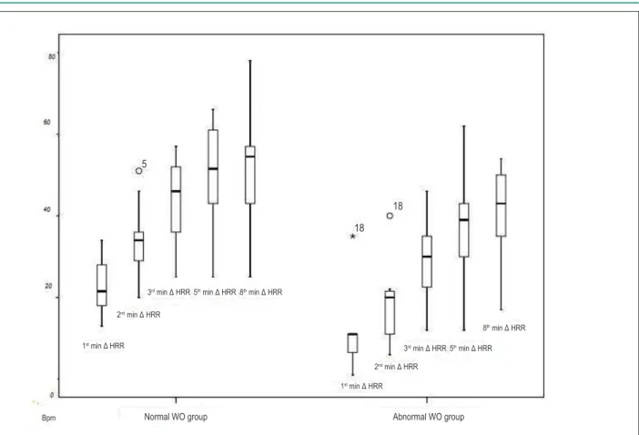
The groups had no significant differences at rest. In HR at peak exercise there was no significant difference between G1 and G2, the latter showing a lower absolute value. In recovery, there were no significant differences in the absolute values of HRR, but the G2 had a smaller variation of HRR (Δ HRR) from the 1st to the 8th minute, as observed in Table 2. Figure 1 compares the variation of HRR post-exertion between the groups.
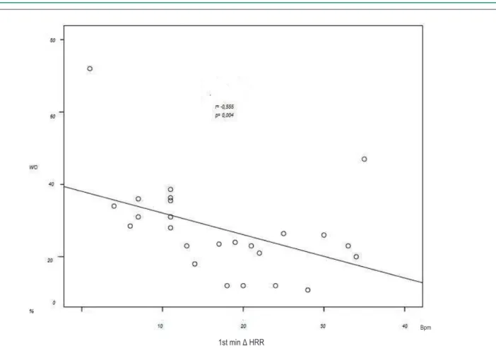
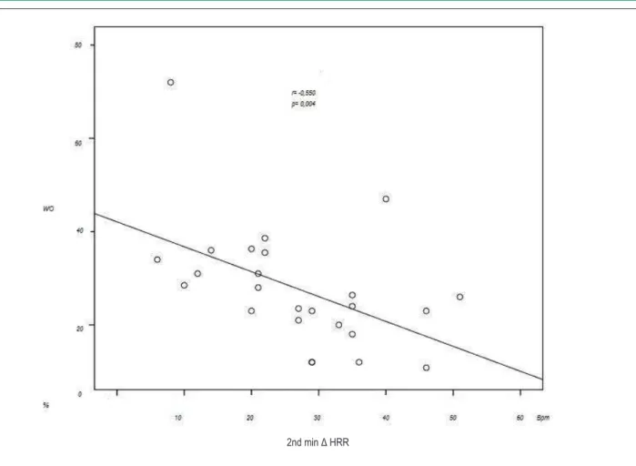
When we evaluated the association between WO and the variation of HRR using the correlation coefficient of Spearman, we observed a significant negative correlation between the WO and 1st minute Δ HRR (r = -0.555; p = 0.004) and 2nd minute Δ HRR (r = -0.550, p = 0.004). No statistical significance was observed in the 3rd to the 8th minute of recovery. Figure 2 shows the correlation between WO and 1st minute ΔHRR, while figure 3 shows the correlation between WO and 2nd minute ΔHRR.
Because of possible factors influencing the behavior of HRR (e.g., heart failure etiology and drug administration), we performed a multivariate analysis, putting the WO as the dependent variable. We found that the 2nd minute Δ HRR was the variable that best correlated with WO: r = 0.246, p = 0.012.
Discussion
Table 2 - Variables at the beginning of exercise and post-exercise recovery
G1 (N =14) WO < 27%
G2 (N =11)
WO ≥ 27% p
SBP at rest (mmHg) 119 (107.75 – 130.25) 104 (83 – 125) 0.311
DBP at rest (mmHg) 80 (70.75 – 89.25) 72 (68 – 76) 0.044
HR at rest (bpm) 75 (69.87 – 81.12) 73 (67 -79) 0.741
HR peak exercise (bpm) 142 (127.25 – 156.75) 121 (108 – 134) 0.009
1st min HRR (bpm) 115.5 (101.75 – 129.25) 107 (96 – 118) 0.258
1st min Δ HRR (bpm) 21.50 (16.12 – 26.87) 11.00 (8.5 – 13.5) 0.001
2nd min HRR (bpm) 108 (96.75 – 119.25) 100 (88 – 112) 0.528
2nd min Δ HRR (bpm) 34 (29 – 39) 20 (14 – 26) 0.001
3rd min HRR (bpm) 95.5 (85.12 – 105.87) 88 (80 – 96) 0.38
3rd min Δ HRR (bpm) 46 (37.87 – 54.12) 30.0 (22.0 – 38.0) 0.005
5th min HRR (bpm) 91.5 (81.62 – 101.3) 86 (78.5 – 93.5) 0.169
5th min Δ HRR (bpm) 51.5 (42.0 – 61.0) 39.0 (31.5 – 46.5) 0.013
8th min HRR (bpm) 90.5 (82.37 – 98.62) 82 (75 -89) 0.037
8th min Δ HRR (bpm) 54.5 (46.5 – 62.5) 43 (34 – 52) 0.037
Results expressed as median and interquatil range; G - group; N - number; WO - washout rate; SBP - systolic blood pressure; DBP - diastolic blood pressure; mmHg - millimeters of mercury; HR - heart rate; bpm - beats per minute; HRR - heart rate recovery; min- minute; Δ HRR - variation in heart rate recovery.
Table 1 - Characteristics of the groups
G1 (N =14) WO < 27%
G2 (N =11)
WO ≥ 27% p
Age (years) 55 (45,87 – 64,12) 60 (47,5 – 72,5) 0,742
Gender (M/F) 9/5 7/4 0,974
IMC (Kg/m2) 26,7 (22,79-30,46) 26,9 (21,25–32,55) 0,427
Etiology (%) 0.648
Ischemic 7,15 18,18
Hypertensive 78,57 45,45
Others 14,28 36,37
Medications (%)
Beta-blocker (Carvedilol) 100 100 1
ACEI/ARB II 71,42 63,63 0,685
Spironolactone 100 90,90 0,259
Diuretic drugs 92,85 90,90 0,861
Digital 42,85 54,54 0,569
LVEF (%) 33 (25,12–40,87) 36 (31 -41) 0,826
30min H-M 1,77 (1,57-1,96) 1,70 (1,47-1,93) 0,827
4h H-M 1,76 (1,56-1,95) 1,58 (1,44-1,71) 0,112
WO % 22 (17,18 – 26,81) 35,5 (31,7–39,3) <0,001
Figure 1 - Comparison of heart rate variation in early and late recovery. Δ HRR - variation in heart rate recovery in relation to heart rate at peak exercise; min - minutes; BPM - beats per minute.
1st min Δ HRR
1st min Δ HRR
2nd min Δ HRR
2nd min Δ HRR
3rd min Δ HRR
3rd min Δ HRR
5th min Δ HRR8th min Δ HRR
5th min Δ HRR
8th min Δ HRR
Normal WO group Abnormal WO group
Bpm
5
18 18
of early and late HRR, demonstrating that both components of the autonomic nervous system are altered after exercise, and an association between these two major variables of great prognostic importance26,29. The presence of a negative correlation between the WO and the HRR from the 1st to the 2nd minute of recovery leads us to agree that increased sympathetic activity at rest (determined by abnormal WO) contributes to changes in HRR, especially in the 2nd minute, as observed after a multivariate analysis whose mechanisms are the parasympathetic re-entry along with sympathetic withdrawal1,30,31.
In a previous study, we evaluated, in heart failure, the influence of adrenergic hyperactivity in the variables during exercise, showing that patients with abnormal WO had a lower functional capacity, and lower inotropic and chronotropic response during exercise compared to patients with normal WO32. To date, there is no description in the literature of the association between the abnormal behavior of HRR post-exercise and abnormalities in cardiac adrenergic innervation by I123 MIBG.
Treatment with beta-blockers improves sympathetic hyperactivity33, but some patients are resistant to beta-blocker therapy, perpetuating this state of adrenergic hyperactivity. In our study, we found that patients on beta-blockers still in a state of adrenergic hyperactivity have an abnormal HRR compared to patients without this characteristic. A program of supervised exercise could be a therapeutic option for this group of patients with heart failure who have adrenergic hyperactivity even on
use of beta-blockers. The dynamic exercise improves vagal activity and adrenergic hyperactivity7,34.
arrhythmias detected by studying the ICD implanted in patients with heart failure.
Exercise testing is a simple test widely available that can promote important information about the autonomic function of more complex and less available imaging tests that have been employed to evaluate. An important suggestion would be to use exercise testing as a screening method to assess which patients with heart failure will be more likely to have altered adrenergic innervation to select patients who will most benefit from I123 MIBG scintigraphy.
Limitations of the study
The main limitation of this study was the small number of patients. However, through a pilot study with 16 patients, a sample size calculation was performed and the number of 11 patients per group has a 90% statistical power to identify 47.6% difference in 1st minute HRR, and a statistical power of 90% to identify 39.93% difference in 2nd minute HRR between groups.
Another significant limitation was that we failed to perform cardiopulmonary exercise testing (CPET) to attest that the patients performed maximal testing. However, as described above, only those patients who completed the test to exhaustion participated in the study (grade 10 in the BORG’s modified rating of perceived
fatigue). CPET was not performed because we did not have this device in our institution at the time of this study.
The evaluation of post-exercise HRR, however, can be made with conventional ET and does not require complex methodologies for analysis. Since the ET is a simple and ubiquitous tool, we believe that information derived from this examination may be useful for cardiologists involved in clinical practice for patients with heart failure, since the information obtained from this simple test correlated with the sophisticated analysis of cardiac innervation.
Conclusion
Patients with heart failure and abnormal WO had slower HRR than normal WO patients, both in early and late phases. This slow recovery in patients with adrenergic hyperactivity state at rest suggests that sympathetic changes at rest, typical of heart failure, may contribute to the autonomic changes observed during recovery after exertion.
Potential Conflict of Interest
No potential conflict of interest relevant to this article was reported.
Figure 2 - Correlation of washout rate and heart rate variation in the 1st minute of recovery. WO - washout rate; Δ HRR - heart rate recovery variation; min- minutes; BPM - beats per minute.
Bpm
Sources of Funding
This study was partially funded by FAPERJ and CNPq.
Study Association
This article is part of the thesis of Doctoral submitted by Leandro Rocha Messias, from Universidade Federal Fluminense. Figure 3 - Correlation between the washout rate and heart rate variation in the 2nd minute of recovery. WO - washout rate; Δ HRR - variation in heart rate recovery;
min - minutes; BPM - beats per minute.
2nd min Δ HRR
References
1. Chapleau MW, Sabharwal R. Methods of assessing vagus nerve activity and reflexes. Heart Fail Rev. 2011; 16(2):109-27.
2. Kannankeril P, Le F, Kadish A, Goldberger J. Parasympathetic effects on heart rate recovery after exercise. J Investig Med. 2004;52(6):394-401. 3. Lahiri MK, Kannankeril PJ, Goldberger JJ. Assessment of autonomic function
in cardiovascular disease physiological basis and prognostic implications. J Am Coll Cardiol. 2008; 51(18):1725-33.
4. Myers J, Tan SY, Abella J, Aleti V, Froelicher VF. Comparison of the chronotropic response to exercise and heart rate recovery in predicting cardiovascular mortality. Eur J Cardiovasc Prev Rehabil. 2007; 14(2):215-21. 5. Imai K, Sato H, Hori M, Kusuoka H, Ozaki H, Yokoyama H, et al.. Vagally mediated heart rate recovery after exercise is accelerated in athletes but blunted in patients with chronic heart failure. J Am Coll Cardiol. 1994; 24(6):1529-35.
6. Lauer MS. Autonomic function and prognosis. Cleve Clin J Med.2009; 76( Suppl 2): S18-22.
7. Freeman JV, Dewey FE, Hadley DM, Myers J, Froelicher VF. Autonomic Nervous system interaction with the cardiovascular system during exercise. Prog Cardiovasc Dis. 2006; 48(5):342-62.
8. Arena R, Guzzi M, Myers J, Peberdy MA. Prognostic value of heart recovery in patients with heart failure. Am Heart J. 2006; 151 (4):851.e7-13.
9. Serra SM, Costa RV, Teixeira de Castro RR, Xavier SS, Nobrega AC. Cholinergic stimulation improves autonomic and hemodynamic profile during dynamic exercise in patients with heart failure. J Cardiac Fail. 2009; 15(2):124-9.
11. Almeida FK, Gross JL, Rodrigues TC. Complicações microvasculares e ddisfunção autonômica cardíaca em pacientes com Diabete Melito Tipo 1. Arq Bras Cardiol. 2011; 96(6):484-9.
12. Cole CR, Foody JM, Blackstone EH, Lauer MS. Heart rate recovery after submaximal exercise testing as a predictor of mortality in a cardiovascularly healthy cohort. Ann Intern Med. 2000; 132(7):552-5. 13. Watanabe J, Thamilarasan M, Blackstone EH, Thomas JD, Lauer MS. Heart rate recovery immediately after treadmill exercise and left ventricular systolic dysfunction as predictors of mortality: the case of stress echocardiography. Circulation. 2001; 104(16):1911- 6. 14. Vivekananthan DP, Blackstone EH, Pothier CE, Lauer MS. Heart rate
recovery after exercise is a predictor of mortality, independent of the angiographic severity of coronary disease. J Am Coll Cardiol. 2003; 42(5):831 - 8.
15. Turpeinen AK, Vanninen E, Magga J, Tuomainen P, Kuusisto J, Sipola P, et al. Cardiac sympathetic activity is associated with inflammation and neurohumoral activation in patients with idiopathic dilated cardiomyopathy. Clin Physiol Funct Imaging. 2009; 29(6):414-9. 16. Agostini D, Carrio I, Verberne HJ. How to use myocardial 123I-MIBG
scintigraphy in chronic heart failure. Eur J Nucl Med Mol Imaging. 2009; 36(4): 555-9.
17. Cohen-Solal A, Esanu Y, Logeart D, Pessione F, Dubois C, Dreyfus G, et al. Cardiac metaiodobenzylguanidine uptake in patients with moderate chronic heart failure: relationship with peak oxygen uptake and prognosis. J Am Coll Cardiol. 1999: 33(3); 759-66.
18. Diakakis GF, Parthenakis FI, Patrianakos AP, Koukourai SI, Stathaki MI, Karkavitsas NS, et al. Myocardial Sympathetic Innervation in Patients with Impaired Glucose Tolerance: Relationship to Subclinical Inflammation. Cardiovasc Pathol. 2008: 17(3) :172-7.
19. Dae MW, De Marco T, Botvinick EH, O’Connell JW, Hattner RS, Huberty JP, et al. Scintigraphic assessment of MIBG uptake in globally denervated human and canine hearts-implications for clinical studies. J Nucl Med. 1992; 33(8):1444-50.
20. Ji SY, Travin MI. Radionuclide imaging of cardiac autonomic innervation. J Nucl Cardiol. 2010; 17(4):655-66.
21. Ando MA, Yamamoto T, Hino A, Sato T, Nakamura Y, Matsuzaki M. Norepinephrine spillover during exercise as a novel parameter to evaluate the severity of heart failure. J Nucl Cardiol. 2010; 17(5):968-73. 22. Verberne HJ, Brewster LM, Somsen A, van Eck-Smit BL. Prognostic value of myocardial 123imetaiodobenzylguanidine (MIBG) parameters in patients with heart failure: a systematic review. Eur Heart J. 2008; 29(9):1147-59.
23. Carrio´ I, CowieMC, Yamazaki J, Udelson J, Camici PG. Cardiac sympathetic imaging with MIBG in heart failure. JACC Cardiovasc Imaging. 2010; 3(1): 92–100.
24. McKee PA, Castelli WP, McNamara PM, Kannel WB. The natural history of congestive heart failure: The Framingham Study. N Engl J Med. 1971; 285(26):1441-6.
25. Scanlon PJ, Faxon DP, Audet AM, Carabello B, Dehmer GJ, Eagle KA, et al. ACC/AHA Guideline for coronary angiography. J Am Coll Cardiol. 1999; 33(6):1756-824.
26. Ogita H, Shimonagata T, Fukunami M, Kumagai K, Yamada T, Asano Y,et al. Prognostic significance of cardiac 123I metaiodobenzylguanidine imaging for
mortality and morbidity in patients with chronic heart failure: a prospective study. Heart. 2001; 86(6): 656-60.
27. Wilson RC, Jones PH. A comparison of the visual analogue scale and modified BORG scale for the measurement of dyspnoea during exercise. Clin Sci (Lond) 1989; 76(3): 277-82.
28. Kallistratos MS, Dritsas A, Laoutaris A, Cokkinos DV. Chronotropic and neurohumoral markers for the evaluation of functional capacity in patients with impaired left ventricular function. Hellenic J Cardiol. 2008; 48(1): 26-32.
29. Arena R, Myers J, Abella J, Peberdy MA, Bensimhon D, Chase P, et al. The Prognostic value of the heart rate response during exercise and recovery in patients with heart failure: influence of beta-blockade. Int J Cardiol. 2010; 138(2):166-73.
30. Sheppard R, Racine N, Roof A, Ducharme A, Blanchet M, White M. Heart rate recovery – a potential marker of clinical outcomes in heart failure patients receiving beta-blocker therapy. Can J Cardiol. 2007; 23(14):1135-8. 31. Nishiyama Y, Morita H, Harada H, Koga Y,et al. Systolic blood pressure response to exercise as a predictor of mortality in patients with chronic heart failure. Int Heart J. 2010; 51(2):111-5.
32. Rocha Messias L, de Queiroz Carreira MA, Ribeiro de Miranda SM, Cunha de Azevedo J, Ambrosio Gava I, Campos Rodrigues R, et al. Relação entre imagem adrenérgica cardíaca e teste ergométrico na insuficiência cardíaca. Arq Bras Cardiol .2011; 96(5):370-5.
33. Kasama S, Toyama T, Hatori T, Sumino H, Kumakura H, Takayama Y, et al. Evaluation of cardiac sympathetic nerve activity and left ventricular remodelling in patients with dilated cardiomyopathy on the treatment containing Carvedilol. Eur Heart J. 2007; 28(8):989-95.
34. Bueno CR Jr, Ferreira JC, Pereira MG, Bacuraw AV, Brum PC. Aerobic exercise training improves skeletal muscle function and Ca2+ handling-related protein expression in sympathetic hyperactivity-induced heart failure. J Appl Physiol. 2010; 109(3):702-9.
35. Yamada T, Shimonagata T, Fukunami M, Comparison if the value of cardiac iodine-123 metaiodobenzylguanidine imaging and heart rate variability in patients with chronic heart failure: a prospective study. J Am Coll Cardiol. 2003; 41(2):231-8.
36. Ta m a k i S , Ya m a d a T, O k u y a m a Y. C a r d i a c I o d i n e - 1 2 3 Metaiodobenzylguanidine imaging predicts sudden cardiac death independently of left ventricular ejection fraction in patients with chronic heart failure and left ventricular systolic dysfunction. J Am Coll Cardiol. 2009;53(5):426–35.