INTRODUCTION
Microbial keratitis is commonly diagnosed worldwide, and conti-nues to cause significant ocular morbidity, requiring prompt and ap-propriate treatment(1,2). A large retrospective cohort recently reported an incidence of ulcerative keratitis of 27.6/100,000 person- years (95% confidence interval, 24.6-30.9)(3).
Predisposing factors for microbial keratitis described in the lite-rature, include: previous ocular surgery, contact lens use, trauma and metal fo reign body, surgical sutures, ocular surface disease, topical corticosteroid use, herpetic keratitis, lid misalignment, and systemic
diseases such as diabetes and smoking(4-16).Predictive factors for out-come such as initial visual acuity at admission have not been widely described in previous studies.
Previous reports describe a variability of etiological agents in diff erent centers around the world(2,4,5,7,8,13,14,16). Empirical, broadspec-trum treatment of keratitis is a strategy selected by many ophthal-mologists(17-21). However the increased resistence of the causative mi croorganism to currently available anti-infective drugs requires ongoing assessment of the trends of clinical and microbiological evaluation of the patients(22).
Clinical characteristics and outcomes of patients admitted with presumed microbial
keratitis to a tertiary medical center in Israel
Características clínicas e desfechos dos pacientes internados com diagnóstico de ceratite microbiana em
um centro terciário em Israel
Fabio Lavinsky1, noah avni-Zauberman1, irina s barequet1
Submitted for publication: May 21, 2012 Accepted for publication: May 29, 2013
Study carried out at Goldschleger Eye Institute, Sheba Medical Center, Tel Aviv University Sackler, Faculty of Medicine, Tel Hashomer, Israel.
1 Physician, Goldschleger Eye Institute, Sheba Medical Center, Tel Aviv University Sackler, Faculty of
Medicine, Tel Hashomer, Israel.
Funding: No specific financial support was available for this study.
Disclosure of potential conflicts of interest: F.Lavinsky, None; N.Avni-Zauberman, None; I.S.Barequet, None.
Correspondence address: Fabio Lavinsky. Rua Quintino Bocaiúva, 673 - 3o andar - Porto Alegre
(RS) - 90440-051 - Brazil - E-mail: [email protected] ABSTRACT
Purposes: Microbial keratitis is commonly diagnosed worldwide, and continues to cause significant ocular morbidity, requiring prompt and appropriate treatment. The objective of this study is to describe the clinical characteristics and outcomes of patients with presumed microbial keratitis admitted to The Goldschleger Eye Institute, Sheba Medical Center, Tel Aviv University, Tel Hashomer, Israel. Methods: A cross-sectional study was conducted, in which the medical records of patients with presumed microbial keratitis admitted during a period of 3 years were reviewed.
Results: Keratitis was diagnosed in 276 patients (51% males and 48.9% females). The mean age was 39.29 ± 22.30 years. The hospital length of stay ranged from 1 to 65 days (mean 5.69 ± 5.508). Fortified antibiotics were still used at discharge in 72% of the cases. Overall visual acuity improved significantly from the time of admission to the 1st-week follow up visit showing a p<0.001 on the Wilcoxon signed ranks test. Contact lens wearing was present in 36.1% of the patients, although there was no significant relation with severity of the presentation and visual outcome (p>0.05). The degree of hypopyon and cells in the anterior chamber was significantly related to the hospital length of stay (r Spearman=0.31; p<0.001 and r Spearman=0.21; p<.001, respectively) as well as to a worse visual outcome (r Spearman=0.32; p<0.01 and r Spearman=0.18; p=0.01, respectively). Of all patients, 2.3% required an urgent therapeutic penetrating keratoplasty, and 1% underwent evisceration. There was no enucleation.
Conclusion: Treating keratitis aggressively and assuring patient compliance is imperative for a good final visual outcome. Inpatient treatment may have a po -sitive impact on this outcome.
Keywords: Cornea; Keratitis/diagnosis; Eye infection, bacterial; Eye infection, fungal; Prognosis; Cross-sectional studies; Tertiary healthcare; Israel
RESUMO
Objetivos: Ceratite microbiana é comumente diagnosticada em todo mundo e ainda continua a causar uma significante morbidade ocular. É necessário tratá-la de forma imediata e apropriada. O objetivo deste estudo é descrever as características clínicas e os desfechos dos pacientes com ceratite microbiana presumida que foram internados no Goldschlager Eye Institute, Sheba Medical Center, Tel Aviv University, Israel. Métodos: Um estudo transversal foi realizado onde arquivos hospitalares dos pacien-tes internados com ceratite microbiana presumida durante um periodo de três anos foram analisados e revisados.
Resultados: Ceratite foi diagnosticada em 276 pacientes (51% masculinos e 48,9% femininos). A média de idade foi 39,29 ± 22,30 anos. A duração da internação foi de 1 a 65 dias (média 5,69 ± 5,508). Antibióticos fortificados permaneceram usados na alta em 72% dos casos. A acuidade visual do seguimento da primeira semana após a alta em relação a internação melhorou na media de forma estatisticamente significativa (p<0,001 usando Wilcoxon signed ranks test). O uso de lentes de contato estava presente em 36,1% dos pacientes, porém não houve relação estatisticamente significativa entre a gravidade da apresentação clínica e a acuidade visual nestes pacientes (p>0,05). O grau de hipópio e células na câmara anterior foram estatisticamente significativos em relação ao tempo de internação (r Spearman=0,0.31; p<0,001 and r Spearman=0,21; p<0,001, respectivamente) e para a acuidade visual (r Spearman=0,32; p<0,01 e r Spearman=0,18; p=0,01, respectivamente). De todos os pacientes, apenas 2,3% necessitaram ceratoplastia penetrante urgente e 1% necessitaram evisceração. Não houve enucleações.
Conclusões: Tratar a ceratite de forma agressiva e garantir a aderência do paciente ao tratamento é imperativo para o bom resultado visual final. O tratamento internado pode ter um impacto positivo neste desfecho.
An earlier survey from a decade ago of the causative agents of ophthalmic infections in an Israeli ophthalmology service showed that coagulasepositive staphylococcus was the most common iso -late from corneal ulcers (33.3%)(23). The combination of warm and humid climate along with the popularity of the contact lens wear may predispose to unique features of microbial keratitis in this re-gion. Therefore, in the current study, we analyzed the demographic and clinical characteristics, the risk factors and the clinical outcomes of patients with keratitis admitted to our department that serves as primary, secondary and tertiary center.
METHODS
In this cross-sectional study, records of all patients admitted to our department with a diagnosis of keratitis over a period of 24 months were analyzed. The department serves as a primary, secondary and tertiary referral center. The admitting policy includes hospitalization when the infiltrate is more severe than very mild. Patients are dis-charged when the infiltrate is responding to treatment, the epithelial defect is closed, and there is no anterior chamber reaction.
Cases were excluded from the analysis if no follow-up visit was registered on the records. The review of the charts was authorized by the hospital directory. The legislation rules of the ethical committee at our institution did not require at that time approval for retrospective data analysis. Data was abstracted for age, gender, hospital length of stay, and season of admission. Predisposing factors such as contact lens use, previous ocular surgery, trauma and foreign body were also recorded. Visual acuity was measured with Snellen charts with the best correction obtained with spectacles or pinhole. The size of the infiltrate was ranked from 1 to 3, where 1 corresponds to an infiltrate of up to 1 mm of diameter, 2 refers to 1 to 2 mm and 3 means over 2 mm. Anterior chamber reaction was measured according to stan-dard parameters of the ophthalmological practice. Cells and flare were measured in a 1-4 scale and hypopyon was measured in millimeters. In addition, the microbiological profile was accessed: the results of cultures of patients that underwent scrapings were analyzed and their relationship with visual outcome was assessed and compared with the population that has not undergone scrapings. The initial antibiotic treatment was also reviewed. The fact that patients are discharged with the same criteria validates the analysis performed using visual acuity at the 7-day post-discharge follow-up visit as an outcome.
The following risk factors for keratitis were analyzed: previous systemic diseases, previous ocular diseases, previous ocular surgeries, wearing of contact lenses, trauma and foreign body.
The statistical analysis was performed using the SPSSTM software. The statistical methods used were the chi-square test, univariate analysis of variance (ANOVA), the Wilcoxon signed ranks test and Spearman’s rho.
RESULTS
The review of the charts of patients admitted with the diagnosis of keratitis identified 276 patients (51% males and 48.9% females). The mean age was 39.29 ± 22.30 years, with a wide range between one month old and 92 years old (Figure 1).
The hospital length of stay ranged from 1 to 65 days (mean 5.69 ± 5.508). The discharge criteria were healed epithelial defect, decreased inflammation or infection signs and absence of anterior chamber reac tion. Fortified antibiotics were continued after discharge in 72% of the cases.
The mean visual acuity at admission was 0.60 logMAR (n=272). The mean visual acuity at the first follow-up visit at the outpatient clinic was 0.42 logMAR (n=223). Comparison of visual acuity of the patients that had assessments both at admission and follow-up visit showed a statically significant improvement. (p=0.0001 Wilcoxon signed ranks test).
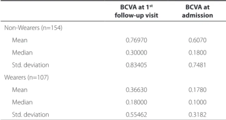
The use of contact lenses was noted in 110 patients (36.1%), their average age was 25.9 years-old (range 0-75 years-old) and 61.46% were females. The inpatient admission period was longer in non-con-tact lens wearers with a median of 5 days versus 4 days in connon-con-tact lens wearers (p<0.003 Mann-Whitney test). The presence of anterior chamber reaction (fibrin, cells and hypopyon) was not significantly associated with contact lens wearing. The 7 day post-discharge visual acuity was better in wearers than in non-wearers (p<0.001) (Table 1). Assessment of age as a prognostic factor showed that patients over 34 years old had a significantly longer hospital stay as well as a worse visual acuity at the 7-day post-discharge follow-up visit (p=0.001 Mann-Whitney test).
There were also 28 (9.2%) cases of keratitis associated with fo reign bodies and 31 (11.2%) cases related to previous intraocular surgeries. This latter group had a worse clinical course than the former. The patients were older (mean age 68.9) and their hospital length of stay was longer (mean 8.7 days). The improvement of the mean visual acuity was significantly lower than in the overall population: 1.62 logMAR at admission to 1.47 logMAR at the first follow-up against 0.43 logMAR and 0.25 logMAR in the overall population (p<0.01 on the Mann-Whitney and Wilcoxon tests).
Figure1. Age distribution of the population.
Table 1. Visual acuity outcome in wearers and non-wearers of con-tact lenses
BCVA at 1st
follow-up visit
BCVA at admission
Non-Wearers (n=154)
Mean 0.76970 0.6070
Median 0.30000 0.1800
Std. deviation 0.83405 0.7481
Wearers (n=107)
Mean 0.36630 0.1780
Median 0.18000 0.1000
Std. deviation 0.55462 0.3182
The size of the infiltrate at admission was 149 (48.9%) in grade 1 (up to 1 mm), 46 (15.1%) in grade 2 (from 1 to 2 mm), and 50 (16.4%) in category 3 (over 2 mm). In 19.6% of the cases records did not clearly defined the grade of the infiltrate. Patients with large infiltrates (grade 3) had a worse visual acuity both at presentation and at the 7-day follow-up visit compared with the other categories (p<0.0001Spearman´s rho).
Anterior chamber reaction was frequently noted at admission: 41.6% of the cases had cells (>+1), 19.8%. had fibrin and 11.1% had hypopyon. The amount of cells and the measurement of hypopyon were related to a worse prognosis. There was a statiscally significantly relationship between length of hospitalization and visual acuity with positive inflammatory signs at admission. (p=0.001) (Figure 2).
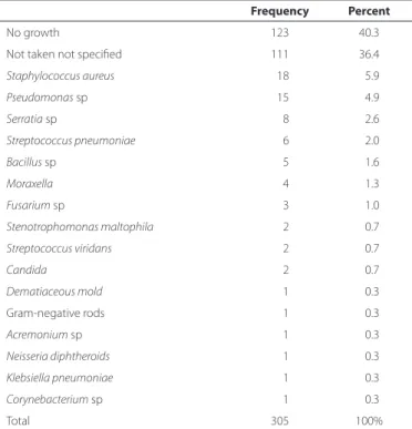
Cultures were obtained from 194 cases. In 111 (36.6%) cases an -tibiotic treatment was used prior to admission. After admission, pa tients were initially treated empirically and changes were made accordingly. The most frequent antibiotic regimen included com-bination of fortified antibiotics such as cefazolin (50 mg/mL) with gen tamicin (13.6 mg/mL) (23%) or vancomycin (33 mg/mL) with cefta zidime (40 mg/mL) (29.2%).
From the cultures taken (194 cases), only 36% (n=71) were posi-tive. The microbiological profile of our population is shown in table 2. Patients with positive cultures had a significantly worse baseline visual acuity as compared with cases with negative cultures and with the group which cultures were not taken (p<0.0001)
The improvement of visual acuity at the 7-day post discharge follow-up visit was statistically significantly smaller in the positive-cul ture group as compared with the negative-positive-culture group and with the no-culture group (p<0.0001) (Figure 3). The hospital length of stay of the positive-culture patients was longer: mean 7.37 days (p=0.001). The rate of urgent surgical intervention was low: tarsorrhaphy in 3.7% (n=10), therapeutic penetrating keratoplasty (PK) in 2.3% (n=7), vitrectomy due to endophthalmitis in 1% (n=3) and evisceration in 1% (n=3).
The impact of previous ocular surgeries such as PK, extracapsular cataract extraction (ECCE) or combined PK + ECCE (n=31) was eva -luated. The mean age of this group was higher as compared the non-previous surgery group (68.96 years ± 20.0 versus 39.29 years ± 22.3 years, respectively).Their mean visual acuity at admission and at the 7-post discharge follow-up visit were statistically significantly worse in the previous surgery group (1.62 logMAR and 1.47 logMAR respectively) than in the non-previous surgery group (0.43 logMAR
and 0.25 logMAR respectively respectively). (p<0.01 Mann-Whitney and Wilcoxon signed ranks tests). The rate of positive cultures were higher in the previous surgery group (61%) as compared with patiens that have not undergone surgery. The rate of urgent surgical inter-vention was significantly higher in the previous surgery group (30%) as compared with the non-previous surgery group (8.7%) (p<0.01). The most common urgent surgical intervention in the previous surgery group was the need for urgent PK (14%) against 0.4% in the non-pre vious surgery group (p<0.01).
DISCUSSION
Microbial keratitis is an ophthalmological emergency that requi-res immediate treatment. Our study evalueted characteristics and
Figure 2. Correlation between anterior chamber reaction and hospitalization days.
Table 2. Culture results
Frequency Percent
No growth 123 40.3
Not taken not specified 111 36.4
Staphylococcus aureus 018 05.9
Pseudomonas sp 015 04.9
Serratia sp 008 02.6
Streptococcus pneumoniae 006 02.0
Bacillus sp 005 01.6
Moraxella 004 01.3
Fusarium sp 003 01.0
Stenotrophomonas maltophila 002 00.7
Streptococcus viridans 002 00.7
Candida 002 00.7
Dematiaceous mold 001 00.3
Gram-negative rods 001 00.3
Acremonium sp 001 00.3
Neisseria diphtheroids 001 00.3
Klebsiella pneumoniae 001 00.3
Corynebacterium sp 001 00.3
Total 305 100%
outcomes of 276 consecutive patients admitted with a diagnosis of presumed keratitis. The early diagnosis and the intensive inpatient treatment may have resulted into the overall clinical and visual im-provement seen in our population.
Daniell et al.(18) described different strategies for approaching pa -tients diagnosed with presumed microbial keratitis. Pa-tients with small peripheral lesions could be treated empirically with topical fluorqui-nolones, whereas the ones with central larger lesions should undergo scrapings for culture and topical quinolone associated with a cepha-losporin should be started. In our department, taking cultures and the initial treatment were based on the clinical judgment of the attending ophthalmologist who examined the patient at the emergency ward.
Some authors(22) studied a model to determine the impact of mi nimal inhibitory concentration (MIC) on the clinical outcome. It in-cluded patients who received monotherapy with a fluoroquinolone who had no subsequent change in their treatment and whose ulcers healed without surgical intervention. He found significant linear as-sociations between clinical outcome and MIC for Pseudomonas spp. (P=0.047), Staphylococcus aureus (P=0.04), and Enterobacteriaceae
(P=0.045), but not for Streptococcus spp. (P=0.85) and coagulase- negative staphylococci (CNS) (P=0.88).
In the population we studied, 36.6% were already using some kind of topical treatment that failed. The treatment approach was the use of fortified topical antibiotics given every hour at the beginning, which included either cefazolin and gentamicin or vancomycin and ceftazidime. Treatment modifications were made according to cultu-re cultu-results and clinical outcome on a case by case basis. Hospitalization assured treatment compliance until stabilization and may be related to the good visual outcome and low complication rate seen in our population.
The risk factors in our population are compatible with the ones described in the literature. Contact lens wearing was the major pre-disposing condition in our population (36%), comparable with the prevalence found in an Australian study (33.7%)(10) and in a Taiwanese study (44.3%)(2).
In our study, contact lens wearers had a significantly better final visual outcome. This fact may be due to their younger age and to early ophthalmological examination performed in this population.
Age and clinical presentation influenced the hospital length of stay. Patients older than 34 years stayed longer (mean of 6.88 days against 4.56 days of younger patients) and their final visual acuity was also significantly worse than those patients under 34 years old. Parmar et al., showed that the elderly tend to have more severe presentations of keratitis with a worse outcome, suggesting that this may be due to the lower immunocompetence of this age group(12). The high preva-lence of patients aged 18 to 21 years in our study group may be due to the referrals from the military medical corps to our tertiary center. The recruits and soldiers can seek medical attention promptly, thus their clinical presentation is usually mild.
The best-corrected visual acuity (BCVA) measured at admission improved significantly compared with the one measured at the first follow-up visit, 1 week after discharge with a closed ulcer and no an-terior chamber reaction. Other studies showed overall visual loss(2,4,7). By analyzing the clinical characteristics at presentation we found that ulcers larger than 2 mm and with hypopyon greater than 1 mm were significantly related to worse visual acuity at admission and at the first follow-up visit. The hospital length of stay was longer in the presence of those findings. This underlines that seeking an ophthal-mologist as soon as symptoms appear has a tremendous impact on the final visual outcome and on the public health expenses on hospitalization and antibiotic treatment. However, other authors(9) demonstrated in their study of an Australian population that the cost to treat was significantly associated only with clinical severity and type of pathogen, not with delay in presentation.
In our series, cultures were obtained in194 of the patients admit-ted (70.3%). The reasons for not obtaining cultures by scrapings
inclu-ded: uncooperative patients, ulcers that are peripheral and small and without anterior chamber reaction, keratitis may be treated empiri-cally. The UK ofloxacin study suggested that the microbiological in-vestigation of ulcers smaller than 2 mm is probably not worthwhile(24). Another study from a Nepalese population by Feilmeir et al., de monstrated that microorganisms were grown from 40% of the corneal scrapings. Pure bacterial cultures were obtained from 39% of the positive scrapings and pure fungal cultures were obtained from 61%(25).
The most prevalent microorganism in our cultures was coagula-se-positive Staphylococcus (25.3% of positive cultures), followed by Pseudomonas (21.1%). These results were compatible with the study of the Israeli microbiological profile of ocular infections also showed that coagulase-positive Staphylococcus was also the most prevalent agent in corneal isolates(23).
Patients with positive cultures (35%) had a longer hospital stay (mean of 7.37 days) and a worse visual prognosis.
Previous ocular surgery has been described as a predisposing fac-tor for microbial keratitis(2,26).In our population, patients with previous ECCE, PK or combined surgery were older and had a significantly worse visual acuity either at admission or at the 7-day follow up visit. The rate of positive cultures was significantly higher in this group (61%), and Gram-positive bacteria were the most common etiologic agents isolated from the scrapings. Moreover, the need of secondary intervention such as urgent PK or evisceration was significantly higher in this subgroup. However, Green et al.(27) demonstrated in a longitudinal study that there is a trend towards decreased incidence of keratitis in patients with previous surgeries.
CONCLUSION
There are some limitations to our study. The data is based on retrospective charts analyzing only patients with keratitis that were addmited to the ward. The decision to admit the patient was based on clinical appearance, on previous treatment failure in the commu-nity and on the evaluation of patient’s possible limitations to comply with the treatment, therefore some mild cases may not have inclu-ded. The ones discharged at the emergency room were not incluinclu-ded. There are some patients that missed the follow-up and were also excluded from the analysis. Adittional prospective controlled studies in this geographic area are needed to confirm our results.
Nevertheless, this present study has a large number of patients studied is able to demonstrate clinical trends such as microbial ke ratitis must be treated aggressively and promptly to achieve a good visual outcome. Furthermore, age, anterior chamber cells and hypopyon, positive cultures and previous surgery were risk factors related to lengthier hospitalization and a worse BCVA at the 7-day follow-up visit.
REFERENCES
1. Blanton CL, Rapuano CJ, Cohen EJ, Laibson PR. Initial treatment of microbial keratitis. CLAO J. 1996;22(2):136-40. Comment in: CLAO J. 1996;22(4):222.
2. Fong CF, Tseng CH, Hu FR, Wang IJ, Chen WL, Hou YC. Clinical characteristics of micro-bial keratitis in a university hospital in Taiwan. Am J Ophthalmol. 2004;137(2):329-36. 3. Jeng BH, Gritz DC, Kumar AB, Holsclaw DS, Porco TC, Smith SD, et al. Epidemiology
of ulcerative keratitis in Northern California. Arch Ophthalmol [Internet]. 2010 [cited 2012 Jul 21];128(8):1022-8. Available from: http://archopht.jamanetwork.com/article. aspx?articleid=426096
4. al-Samarrai AR, Sunba MS. Bacterial corneal ulcers among Arabs in Kuwait. Ophthal-mic Res. 1989;21(3):278-84.
5. Carmichael TR, Wolpert M, Koornhof HJ. Corneal ulceration at an urban African hos-pital. Br J Ophthalmol. 1985;69(12):920-6.
6. Cheung J, Slomovic AR. Microbial etiology and predisposing factors among patients hospitalized for corneal ulceration. Can J Ophthalmol. 1995;30(5):251-5.
8. Katz NN, Wadud SA, Ayazuddin M. Corneal ulcer disease in Bangladesh. Ann Ophthal-mol. 1983;15(9):834-6.
9. Keay L, Edwards K, Brian G, Stapleton F. Surveillance of contact lens related microbial keratitis in Australia and New Zealand: multi-source case-capture and cost-effective-ness. Ophthalmic Epidemiol. 2007;14(6):343-50.
10. Keay L, Edwards K, Naduvilath T, Taylor HR, Snibson GR, Forde K, et al. Microbial keratitis predisposing factors and morbidity. Ophthalmology. 2006;113(1):109-16. Com ment in: Ophthalmology. 2006;113(11):2115-6; author reply 2116.
11. Kunimoto DY, Sharma S, Garg P, Gopinathan U, Miller D, Rao GN. Corneal ulceration in the elderly in Hyderabad, south India. Br J Ophthalmol [Internet]. 2000[cited 2011 Mar 19];84(1):54-9. Available from: http://bjo.bmj.com/content/84/1/54.long 12. Parmar P, Salman A, Kalavathy CM, Kaliamurthy J, Thomas PA, Jesudasan CA. Microbial
keratitis at extremes of age. Cornea. 2006;25(2):153-8.
13. Upadhyay MP, Karmacharya PC, Koirala S, Tuladhar NR, Bryan LE, Smolin G, et al. Epi-demiologic characteristics, predisposing factors, and etiologic diagnosis of corneal ulceration in Nepal. Am J Ophthalmol. 1991;111(1):92-9.
14. van der Meulen IJ, van Rooij J, Nieuwendaal CP, Van Cleijnenbreugel H, Geerards AJ, Remeijer L. Age-related risk factors, culture outcomes, and prognosis in patients admitted with infectious keratitis to two Dutch tertiary referral centers. Cornea. 2008; 27(5):539-44.
15. Varaprasathan G, Miller K, Lietman T, Whitcher JP, Cevallos V, Okumoto M, et al. Trends in the etiology of infectious corneal ulcers at the F. I. Proctor Foundation. Cornea. 2004;23(4):360-4.
16. Wong T, Ormonde S, Gamble G, McGhee CN. Severe infective keratitis leading to hos-pital admission in New Zealand. Br J Ophthalmol [Internet]. 2003[cited 2012 Dec 21]; 87(9):1103-8. Available from: http://bjo.bmj.com/content/87/9/1103.long
17. Bharathi MJ, Ramakrishnan R, Meenakshi R, Mittal S, Shivakumar C, Srinivasan M. Microbiological diagnosis of infective keratitis: comparative evaluation of direct
microscopy and culture results. Br J Ophthalmol [Internet]. 2006[cited 2011 Feb 12]; 90(10):1271-6. Available from: http://bjo.bmj.com/content/90/10/1271.long 18. Daniell M, Mills R, Morlet N. Microbial keratitis: what’s the preferred initial therapy?
Br J Ophthalmol. 2003;87(9):1167. Comment in: Br J Ophthalmol. 2003; 87(9):1167-9. Br J Opthalmol. 2003;87(9):1172-4. Br J Ophthalmol. 2003;87(9):1169-72.
19. Jones DB. Decision-making in the management of microbial keratitis. Ophthalmolo-gy. 1981;88(8):814-20.
20. Pharmakakis NM, Andrikopoulos GK, Papadopoulos GE, Petropoulos IK, Kolonitsiou FI, Koliopoulos JX. Does identification of the causal organism of corneal ulcers influence the outcome? Eur J Ophthalmol. 2003;13(1):11-7.
21. Rao NA. A laboratory approach to rapid diagnosis of ocular infections and prospects for the future. Am J Ophthalmol. 1989;107(3):283-91.
22. Kaye S, Tuft S, Neal T, Tole D, Leeming J, Figueiredo F, et al. Bacterial susceptibility to topical antimicrobials and clinical outcome in bacterial keratitis. Invest Ophthalmol Vis Sci [Internet]. 2010[cited 2012 Oct 21];51(1):362-8. Comment in: Invest Ophthalmol Vis Sci. 2010;51(12): 6902-3; author reply 6903. Available from: http://www.iovs.org/content/51/1/362.long 23. Mezer E, Gelfand YA, Lotan R, Tamir A, Miller B. Bacteriological profile of ophthalmic
infections in an Israeli hospital. Eur J Ophthalmol. 1999;9(2):120-4.
24. Ofloxacin monotherapy for the primary treatment of microbial keratitis: a double-mas ked, randomized, controlled trial with conventional dual therapy. The Ofloxacin Study Group. Ophthalmology. 1997;104(11):1902-9.
25. Feilmeier MR, Sivaraman KR, Oliva M, Tabin GC, Gurung R. Etiologic diagnosis of corneal ulceration at a tertiary eye center in Kathmandu, Nepal. Cornea. 2010;29(12):1380-5. 26. Llovet F, de Rojas V, Interlandi E, Martin C, Cobo-Soriano R, Ortega-Usobiaga J, et al.
Infectious keratitis in 204 586 LASIK procedures. Ophthalmology. 2010;117(2):232-8 e1-4. Comment in: Ophthalmology. 2011;118(2):425; author reply 425.e1