1
Original Article
Maximal Heart Rate in Exercise Tests on Treadmill and
in a Cycloergometer of Lower Limbs
Claudio Gil Soares de Araújo e Vivian Liane Mattos Pinto
Clínica de Medicina do Exercício - CLINIMEX and Universidade Gama Filho - Rio de Janeiro, RJ - Brazil
Mailing Address: Claudio Gil Soares de Araújo - Rua Siqueira Campos, 93/101 - 22031-070 - Rio de Janeiro, RJ - Brazil E-mails: [email protected] and [email protected]
Received for publishing on 07/13/2004 Accepted on 01/20/2005
Objective
To compare, retrospectively, the values of maximum heart rate (MHR) and the decrease of the heart rate at the first minute of recovery, which were obtained in an exercise test (ET) performed in two different ergometers and at different moments.
Methods
Sixty individuals (from 29 to 80 years old), submitted to cardiopulmonary ET in a cycle of lower limbs (CLL) in our labo-ratory and who had previous ET (up to 36 months) in a treadmill (TRM) in other laboratories, under identical conditions of medi-cations of negative chronotropic action.
Results
MHR was similar in CLL: 156±3 and TRM: 154±2 bpm (p=0.125), whereas dHR was higher in CLL: 33±2, EST: 26±3 bpm (mean ± standard error of the mean) (p<0.001). In hemo-dynamic variables studied, the systolic blood pressure and the double product were higher in the ET-CLL (p<0.001). The e-lectrocardiogram (ECG) was similar in both ETs, except due to more frequent supraventricular arrhythmias in CLL.
Conclusion
a) With some diligence from the examiner and previous knowledge of MHR in a previous ET it is possible to obtain high levels of MHR in an ET-CLL; b) interrupting the MHR-based ET forecast through equations tends to lead to sub-maximum efforts; c) dHR differs in active and passive recoveries; d) new ways to analyze the HR behavior under exercise, which is not only the value of MHR, are necessary to characterize an ET as maximum.
Key words
maximal heart rate, exercise test, ergometer, heart rate recovery
The decision of using a maximum or sub-maximum exercise test depends greatly on the reasons to perform the test, the type of individual to be tested and the availability of equipment and proper human resources. For a better clinical result, an exercise test (ET) must be preferably maximum, thus allowing for obtaining a greater quantity of relevant information1-3.
There are different protocols and procedures for the performance of an ET4-7. However, there are always some practical and real
difficulties to determine when an individual has effectively reached his/her maximum. Probably due to its easiness of measurement, the heart rate (HR) has been one of the most frequently used clinical variables used to characterize the obtention of a maximum effort in a conventional ET (without the measurement of exhaled gases), from the comparison with maximum values forecast due to age, when it is called maximal HR (MHR)8. The statistic
theo-ry tells us that at least 50% from healthy individuals must reach values equal to or higher than those forecast through a regression equation, which makes this criterion very limited and probably not valid in many day-to-day clinical situations.
More recently, studies from Cleveland Clinic have shown an important prognostic clinic meaning for MHR9,10 and for the
de-crease of HR at the first minute of recovery (dHR)11, making even
more important that ET is taken to real maximum levels and not early interrupted in theoretically forecast values of MHR.
Along the last years our research group has been trying to dis-cuss many of the aspects connected to heart rate and exercise12-15.
Theoretically it is possible to expect that ETs performed with different protocols and in different ergometers can show non-coincident results of MHR. For instance, Araújo et al.13, through the performance of
nine different protocols of maximum test, tried to identify which one would provide the highest MHR, by having found that it would happen in a ET on a treadmill (TRM) in progressive increment and continuous nature protocols. That same theme was also analyzed by Myers et al.5, who not only compared the MHR but also the hemodynamic
responses and gases exchanges among different ergometers and diffe-rent protocols, with the aim of establish a protocol that would pro-vide better suitability and accuracy of measurements.
Concerning the measurement of physiologic variables and the proper use of the type of ergometer, although some studies16,17
suggest similar results for MHR and the maximum consumption of oxygen in the TRM and in the cycloergometer of lower (CLL), most works point out to greater values for those variables in the TRM18-21.
2
one of the reasons alleged to the possibility of reaching a higher MHR at the end of the exercise. By considering the hemodymamic and metabolic differences associated to the performance of a ma-ximum exercise in TRM and in CLL, either by the body position or by the muscular groups involved, the differences favorable to TRM in MHR would not be strange. However, it is possible that other reasons, other than those already mentioned, could interfere in such matter. For example, the previous knowledge of the value of MHR in a previous ET and the motivation of the examiner in taking the individual to a relatively maximum exercise can be as or more important than the type of ergometer in which the ET is performed.
So, our study tried to approach that matter in an original way, by trying to verify whether an experienced and motivated examiner could obtain an MHR in an ET, performed in CLL, that would be at least equal to one from a relatively recent ET, carried out on a TRM in another laboratory, whose result was known by him/her.
Methods
All ETs from our laboratory, performed by a single physician, between January 2002 and August 2003 were retrospectively analyzed. For sample definition, the individuals should have: a) made available the electrocardiographic results, reports and tracings of a maximum ET, without early interruption due to clinical crite-rion, in a TRM performed in the last three years (50% of them in the last 12 months), b) been submitted to a maximum exercise cardiopulmonary test in CLL; c) be adults and be under identical conditions of use of medications of negative chronotropic action at the time of the two ETs. By taking those criteria under consi-deration, it was possible to select data from 60 individuals with age ranging from 29 to 80 years old (56±10), being 43 men. On table I we can see more detailed the profile of the sample anal-yzed in accordance to its clinical conditions. So, we can notice that most of the sample consists of individuals carriers of risk factors, and many of them with coronary events. Among those 60 individuals, 25% had a regular use of ß-blockers.
After anamnesis and clinical examination, the individuals were submitted to an exercise cardiopulmonary test, monitored by a digital electrocardiograph with specific software (ErgoPC Elite version 3.2.1.5, Micromed, Brazil), in a ramp individualized pro-tocol, with an average length between 8 to 12 minutes, which made possible an increase of load in accordance to the individual’s condition, in a electromagnetic braking CLL Ergociser EC 1600 (Cateye, Japan). For the measurement and analysis of exhaled gases, a metabolic analyzer VO2000, (MedGraphics, United States) was used.
All ETs in CLL were carried out by a single and experienced examiner, who previously knew the MHR obtained in ET on TRM. In addition to MHR, the dHR was also analyzed, which means the MHR subtracted from the HR at the first post-exercise minute. The ways of maximum exercise recovering differed in both ETs, and in the TRM, made through an active recovery (walking at slow speeds between 2.5 and 3.5 km/h without incline), whereas in the Et in CLL, the measurement was done under passive recovery, at supine position, which was quickly adopted after the procedure (generally, 20 to 40 seconds after the interruption of the exercise).
In addition to HR measurements, other variables were also analyzed and compared in the two ETs. In the hemodynamic aspect, the values of the systolic blood pressure (SBP), diastolic blood pressure (DBP) and rate-pressure product (RPP) - SBP multiplied by HR - (RPP) reached at the last minute of exercise were taken into consideration. The tracings of electrocardiogram (ECG) were examined for the identification of the presence or absence of changes in the segment ST, being recorded the presence of infraunlevelling of the segment ST lower or higher than 0.1 mV at 80 ms from point J and the occurrence of heart arrhythmias, which were simply classified as supraventricular, ventricular or mixed. Finally, the total time of exercise in ET was also marked down. Before the ET in CLL, the individuals signed an informed consent term, in which the use of data for research was specifically authorized. The study protocol was institutionally approved.
For the comparison between the data from the two different both ETs, the matching t-test for continuous variables and the chi-square test for dichotomous variables. The association between data was assessed through linear and regression correlation, as suitable. Afterwards, the sample was subdivided in terms of clinical condition in apparently healthy and coronary artery disease carriers. In all cases, the threshold of 5% as significance level and the confidence interval of 95% were adopted.
Results
The ETs in the CLL lasted between 5 and 15 minutes, with a median of 10 minutes, which is exactly the time recommended for ET protocols aiming at reaching the maximum consumption of oxygen. Despite a slightly higher tendency for MHR in CLL, there were no significant differences for the MHR obtained in ETs in TRM and in CLL, respectively, 154±2 vs. 156±3 bpm (mean±standard error) (p=0.125; CI95%=-5.48 to 1.45). In 57% of individuals, higher values of MHR happened in CLL. As shown in figure 1, a good association between MHR measured in the two ETs in the different ergometers (r=0.79; p<0.001; IC95%=-4.25 to -1.11) was observed.
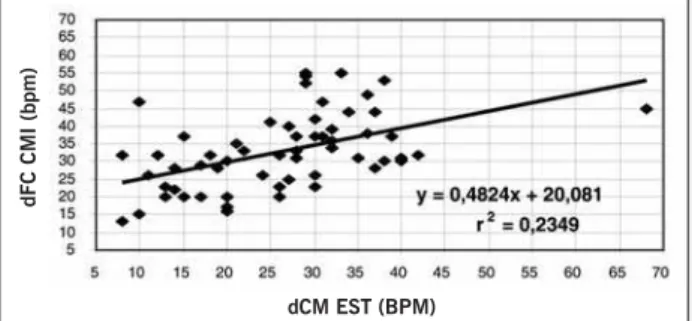
When we analyzed the dHR, the ET performed in CLL showed a sharper decrease of HR at the first minute post-exercise in CLL than in TRM, respectively, 33±1.4 vs. 26±1.4 bpm (p<0.001; CI95%=-9.43 to -3.83), with a significant association, but relatively lower between the two responses (r=0.48; p<0.01; CI95%=-7.35 to -2.66) (figure 2). At the same time, the SBP also recorded higher levels in CLL -204±3 vs. 187±4 mmHg (p<0.001; CI95%=-24.2 to -9.69), whereas DBP showed a similar behavior at the last minute of exercise in both ETs. Starting from higher values of HR and, significantly higher ones of SBP, it
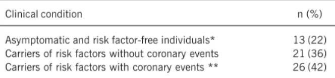
Table I - Profile of the sample based in its clinical condition (n=60)
Clinical condition n (%)
Asymptomatic and risk factor-free individuals* 13 (22) Carriers of risk factors without coronary events 21 (36) Carriers of risk factors with coronary events ** 26 (42)
3
is natural that the maximum values of RPP were also significantly higher in CLL -31496±809, vs. 28814±753 bpm. mmHg (p= 0.001; CI95%=-4252 to -1112), as can be observed on table II. The effective length of time of exercise for the maximum to be reached was very similar in the two ETs - CLL: 9.9±0.3 vs. TRM: 9.8±0.4 min (p=0.087; CI95%=-0.76 to 0.65).
The clinical condition-stratified analysis showed, except for DBP, a trend to higher values in hemodynamic variables for asym-ptomatic individuals. There is a very interesting tendency to a greater difference in favor of CLL in MHR in asymptomatic indivi-duals (TRM: 163±4 vs. CLL: 169±4 bpm, p=0.348) than in those carriers of signs of cardiovascular disease (TRM: 152±3 vs. CLL: 154±7, p=0.737). A similar phenomenon took place in relation to dHR, with asymptomatic individuals showing greater similarity between the two ergometers (TRM: 26±3 vs. CLL: 33±2, p=0.085; CI95%=-14.62 to 1.079), whereas for the coronary artery disease carriers the decrease speed of post-exercise HR was much higher in CLL than in TRM (TRM: 26±1 vs. CLL: 33±2 bpm, p=0.001; CI95%=-10.49 to 2.69).
Regarding exercise ECG, 33% of individuals did not show chan-ges in the segment ST (even minimal, i.e., fast ascendant with 0.5 to 1 mm at 80 ms from the origin) or any cardiac arrhythmias in the two ETs. However, by analyzing separately per procedure, in CLL and in TRM, 50% of individuals did not show changes of ECG. So, we had individuals who showed changes in only one procedure. There was a trend so that the changes in segment ST were more frequent in ETs in TRM (p=0.066), whereas cardiac arrhythmias were more usually found in ETs in CLL (p=0.003), notably the supraventricular (18 vs. 3 cases).
Discussion
Initially we are going to discuss some specific characteristics and potential limitations in the present study. Different from other studies in the literature, the two ETs were not performed in random order and with a minimum time interval, as for example, two or three days. Besides, the ETs in TRM were carried out in many laboratories and by many different examiners, whereas the ETs in CLL were performed by a single and experienced examiner, who previously knew the MHR obtained in the ET in TRM, which somehow could have facilitated the obtainment of a higher MHR in some cases of ET in CLL. It is also possible that the obtainment of measurements of exhaled gases during all ETs in CLL and in only 10% of ETs in TRM can be influenced in the obtainment of a truly maximum test, either due to additional difficulties caused by the mouthpiece or for the
reading of ventilation variables and indexes that favor the identification of a truly maximum exercise. With those considerations shown, some of the comparisons made, notably those of ECG, were limited. Ho-wever, it is important to mention that this study involved a retrospective data analysis, without any intention in contributing or influencing the results obtained. When the examiner has an individual to be submitted to an ET, who brings the results from similar examinations, previous-ly performed in that or another laboratory, is a common situation in medical practice.
Theoretically it is possible to think that CLL is shown with physical coordination that not all individuals have and that for being restricted to a limited muscular mass, it would have a tendency to be early interrupted with a great component of peri-pheral fatigue, without the maximum cardiovascular performance being reached13,22, which leads us to conclude that the individual
would finish the ET-CLL earlier than the ET-TRM, thus showing lower values for MHR. However, despite all these difficulties shown and as opposed to would be theoretically expected, the individuals in our study reached an MHR in CLL similar and even slightly, though without statistic meaning, higher than those obtained in TRM. In practice, it was more usual to obtain higher values in ET-CLL – 57% vs. 43% – than in ET-TRM.
We could also speculate on the clinical condition of the indi-viduals not being similar, after one to three years from ET-TRM, when the ET-CLL was performed. However, by analyzing logically, some factors that could influence the results, as the forecast value of MHR and the presence or progression of CAD, in fact they direct in the opposite way, which means, impairing the result from ET-CLL carried out in our laboratory, as the MHR decreases linearly with the age23, between 5 to 10 heartbeats for
each decade24,25. Besides, individuals with CAD diagnosis tend to
show a progressive record of the disease or, analyzing positively, a stabilization of the clinical features, contributing for a ET in CLL with lower values than those obtained in ET in TRM, which was not found in our results.
Our study did not control systematically the level of aerobic training, although we know that none of the individuals tested in our laboratory participated in supervised exercise programs. However, that does not seem to be relevant, from the fact that MHR does not seem to vary appreciably in accordance to the level of physical training17,26,27.
In our results, we verified similar values for MHR for the two ETs carried out in different ergometers, which should not happen under physiological conditions recommended for a maximum ET, in which higher values should have been found for TRM. When
dFC CMI (bpm)
dCM EST (BPM)
Fig. 2 - Decrease of heart rate at the first minute of recovery (dHR) in exercise tests performed on a treadmill (TRM) and cycloergometer of lower limbs (CLL) (n=60). Fig. 1 - Maximum heart rate (MHR) in exercise tests performed on a treadmill
(TRM) and in cycloergometer of lower limbs (CLL) (n=60).
FCM CMI (bpm)
4
we analyze the results in terms of clinical condition, we could note that the coronary artery disease carriers showed lower values for MHR in both ergometers, which probably is due to the regular use of medications of negative chronotropic action, as well as the presence of cardiovascular diseases, which tend to compromise the chronotropic response to exercise, which has been showing to bring about a greater mortality risk9,28,29.
Regarding the decrease of HR in the beginning of recovery, we were able to notice a higher speed in ET in CLL. However, that probably happened due to the fact that the recovery in CLL being performed passively and in supine position, and not in active way, in orthostatic position, as typically observed in ETs carried out in TRM. Recently, Gaibazzi et al.30 observed a faster decrease
of HR at the 1st minute of recovery in individuals tested in CLL and
with a similar way of post-exercise recovery, which suggests that may be a real difference between the two ETs performed in the two types of ergometers. That matter surely deserves further studies with a thoroughly controlled methodology for its better clarifying. Another point to be emphasized was the presence of higher levels of SBP in CLL. An interesting theoretical possibility is that the difference in the values is due only to a greater facility, in terms of greater stability of the arm position of the assessed indivi-dual and the lower noise from the ergometer, and a better accuracy of reading (every 2 mmHg) during the ET in CLL, when compared to TRM, and not due to a real difference. Notwithstanding, the finding of higher systolic levels due to exercise in CLL is corrobo-rated by the data of Wicks et al.19 and of Niederberger et al.31.
The greatest rise of SBP contributed expressively for the values of DP were significantly higher in CLL, especially for asymptomatic individuals, when compared to coronary artery disease carriers. For the DBP, the results found did not differ between the ergometers and between the subgroups per clinical condition either.
The interpretation of ECG results must be carefully approached. In fact, there are real possibilities that the clinical condition of the patients was not exactly the same in both ETs, in terms of the period of time elapsed between them and the non-randomization of the sequence of ETs. Nevertheless, it seem peculiar that a tendency to greater changes in ST had happened in ET in TRM and that the arrhythmias were more frequent in ET in CLL carried out months or even some years later. From our point of view, considering the excellent level of socioeconomic condition and clinical follow-up of our individuals, it is possible that their clinical condition and, more specifically, the cardiovascular condition had been optimized after casual changes observed in the ET-TRM, which would result in less myocardial ischemia electrocardiogra-phically identified in the ET carried out afterwards in CLL. In our opinion, concerning cardiac arrhythmias, there are two plausible
explanations. The first refers to the fact that the cardiac vagal tonus tends to diminish with the age32 and, therefore, the incidence
of arrhythmias tends to increase along the years. The second possibility is that the examiners from other laboratories have not reported or valued in their reports and have not either recorded isolated and low frequency supraventricular extrasystoles along the ET. As saving digitally all rhythm tracings of ETs is a routine in our laboratory, we maybe have a greater possibility of identifying such arrhythmias. Notwithstanding such comments, we observed three cases of supraventricular tachycardia, two of short duration and one sustained for 65 seconds, in ETs in CLL, in individuals who did not have that pattern of arrhythmias due to exercise (two of them had rare and isolated supraventricular extrasystoles and the other did not show any arrhythmia in the previous ET in TRM). Generally, there are controversies in literature on this theme19-21 that should be observed in future studies, as our study,
due to its delineation, does not allow for answering.
Finally, it is worthy trying to explain the reasons for which we had a relatively high MHR in ET in CLL in individuals studied in our laboratory. Maybe the most plausible explanation is the moti-vation of the examiner in obtaining a truly maximum ET, by using the MHR previously obtained in an ET in TRM as reference to be overcome and without considering casual values forecast through equations for MHR, as a criterion for interruption. In practice, 30% of our individuals (40% of those who did not use ß-blockers) exceeded the value of MHR forecast by the most commonly used equation of 220 – age in the ET in CLL. It is possible that other laboratories and examiners did not have the same diligence and motivation, leading to ETs casually interrupted in submaximum levels. In this moment, a probably relevant mater stays without answer, would our individuals, with the same examiner, have reached higher levels of MHR in ET-TRM? Besides, it is possible to consider the possibility that the patients could have felt safer to reach their true maximum, in a Et in which they had total control over the moment of end, as in the CLL they would only need to stop pedaling, whereas in the TRM, they would probably feel depending on the physician to finish the exam and decrease the speed. Finally, our data suggest that other ways of analysis of behavior of HR during exercise, that not only the MHR obtained, must be researched, aiming at the clinical application for the characterization of a new physical exercise or ET as maximum.
Considering the results obtained, it is possible to suggest that: the previous knowledge of the MHR obtained in a prior ET to distinguish from the MHR to be reached in a new ET seems to be useful; we should not use the MHR forecast through regression equations as a criterion to characterize an ET as truly maximum; with the duly diligence from the examiner it is possible to reach
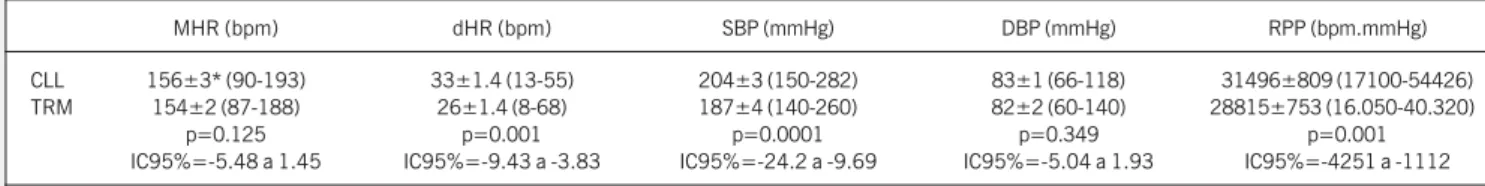
Table II - Hemodynamic variables in the maximum exercise test in cycloergometer (CLL) and treadmill (TRM) (n=60)
MHR (bpm) dHR (bpm) SBP (mmHg) DBP (mmHg) RPP (bpm.mmHg)
CLL 156±3* (90-193) 33±1.4 (13-55) 204±3 (150-282) 83±1 (66-118) 31496±809 (17100-54426) TRM 154±2 (87-188) 26±1.4 (8-68) 187±4 (140-260) 82±2 (60-140) 28815±753 (16.050-40.320)
p=0.125 p=0.001 p=0.0001 p=0.349 p=0.001
IC95%=-5.48 a 1.45 IC95%=-9.43 a -3.83 IC95%=-24.2 a -9.69 IC95%=-5.04 a 1.93 IC95%=-4251 a -1112
5
suitable MHR values in ETs carried out in CLL; the behavior of dHR is not directly comparable when the ETs are performed in CLL and TRM with different forms of posture (standing versus laying down) and of status (light exercise versus rest); values of
SBP and, consequently, of RPP, seem to be higher in ETs performed in CLL; there can be differences in the behavior of the segment ST and the occurrence of arrhythmias, notably supraventricular, when the results from ETs obtained in CLL and TRM are compared.
1 Elhendy A, Mahoney DW, Khandheria BK, et al. Prognostic significance of impair-ment of heart rate response to exercise: impact of left ventricular function and myocardial ischemia. J Am Coll Cardiol 2003; 42: 823-30.
2 Vacanti LJ, Sespedes LB, Sarpi MO. O teste ergométrico é útil, seguro e eficaz, mesmo em indivíduos muito idosos, com 75 anos ou mais. Arq Bras Cardiol 2004; 82: 147-50.
3 Guimarães JI, Stein R, Vilasboas F, et al. Normatização de Técnicas e Equipamen-tos para a Realização de Exames em Ergometria e Ergoespirometria. Arquivos Bra-sileiros de Cardiologia 2003; 80: 458-64.
4 Naughton J, Patterson J, Fox SM, 3rd. Exercise tests in patients with chronic dis-ease. J Chronic Dis 1971; 24: 519-22.
5 Myers J, Buchanan N, Walsh D, et al. Comparison of the ramp versus standard exercise protocols. J Am Coll Cardiol 1991; 17: 1334-42.
6 Myers J, Bellin D. Ramp exercise protocols for clinical and cardiopulmonary exerci-se testing. Sports Med 2000; 30: 23-9.
7 Bruce RA. Exercise testing of patients with coronary heart disease. Principles and normal standards for evaluation. Ann Clin Res 1971; 3: 323-32.
8 Kindermann M, Schwaab B, Finkler N, et al. Defining the optimum upper heart ra-te limit during exercise: a study in pacemaker patients with heart failure. Eur Heart J 2002; 23: 1301-8.
9 Lauer MS, Francis GS, Okin PM, et al. Impaired chronotropic response to exercise stress testing as a predictor of mortality. Jama 1999; 281: 524-9.
10 Lauer MS, Okin PM, Larson MG, et al. Impaired heart rate response to graded exercise. Prognostic implications of chronotropic incompetence in the Framingham Heart Study. Circulation 1996; 93: 1520-6.
11 Cole CR, Blackstone EH, Pashkow FJ, et al. Heart-rate recovery immediately after exercise as a predictor of mortality. N Engl J Med 1999; 341: 1351-7. 12 Araújo CGS. Fast “on” and “off” heart rate transients at different bicycle exercise
levels. Int J Sports Med 1985; 6: 68-73.
13 Araújo CGS, Bastos MAPM, Pinto NLS, et al. A freqüência cardíaca máxima em no-ve diferentes protocolos de teste máximo. Rev Bras Ciên Esporte 1980; 2: 20-31. 14 Almeida MB, Araújo CGS. Efeitos do treinamento aeróbico sobre a freqüência
car-díaca. Rev Bras Med Esporte 2003; 9: 104-12.
15 Araújo CGS. Respostas cardiorrespiratórias a um exercício submáximo prolongado. Arq Bras Cardiol 1983; 41: 37-45.
16 Nagle F, Balke B, Baptista G, et al. Compatibility of progressive treadmill, bycicle and step tests based on oxygen uptake responses. Med Sci Sports Exerc 1971; 3: 149-54.
References
17 Hermansen L, Saltin B. Oxygen uptake during maximal treadmill and bicycle exer-cise. J Appl Physiol 1969; 26: 31-7.
18 LeMura LM, von Duvillard SP, Cohen SL, et al. Treadmill and cycle ergometry testing in 5- to 6-year-old children. Eur J Appl Physiol 2001; 85: 472-8.
19 Wicks JR, Sutton JR, Oldridge NB, et al. Comparison of the electrocardiographic changes induced by maximam exercise testing with treadmill and cycle ergometer. Circulation 1978; 57: 1066-70.
20 Williams KA, Taillon LA, Carter JE, Jr. Asymptomatic and electrically silent myocar-dial ischemia during upright leg cycle ergometry and treadmill exercise (clandestine myocardial ischemia). Am J Cardiol 1993; 72: 1114-20.
21 Hambrecht RP, Schuler GC, Muth T, et al. Greater diagnostic sensitivity of treadmill versus cycle exercise testing of asymptomatic men with coronary artery disease. Am J Cardiol 1992; 70: 141-6.
22 Araújo CGS. Teste de exercício: terminologia e algumas considerações sobre passado, presente e futuro baseadas em evidências. Rev Bras Med Esporte 2000; 6: 77-84. 23 Lester M, Sheffield LT, Trammell P, et al. The effect of age and athletic training on the maximal heart rate during muscular exercise. Am Heart J 1968; 76: 370-6. 24 Davies CT. Limitations to the prediction of maximum oxygen intake from cardiac
frequency measurements. J Appl Physiol 1968; 24: 700-6.
25 Tanaka H, Monahan KD, Seals DR. Age-predicted maximal heart rate revisited. J Am Coll Cardiol 2001; 37: 153-6.
26 Ekblom B, Astrand PO, Saltin B, et al. Effect of training on circulatory response to exercise. J Appl Physiol 1968; 24: 518-28.
27 Patton JF, Vogel JA. Cross-sectional and longitudinal evaluations of an endurance training program. Med Sci Sports 1977; 9: 100-3.
28 Gauri AJ, Raxwal VK, Roux L, et al. Effects of chronotropic incompetence and beta-blocker use on the exercise treadmill test in men. Am Heart J 2001; 142: 136-41. 29 Dresing TJ, Blackstone EH, Pashkow FJ, et al. Usefulness of impaired chrono-tropic response to exercise as a predictor of mortality, independent of the severity of coronary artery disease. Am J Cardiol 2000; 86: 602-9.
30 Gaibazzi N, Petrucci N, Ziacchi V. One-minute heart rate recovery after cycloergometer exercise testing as a predictor of mortality in a large cohort of exercise test candidates: substantial differences with the treadmill-derived parameter.Ital Heart J2004; 5: 183-8. 31 Niederberger M, Bruce RA, Kusumi F, et al. Disparities in ventilatory and circulatory
responses to bicycle and treadmill exercise. Br Heart J 1974; 36: 377-82. 32 Araújo CGS, Nóbrega ACL, Castro CLB. Vagal activity: effect of age, sex and