CLINICAL SCIENCE
Relationship between plasminogen activator inhibitor
type-1 (PAI-1) gene polymorphisms and osteoporosis
in Turkish women
Merih Ozgen,IDidem Turgut Cosan,IIFulya Doganer,IIAhu Soyocak,IIOnur Armagan,IHasan Veysi Gunes,II Irfan Degirmenci,IIGulsah Ogutler Ozkara,IFezan Sahin MutluIII
IEskis¸ehir Osmangazi University, Faculty of Medicine, Department of Physical Medicine and Rehabilitation, Eskisehir, Turkey.IIEskis¸ehir Osmangazi University, Faculty of Medicine, Department of Medical Biology, Eskisehir, Turkey.IIIEskisehir Osmangazi University, Faculty of Medicine, Department of Biostatistics and Medical Informatics, Eskisehir,Turkey.
OBJECTIVE:The development of osteoporosis is associated with several risk factors, such as genetic structures that affect bone turnover and bone mass. The impact of genetic structures on osteoporosis is not known. Plasminogen activator inhibitor type-1 regulates the bone matrix and bone balance. This study assessed the correlation between plasminogen activator inhibitor type-1 gene 4G/5G polymorphisms and osteoporosis in a population of Turkish women.
METHODS:A total of 195 postmenopausal female patients who were diagnosed with osteoporosis (Group I) based on bone mineral density measurements via dual-energy x-ray absorptiometry and 90 females with no osteoporosis (Group II) were included in this study. Correlations between PAI-1 gene 4G/5G polymorphisms and osteoporosis were investigated through the identification of PAI-1 gene 4G/5G polymorphism genotypes using the polymerase chain reaction.
RESULTS:No significant differences in the genotype and allele frequency of 4G/5G plasminogen activator inhibitor type-1 polymorphisms were observed between the two groups, and both groups exhibited the most frequently observed 4G5G genotype.
CONCLUSION:No correlation between the development of osteoporosis in the female Turkish population and 4G/ 5G plasminogen activator inhibitor type-1 gene polymorphisms was observed.
KEYWORDS: Osteoporosis; Polymorphism; Plasminogen Activator Inhibitor Type-1 (PAI-1) Gene; Bone Mineral Density; Turkish Women.
Ozgen M, Turgut Cosan D, Doganer F, Soyocak A, Armagan O, Gunes HV, et al. Relationship between plasminogen activator inhibitor type-1 (PAI-1) gene polymorphisms and osteoporosis in Turkish women. Clinics. 2012;67(11):1299-1302.
Received for publication onSeptember 14, 2012;First review completed onSeptember 16, 2012;Accepted for publication onSeptember 20, 2012
E-mail: [email protected] Tel.: 0 90 532-5837099
INTRODUCTION
Osteoporosis is a systemic skeletal disease that is characterized by an increase in bone fragility due to a decrease in bone mass and the deterioration of bone microarchitecture (1). This disease is especially prevalent in the elderly population, and it is a significant public health issue that reduces patient functioning and quality of life. An improved understanding of the risk factors for osteoporosis is important for the diagnosis, maintenance, and treatment of this significant disease (2,3).
Several epidemiological and clinical studies have demon-strated the importance of genetics in osteoporosis pathogenesis (4-6). Genetic factors affect bone turnover and can result in the reduction of bone mass to,50-80% (5-7). Gene polymorphisms
may contribute to osteoporosis and impact bone mineral density (4-8).
Plasminogen activator inhibitor-1 (PAI-1) is a 50-kDa, single-chain glycoprotein in the serine protease inhibitor family (9). A plasminogen activation system (PAS) that was initially identified in the fibrinolytic system, and its fundamental inhibitor, PAI-1, regulate the bone matrix and alter bone balance (10). PAI-1 primarily inhibits tissue-type (tPa) and urokinase (uPa) plasminogen activators (11) and reduces extracellular matrix destruction by decreasing the plasmin-mediated activation of matrix metalloprotei-nases (MMPs) (12).
Genetic factors predominantly determine plasma PAI-1 levels (13). The human PAI-1 gene contains various
Copyrightß2012CLINICS– This is an Open Access article distributed under
the terms of the Creative Commons Attribution Non-Commercial License (http:// creativecommons.org/licenses/by-nc/3.0/) which permits unrestricted non-commercial use, distribution, and reproduction in any medium, provided the original work is properly cited.
No potential conflict of interest was reported.
CLINICS 2012;67(11):1299-1302 DOI:10.6061/clinics/2012(11)13
polymorphic loci in approximately 12.22 kb on chromosome 7q22. The 4G/5G insertion/deletion is the most investigated polymorphism, which is 675 base pairs (bp) upstream of the transcriptional start site. This polymorphism regulates the expression of the PAI-1 gene (9,13,14).
The correlation of the PAI-1 4G/5G insertion/deletion polymorphism with several diseases, such as coronary artery disease, asthma, hypertension, stroke, obesity, rheu-matoid arthritis, and osteoarthritis, has been investigated previously (15-21). However, the contribution of PAI-1 insertion/deletion variations (4G/5G) to osteoporosis has not been investigated in the Turkish population. This study investigated the correlation between PAI-1 gene poly-morphisms and osteoporosis in Turkish females.
MATERIALS AND METHODS
Subjects
Postmenopausal females who were admitted to the Osteoporosis Clinic of the Physical Medicine and Rehabi-litation Department of Eskisehir Osmangazi University (Turkey) were informed of the study, and patients who opted for inclusion in the study were evaluated. Patients who were diagnosed with parathyroid, thyroid, liver, and rheumatolo-gical diseases that affect bone metabolism; patients with a history of malignancy or surgically induced menopause; and patients who used drugs affecting bone metabolism (e.g., corticosteroids, anticonvulsants, and heparin) during the clinical and laboratory assessments were excluded from the study. Erythrocyte sedimentation rate, complete blood count, serum alkaline phosphatase, calcium, phosphorous, serum glutamic oxaloacetic transaminase, serum glutamic pyruvic transaminase, gamma-glutamyl transpeptidase, blood urea nitrogen, creatinine, glucose, uric acid, albumin, total protein, urine calcium/creatinine, thyroid-stimulating hormone, para-thyroid hormone, cortisol and vitamin D levels were measured prior to the study. A total of 285 patients satisfied the study criteria and were included in the study. The age, height, weight, and body mass index (BMI) of the participants were evaluated. All participants underwent dual-energy x-ray absorptiometry (DEXA) evaluations, and 195 postmeno-pausal females were diagnosed with osteoporosis based on this assessment (Group I). Ninety patients without osteo-porosis were included in the control group (Group II). All participants provided informed consent in compliance with the study protocol (#2009/229), which was approved by the Ethics Committee of the Medical Faculty of Eskisehir Osmangazi University (Turkey).
Bone mineral density
The participants underwent DEXA scanning using a Hologic QDR 4500 W system (Hologic, Inc., Bedford, USA) to assess bone mineral density (BMD), and the lumbar spine (vertebrae L1-L4) and hip (femur neck) were evaluated. Patients with a mean bone density below 2.5 SD were diagnosed with osteoporosis, as recommended by the World Health Organization (WHO).
Sample collection and determination of PAI-1 genotypes
Genomic DNA isolation was performed using the salt-extraction method in 10 ml of peripheral blood that was collected in EDTA tubes for the analysis of 4G/5G polymorphisms in the promoter region of PAI-1. The
obtained genomic DNA was maintained at 4
˚
C. The PAI-1 polymorphism gene region was amplified in a thermal cycler (Sacem Life Technologies, Peltier-based Thermal Cycler SCM 96G, Turkey) using 25ml of a PCR mixture containing 0.5ml DNA, 10X PCR Buffer (Biolabs, New England), 0.2 mmol/L dNTPs (Sigma, Germany), 1.25 U Taq polymerase (Biolabs, New England), 50 pmol of 4G- or 5G-specific primer, 50 pmol of downstream primer, and 2.5 pmol of upstream primer. The following primers (Metabion, Germany) were used: 5’-GTC TGG ACA CGT GGG GG-3’ for the 5G allele, 5’-GTC TGG ACA CGT GGG GA-3’ for the 4G allele, 5’-TGC AGC CAG CCA CGT GAT TGT CTA G-3’ for the downstream primer, and 5’-AAG CTT TTA CCA TGG TAA CCC CTG GT-3’ for the upstream primer (positive control). The PCR mixture was subjected to 35 cycles for 60 sec at 94˚
C, 30 sec at 54˚
C, and 40 sec at 72˚
C following the initial denaturation for 3 min at 94˚
C. These PCR products were processed in 2% agarose gel and analyzed under UV light (Labwork, Cambridge, United Kingdom). The 4G and 5G alleles were defined according to a 139-bp DNA fragment of the general downstream primer that was produced during the PCR process. Samples that produced a 139-bp band from the 4G primer and that did not produce a 139-bp band from the 5G primer were considered a homozygous 4G genotype. Samples that produced a 139-bp band from the 5G primer but that did not produce a 139-bp band from the 4G primer were considered a homozygous 5G genotype. Samples that produced a 139-bp band from both primers were considered a heterozygous 4G5G genotype.Statistical analysis
The data were evaluated using SPSS Version 20 software (IBM Corp. Armonk, New York, USA). The continuous variables were not normally distributed based on the Shapiro-Wilk test for normality. The Mann-Whitney U test was implemented for the comparison of the two groups. Medians (quartiles) are provided as descriptive statistics. The Pearson chi-square test was conducted for categorical variables. N and % values are provided. A p,0.05 was considered statistically significant.
RESULTS
This study investigated the effect of the PAI-1 gene 4G/ 5G polymorphisms on the development of osteoporosis in Turkish women. The study groups are listed in Table 1. No significant differences in the genotype and allele frequency of the 4G/5G PAI-1 polymorphism were observed between the groups (p= 0.619 andp= 0.361, respectively). However,
the most frequent genotype, 4G5G, was observed in both groups. The 4G5G genotype was 39.49% in Group I and 42.22% in Group II. The 4G and 5G allele frequencies ranged from 47.4 - 52.6% in Group I and 43.3 - 56.7% in Group II.
DISCUSSION
Osteoporosis is characterized by low bone mass, an increase in bone fragility, deterioration in bone microarch-itecture, and an increase in the risk of fracture (1). Some metabolic changes, such as those that occur due to a lack of estrogen, immobilization, metabolic acidosis, hyperpar-athyroidism, and systemic and local inflammatory diseases, affect the osteoclast count and activity associated with bone
PAI-1 gene polymorphism and osteoporosis
Ozgen M et al. CLINICS 2012;67(11):1299-1302
turnover (22). Prostaglandins, insulin-like growth factors (IGFs), interleukins (IL-1, IL-6, and IL-11), tumor necrosis factor (TNF), and several local factors in bone, such as transforming growth factor (TGF), also contribute to the regulation of bone formation and resorption (22,23).
TGF-b is an anabolic factor that increases extracellular matrix production and the expression of various types of collagen and proteoglycans (24,25). TGFb1 polymorphisms may be significantly relevant in BMD and the occurrence of fracture (24,26). PAI-1 is known to have a regulatory effect on matrix components, including TGF-b, matrix c -carbox-yglutamic acid (Gla) protein, and osteocalcin. Therefore, PAI-1 may affect bone matrix biology and significantly regulate bone remodeling (10). PAI-1 levels are regulated by a 4G/5G insertion/deletion polymorphism (13).
This study investigated the relationship of the 4G/5G polymorphism, which regulates PAI-1 as an inhibitor of the plasminogen activator system, with osteoporosis in Turkish women.
A relationship between the PAI-1 4G/5G gene poly-morphism and diseases, such as coronary artery disease, hypertension, stroke, and obesity, has been reported previously, but this polymorphism is not related to asthma, rheumatoid arthritis, and osteoarthritis (15-21). Genetic variations occur in populations. Previous studies have investigated the PAI-1 4G/5G insertion/deletion poly-morphism in the Turkish population, but its relationship with osteoporosis has not been investigated; this relation-ship was examined in our study for the first time.
No differences in the PAI-1 4G/5G genotype were observed between the postmenopausal osteoporotic patients and the healthy control group. The role of common variations of COLIA-1, TGFb-1, and PAI-1 genes in early postmenopausal osteoporotic Caucasians and healthy women was previously investigated by Hubacek et al., who observed no significant difference in the PAI-1 4G/5G genotype between osteoporotic patients and the healthy control group, which is consistent with our study. However, the 4G4G genotype was more common in the osteoporotic patient group compared with the control group (27).
Our results suggest that the 4G/5G PAI-1 polymorphism cannot be used as a marker for the development of osteoporosis in Turkish women. However, this result may not be applicable to all populations when gene pools, lifestyles, and gene-environment interactions in various populations are considered. Therefore, multi-centered stu-dies on different populations and in different gene regions in larger samples are required to establish the correlation between the 4G/5G PAI-1 polymorphism and osteoporosis.
AUTHOR CONTRIBUTIONS
Ozgen M was responsible for the study design, evaluation and collection of clinical data, manuscript writing, and critical review. Turgut Cosan D was responsible for the study design, molecular biological analysis, genetic counseling, manuscript writing, and critical review. Doganer F and Soyocak A contributed to the molecular biological analysis, genetic counseling, manuscript writing, and critical review. Gunes HV and Degirmenci I contributed to the genetic counseling, manuscript writing, and critical review. Armagan O contributed to the manuscript writing and critical review. Ogutler Ozkara G contributed to the evaluation and collection of clinical data. Sahin Mutlu F contributed to the statistical analysis.
REFERENCES
1. Kanis JA, Melton LJ, Christiansen C, Johnston CC, Khaltaev N. The diagnosis of osteoporosis. J Bone Miner Res. 1994;9(8):1137-41. 2. Dontas IA, Yiannakopoulos CK. Risk factors and prevention of
osteoporosis-related fractures. Journal of Musculoskeletal & Neuronal Interactions. 2007;7(3):268-72.
3. Borer KT. Physical activity in the prevention and amelioration of osteoporosis in women: interaction of mechanical, hormonal and dietary factor. Sports Medicine. 2005;35(9):779-830, http://dx.doi.org/10.2165/ 00007256-200535090-00004.
4. Eisman JA. Genetics of osteoporosis. Endocr Rev. 1999;20(6):788-804, http://dx.doi.org/10.1210/er.20.6.788.
5. Ralston SH. Genetic Control of Susceptibility to Osteoporosis. The Journal of Clinical Endocrinology & Metabolism. 2002;87(6):2460-6, http://dx.doi.org/10.1210/jc.87.6.2460.
6. Uitterlinden AG, Van Meurs JB, Rivadeneira F, Pols HA. Identifying genetic risk factors for osteoporosis. J Musculoskelet Neuronal Interact. 2006;6(1):16-26.
7. Ferrari S. Human genetics of osteoporosis. Best Pract Res Clin Endocrinol Metab. 2008;22(5):723-35, http://dx.doi.org/10.1016/j.beem.2008.08.007. 8. Mitchell BD, Yerges-Armstrong LM. The genetics of bone loss: challenges and prospects. J Clin Endocrinol Metab. 2011;96(5):1258-68, http://dx.doi.org/10.1210/jc.2010-2865.
9. Ha H, Oh EY, Lee HB. The role of plasminogen activator inhibitor 1 in renal and cardiovascular diseases. Nat Rev Nephrol. 2009;5(4):203-11, http://dx.doi.org/10.1038/nrneph.2009.15.
10. Nordstrom SM, Carleton SM, Carson WL, Eren M, Phillips CL, Vaughan DE. Transgenic over-expression of plasminogen activator inhibitor-1 results in age-dependent and gender-specific increases in bone strength and mineralization. Bone. 2007;41(6):995-1004, http://dx.doi.org/ 10.1016/j.bone.2007.08.020.
11. Vassalli JD, Sappino AP, Belin D. The plasminogen activator/plasmin system. J Clin Invest. 1991;88(4):1067-72, http://dx.doi.org/10.1172/ JCI115405.
12. Wang X, Lee SR, Arai K, Tsuji K, Rebeck GW, Lo EH. Lipoprotein receptor-mediated induction of matrix metalloproteinase by tissue plasminogen activator. Nat Med. 2003;9(10):1313-7, http://dx.doi.org/ 10.1038/nm926.
13. Dawson S, Hamsten A, Wiman B, Henney A, Humphries S. Genetic variation at the plasminogen activator inhibitor-1 locus is associated with altered levels of plasma plasminogen activator inhibitor-1 activity. Arterioscler Thromb. 1991;11(1):183-90, http://dx.doi.org/10.1161/ 01.ATV.11.1.183.
14. Dawson SJ, Wiman B, Hamsten A, Green F, Humphries SE, Henney AM. The two allele sequences of a common polymorphism in the promoter of the plasminogen activator inhibitor-1 (PAI-1) gene respond differently to interleukin-I in HepG2 cells. J Biol Chem. 1993;268(15):10739-45. 15. Margaglione M, Cappucci G, Colaizzo D, Giuliani N, Vecchione G,
Grandone E, et al. The PAI-1 gene locus 4G/5G polymorphism is associated with a family history of coronary artery disease. Arterioscler Thromb Vasc Biol. 1998;18(2):152-6, http://dx.doi.org/10.1161/ 01.ATV.18.2.152.
16. Cosan D, Kurt E, Kurt H, Degirmenci I, Kuc¸ukarabaci B, Metintas M, et al. Plasminogen activator inhibitor type-1 gene 4G/5G polymorphism
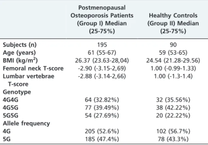
Table 1 -Age, body mass index, femoral neck and lumbar vertebrae T-score averages and 4G/5G PAI-1 gene distribution in both groups.
Postmenopausal Osteoporosis Patients
(Group I) Median (25-75%)
Healthy Controls (Group II) Median
(25-75%)
Subjects (n) 195 90
Age (years) 61 (55-67) 59 (53-65)
BMI (kg/m2) 26.37 (23.63-28,04) 24.54 (21.28-29.56)
Femoral neck T-score -2.90 (-3.15-2,69) 1.00 (-0.99-1.33)
Lumbar vertebrae T-score
-2.88 (-3.14-2,66) 1.00 (-1.3-1.4)
Genotype
4G4G 64 (32.82%) 32 (35.56%)
4G5G 77 (39.49%) 38 (42.22%)
5G5G 54 (27.69%) 20 (22.22%)
Allele frequency
4G 205 (52.6%) 102 (56.7%)
5G 185 (47.4%) 78 (43.3%)
Age, 1-2:p= 0.105; BMI, 1-2:p= 0.171; Femoral neck T-score, 1-2:p,0.001; Lumbar vertebrae T-score, group I - group II:p,0.001; Genotypex2: 0.961
df = 2p= 0.619; Allele frequencyx2: 0.834, df = 1,p= 0.361.
CLINICS 2012;67(11):1299-1302 PAI-1 gene polymorphism and osteoporosis Ozgen M et al.
in Turkish adult patients with asthma. Genet Test Mol Biomarkers. 2009;13(4):543-6, http://dx.doi.org/10.1089/gtmb.2009.0036.
17. Gunes HV, Cosan DT, Ata N, Birdane A, Ustuner MC, Dikmen M, et al. Plasminogen activator inhibitor type-1 gene 4G/5G polymorphism is associated with hypertensive patients in the Turkish population. Genet Test Mol Biomarkers. 2010;14(3):303-5, http://dx.doi.org/10.1089/ gtmb.2009.0199.
18. Kucukarabaci B, Gunes HV, Ozdemir G, Cosan D, Ozbabalik D, Dikmen M, et al. Investigation of association between plasminogen activator inhibitor type-1 (PAI-1) gene 4G/5G polymorphism frequency and plasma PAI-1 enzyme activity in patients with acute stroke. Genet Test. 2008;12(3):443-51, http://dx.doi.org/10.1089/gte.2008.0025.
19. Fernandes KS, Sandrim VC. 4G/5G polymorphism modulates PAI-1 circulating levels in obese women. Molecular Cell Biochemicals. 2012;364(1-2):299-301, http://dx.doi.org/10.1007/s11010-012-1230-1. 20. Mun˜oz-Valle JF, Ruiz-Quezada SL, Orego´n-Romero E,
Navarro-Herna´ndez RE, Castan˜eda-Saucedo E, De la Cruz-Mosso U, et al. PAI-1 mRNA expression and plasma level in rheumatoid arthritis: relation-ship with 4G/5G PAI-1 polymorphism. Rheumatol Int. 2011; (Epub ahead of print).
21. Bayram B, Sayin E, Erkasap N, Onlu H, Ozkurt M, Sahin F, et al. Lack of association between plasminogen activator inhibitor type-1 (PAI-1) gene
4G/5G polymorphism and osteoarthritis. Rheumatol Int. 2012;32(1):259-62, http://dx.doi.org/10.1007/s00296-010-1737-2.
22. Manolagas SC, Jilka RL. Bone marrow, cytokines, and bone remodeling. N Engl J Med. 1995;332(5):305-31.
23. Estai MA, Suhaimi F, Das S, Shuid AN, Mohamed Z, Soelaiman IN. Expression of TGF-b1 in the blood during fracture repair in an estrogen-deficient rat model. Clinics. 2011;66(12):2113-9, http://dx.doi.org/ 10.1590/S1807-59322011001200018.
24. Yamada Y, Miyauchi A, Goto J, Takagi Y, Okuizumi H, Kanematsu M, et al. Association of a polymorphism of the transforming growth factor-beta1 gene with genetic susceptibility to osteoporosis in postmenopausal Japanese women. J Bone Miner Res. 1998;13(10):1569-76.
25. Shuler FD, Georgescu HI, Niyibizi C et al. Increased matrix synthesis following adenoviral transfer of a transforming growth factor beta1 gene into articular chondrocytes. J Orthop Res. 2000;18(4):585-92, http:// dx.doi.org/10.1002/jor.1100180411.
26. Ralston SH. Genetics of osteoporosis. Ann NY Acad Sci. 2010;1192:181-9, http://dx.doi.org/10.1111/j.1749-6632.2009.05317.x.
27. Hubacek JA, Weıchetova M, Bohuslavova R, Skodova Z, Stepan JJ, Ada´mkova V. Genetic polymorphisms of TGF-b, PAI-1, and COL1A-1, and determination of bone mineral density in caucasian females. Endocrine Regulations. 2006;40(3):77-81.
PAI-1 gene polymorphism and osteoporosis
Ozgen M et al. CLINICS 2012;67(11):1299-1302