The 4G/4G Genotype of PAI-1 Polymorphism
Is Associated with Higher Plasma PAI-1
Concentrations and Mortality in Patients with
Severe Sepsis
Leonardo Lorente1
*, María M. Martín2, Juan M. Borreguero-León3, Ysamar Barrios4,
Jordi Solé-Violán5, José Ferreres6, Lorenzo Labarta7, César Díaz8, Alejandro Jiménez9
1Intensive Care Unit, Hospital Universitario de Canarias, La Laguna, Santa Cruz de Tenerife, Spain,
2Intensive Care Unit, Hospital Universitario Nuestra Señora Candelaria, Santa Cruz Tenerife, Spain, 3Laboratory Deparment, Hospital Universitario de Canarias, La Laguna, Tenerife, Spain,4Laboratory Deparment of the Research Unit, Hospital Universitario de Canarias, La Laguna, Santa Cruz de Tenerife, Spain,5Intensive Care Unit, Hospital Universitario Dr. Negrín, Las Palmas de Gran Canaria, Spain,
6Intensive Care Unit, Hospital Clínico Universitario de Valencia, Valencia, Spain,7Intensive Care Unit, Hospital San Jorge, Huesca, Spain,8Intensive Care Unit, Hospital Insular, Las Palmas de Gran Canaria, Spain,9Statistical Deparment of the Research Unit, Hospital Universitario de Canarias, La Laguna, Santa Cruz de Tenerife, Spain
Abstract
Objective
Two studies have reported that patients with the 4G/4G genotype of the plasminogen acti-vator inhibitor-1 (PAI-1) genetic polymorphism had higher plasma PAI-1 concentrations and higher risk of death than those with the 4G/5G or 5G/5G genotypes; one study involved 175 children with meningococcal disease, and the other included 88 adult patients with septic shock. Thus, the objective of this study was to determine whether there is an association be-tween carriage of the 4G/4G genotype, plasma PAI-1 concentrations and mortality in a large series of adult septic patients.
Methods
An observational, prospective, multicenter study was carried out in six Spanish Intensive Care Units including severe septic patients. We determined the PAI-1 4G/5G polymorphism and plasma PAI-1 concentrations in all patients. The end-points of the study were 30-day and 6-month mortality.
Results
We included a total of 260 patients, 82 (31.5%) with 4G/4G, 126 (48.5%) with 4G/5G and 52 (20.0%) with 5G/5G genotype. Multivariate logistic regression analysis showed that the 4G/4G genotype was associated with higher mortality at 30 days (Odds Ratio = 1.95; 95% CI = 1.063– 3.561; p = 0.03) and at 6 months (Odds Ratio = 2.19; 95% CI = 1.221–3.934; p = 0.01), and
OPEN ACCESS
Citation:Lorente L, Martín MM, Borreguero-León JM, Barrios Y, Solé-Violán J, Ferreres J, et al. (2015) The 4G/4G Genotype of PAI-1 Polymorphism Is Associated with Higher Plasma PAI-1 Concentrations and Mortality in Patients with Severe Sepsis. PLoS ONE 10(6): e0129565. doi:10.1371/journal. pone.0129565
Academic Editor:Giovanni Camussi, University of Torino, ITALY
Received:February 25, 2015
Accepted:May 11, 2015
Published:June 11, 2015
Copyright:© 2015 Lorente et al. This is an open access article distributed under the terms of the
Creative Commons Attribution License, which permits unrestricted use, distribution, and reproduction in any medium, provided the original author and source are credited.
Data Availability Statement:All relevant data are within the paper.
that higher plasma PAI-1 concentrations were associated with higher mortality at 30 days (Odds Ratio = 1.01; 95% CI = 1.002–1.022; p = 0.02) at 6 months (Odds Ratio = 1.01; 95% CI = 1.003–1.023; p = 0.01). Multivariate linear regression analysis showed that increased plasma PAI-1 concentrations were associated with the PAI-1 4G/4G genotype (regression co-efficient = 4.82; 95% CI = 3.227 to 6.406; p<0.001).
Conclusions
The major findings of our study, to our knowledge the largest series reporting data about 4G/5G polymorphism of the PAI-1 gene, plasma PAI-1 concentrations and mortality in sep-tic patients, were that sepsep-tic patients with the 4G/4G genotype had higher plasma PAI-1 concentrations and higher risk of death than those with 4G/5G or 5G/5G genotypes.
Introduction
Sepsis represents a systemic response of the immune system to infection, which is a common, expensive, and frequently fatal condition. During sepsis, the pro-coagulant and anti-fibrinolyt-ic pathways lead to manti-fibrinolyt-icrovascular fibrin deposition, resulting in morgan failure, and ulti-mately death. [1].
Plasminogen activator inhibitor-1 (PAI-1) plays an important role in the fibrinolytic re-sponse [2]. Previous studies have found higher plasma PAI-1 concentrations in non-surviving than in surviving septic patients [3–12]. In addition, the association between PAI-1 4G/5G polymorphism and mortality in septic patients has been studied [10–20]. Some authors report that patients with the 4G/4G genotype of the PAI-1 gene had higher mortality rates than pa-tients with other genotypes [10–14]; however, this was not found in other studies [15–20]. A recently published meta-analysis [21], including most of those studies [10–19], found that pa-tients with the 4G/4G genotype of the PAI-1 gene had a higher risk of death than papa-tients with 4G/5G or 5G/5G genotypes. However, only 3 of those studies also measured plasma PAI-1 con-centrations [10–12]. Two studies have reported that patients with the 4G/4G genotype of the PAI-1 gene had higher plasma PAI-1 concentrations and higher risk of death than those with the 4G/5G or 5G/5G genotypes; one study involved 175 children with meningococcal disease [10], and the other included 88 adult patients with septic shock [11]. Another study with 166 adult septic patients reported that patients with the 4G/4G genotype and that patients with higher plasma PAI-1 concentrations had a higher risk of death; however, an association be-tween PAI-1 4G/5G polymorphism and plasma PAI-1 concentrations was not reported [12]. Thus, the objective of this study was to determine whether there is an association between car-riage of the PAI-1 4G/4G genotype, plasma PAI-1 concentrations and mortality in a large series of adult septic patients.
Materials and Methods
Design and Subjects
A multicenter, cohort study was carried out in 260 patients with severe sepsis from six Spanish Intensive Care Units. The study was approved by the Institutional Review Boards of the six participating hospitals: Hospital Universitario de Canarias (La Laguna. Santa Cruz de Tenerife. Spain), Hospital Universitario Nuestra Señora de Candelaria (Santa Cruz de Tenerife. Spain), Hospital Universitario Dr. Negrín (Las Palmas de Gran Canaria. Spain), Hospital Clínico
Universitario de Valencia (Valencia. Spain), Hospital San Jorge (Huesca. Spain) and Hospital Insular (Las Palmas de Gran Canaria. Spain). Written informed consent from the patients or from their family members was obtained.
The diagnosis of severe sepsis was established according to the International Sepsis Defini-tions Conference [22]. Exclusion criteria were: age<18 years, pregnancy, lactation, human
immunodeficiency virus (HIV), white blood cell count<1,000/mm3, solid or hematologic
tu-mour, or immunosuppressive, steroid or radiation therapy.
Variables recorded
The following variables were recorded for each patient: age, sex, diabetes mellitus, ischemic heart disease, chronic obstructive pulmonary disease (COPD), Acute Physiology and Chronic Health Evaluation II (APACHE II) score [23], activated partial thromboplastin time (aPTT), empiric an-timicrobial treatment, bilirubin, bloodstream infection, creatinine, international normalized ratio (INR), lactic acid, leukocytes, microorganism responsible, pressure of arterial oxygen/fraction in-spired of oxygen (PaO2/FiO2), platelets, site of infection, and Sepsis-related Organ Failure
Assess-ment [SOFA] score [24]. The end-points of the study were 30-day and 6-month mortality.
Blood samples
Blood samples were collected from patients at the time severe sepsis was diagnosed. Venous blood samples were placed in citrated plasma tubes and centrifuged within 30 minutes at 1000g for 15 minutes, and frozen at -80°C until the determination of PAI-1 concentration. In addition, venous blood were placed in EDTA-containing tubes and frozen at -80°C until deter-mination of genetic polymorphism of PAI-1.
Gene analysis
Gene analysis was centralized at the Research Unit of the Hospital Universitario de Canarias (La Laguna. Tenerife. Spain). Venous blood samples in EDTA-containing tubes were subjected to DNA purification using proteinase K, phenol-chloroform extraction, and ethanol precipita-tion. Genotyping was performed in a blinded manner, without knowledge of any clinical data. PAI-1 4G/5G polymorphism was determined for all patients using primers and restriction en-donuclease digestion. In addition, 22 neutral markers were genotyped to follow genomic con-trol strategies that would detect spurious associations due to population substructure. The neutral markers chosen were Alu repeats distributed throughout the genome.
Plasma PAI-1 level assay
The assay of plasma PAI-1 concentrations was centralized at the Laboratory Department of the Hospital Universitario de Canarias (La Laguna. Tenerife. Spain). PAI-1 antigen was assayed by specific ELISA (Imubind Plasma PAI-1 Elisa. American Diagnostica, Inc., Stanford, CT, USA) according to the manufacturer's instructions. The assay detects latent (inactive) and active forms of PAI-1 and PAI-1 complexes. The interassay coefficient of variation (CV) was<5%
(n = 20) and the detection limit for the assay was 1 ng/ml.
Statistical Methods
analysis was applied to determine the independent contribution of PAI-1 4G/5G polymor-phism on 30-day and 6-month mortality, after controlling for diabetes mellitus, ischemic heart disease, COPD, age, SOFA score and serum lactic acid concentrations. Odds ratio (OR) and 95% confidence intervals (CI) were calculated as measurement of the clinical impact of the pre-dictor variables. We used linear regression modelling to analyze the relationship between plas-ma PAI-1 concentrations as the dependent variable, and 4G/5G polymorphism, diabetes mellitus, ischemic heart disease and COPD as independent variables. Regression coefficients and 95% CI were calculated as measurement of the clinical impact of the predictor variables. Survival curves at 30 days and 6 months, using 4G/4G vs other genotypes of PAI-1 gen, were plotted using the Kaplan-Meier method and compared by log-rank test. We used Chi-square to test Hardy-Weinberg equilibrium among our genotypes. A P value of less than 0.05 was con-sidered statistically significant. Statistical analyses were performed with SPSS 17.0 (SPSS Inc., Chicago, IL, USA) and NCSS 2000 (Kaysville, Utah).
Results
We included a total of 260 patients with severe sepsis; 82 (31.5%) with genotype 4G/4G, 126 (48.5%) with genotype 4G/5G and 52 (20.0%) with genotype 5G/5G of the PAI-1 gene. We found no significant deviation from Hardy-Weinberg equilibrium among our genotypes (Chi-square = 0.08; p = 0.78)
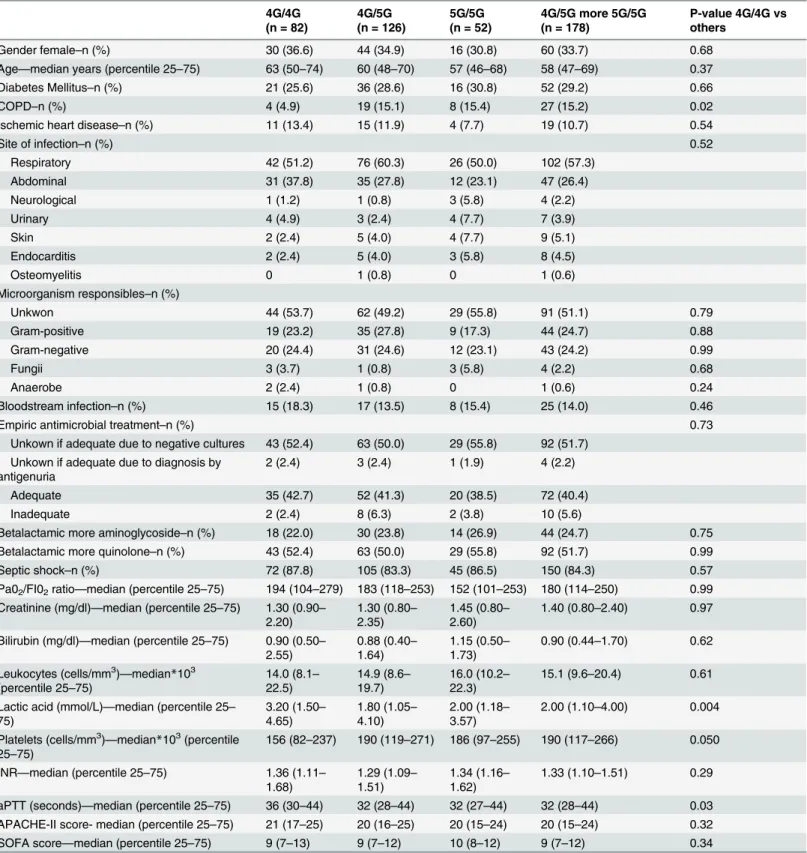
As shown inTable 1, there were no significant differences between different genotypes in gender, age, diabetes mellitus, ischemic heart disease, CRF, site of infection, microorganism re-sponsible, bloodstream infection, septic shock, empiric antimicrobial treatment, PaO2/FIO2,
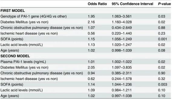
creatinin, bilirubin, leukocytes count, INR, APACHE-II score and SOFA score. However, pa-tients with the 4G/4G genotype showed higher lactatemia, aPTT, and plasma PAI-1 concentra-tions, and lower platelet count and COPD rate than patients with other genotypes. In addition, patients with the 4G/4G genotype showed higher 30-day and 6-month mortality than patients with other genotypes (Table 2). In addition, we have not found statistically significant differ-ences in 30-day (p = 0.53) and 6-month (p = 0.42) mortality rate between the different ICUs. Multiple logistic regression analysis showed that the 4G/4G genotype was associated with higher mortality at 30 days (Odds Ratio = 1.95; 95% CI = 1.063–3.561; p = 0.03) and at 6 months (Odds Ratio = 2.19; 95% CI = 1.221–3.934; p = 0.01), after controlling for diabetes mel-litus, ischemic heart disease, COPD, age, SOFA score and serum lactic acid concentrations (Ta-bles3and4). In addition, multiple logistic regression analysis showed that plasma PAI-1 concentrations were associated with higher mortality at 30 days (Odds Ratio = 1.01; 95% CI = 1.002–1.022; p = 0.02) and at 6 months (Odds Ratio = 1.01; 95% CI = 1.003–1.023; p = 0.01), after controlling for diabetes mellitus, ischemic heart disease, COPD, age, SOFA score and serum lactic acid concentrations (Tables3and4).
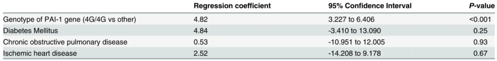
Multivariate linear regression analysis showed that plasma PAI-1 concentrations were asso-ciated with PAI-1 4G/5G polymorphism, after controlling for diabetes mellitus, ischemic heart disease and COPD (regression coefficient = 4.82; 95% CI = 3.227 to 6.406; p<0.001) (Table 5).
Survival analysis showed that patients with the PAI-1 4G/4G genotype presented lower 30-day survival (Chi-square = 8.82; Hazard ratio = 1.9 (95% CI = 1.17–2.95); p = 0.003) and lower 6-month survival square = 8.82; p = 0.003) than those with other genotypes (Chi-square = 11.4; Hazard ratio = 1.9 (95% CI = 1.24–2.88); p<0.001) (Fig 1).
Discussion
Table 1. Characteristics of severe septic patients according to genotype in the PAI-1 gene.
4G/4G (n = 82)
4G/5G (n = 126)
5G/5G (n = 52)
4G/5G more 5G/5G (n = 178)
P-value 4G/4G vs others
Gender female–n (%) 30 (36.6) 44 (34.9) 16 (30.8) 60 (33.7) 0.68
Age—median years (percentile 25–75) 63 (50–74) 60 (48–70) 57 (46–68) 58 (47–69) 0.37
Diabetes Mellitus–n (%) 21 (25.6) 36 (28.6) 16 (30.8) 52 (29.2) 0.66
COPD–n (%) 4 (4.9) 19 (15.1) 8 (15.4) 27 (15.2) 0.02
Ischemic heart disease–n (%) 11 (13.4) 15 (11.9) 4 (7.7) 19 (10.7) 0.54
Site of infection–n (%) 0.52
Respiratory 42 (51.2) 76 (60.3) 26 (50.0) 102 (57.3)
Abdominal 31 (37.8) 35 (27.8) 12 (23.1) 47 (26.4)
Neurological 1 (1.2) 1 (0.8) 3 (5.8) 4 (2.2)
Urinary 4 (4.9) 3 (2.4) 4 (7.7) 7 (3.9)
Skin 2 (2.4) 5 (4.0) 4 (7.7) 9 (5.1)
Endocarditis 2 (2.4) 5 (4.0) 3 (5.8) 8 (4.5)
Osteomyelitis 0 1 (0.8) 0 1 (0.6)
Microorganism responsibles–n (%)
Unkwon 44 (53.7) 62 (49.2) 29 (55.8) 91 (51.1) 0.79
Gram-positive 19 (23.2) 35 (27.8) 9 (17.3) 44 (24.7) 0.88
Gram-negative 20 (24.4) 31 (24.6) 12 (23.1) 43 (24.2) 0.99
Fungii 3 (3.7) 1 (0.8) 3 (5.8) 4 (2.2) 0.68
Anaerobe 2 (2.4) 1 (0.8) 0 1 (0.6) 0.24
Bloodstream infection–n (%) 15 (18.3) 17 (13.5) 8 (15.4) 25 (14.0) 0.46
Empiric antimicrobial treatment–n (%) 0.73
Unkown if adequate due to negative cultures 43 (52.4) 63 (50.0) 29 (55.8) 92 (51.7) Unkown if adequate due to diagnosis by
antigenuria
2 (2.4) 3 (2.4) 1 (1.9) 4 (2.2)
Adequate 35 (42.7) 52 (41.3) 20 (38.5) 72 (40.4)
Inadequate 2 (2.4) 8 (6.3) 2 (3.8) 10 (5.6)
Betalactamic more aminoglycoside–n (%) 18 (22.0) 30 (23.8) 14 (26.9) 44 (24.7) 0.75 Betalactamic more quinolone–n (%) 43 (52.4) 63 (50.0) 29 (55.8) 92 (51.7) 0.99
Septic shock–n (%) 72 (87.8) 105 (83.3) 45 (86.5) 150 (84.3) 0.57
Pa02/FI02ratio—median (percentile 25–75) 194 (104–279) 183 (118–253) 152 (101–253) 180 (114–250) 0.99 Creatinine (mg/dl)—median (percentile 25–75) 1.30 (0.90–
2.20)
1.30 (0.80– 2.35)
1.45 (0.80– 2.60)
1.40 (0.80–2.40) 0.97
Bilirubin (mg/dl)—median (percentile 25–75) 0.90 (0.50– 2.55)
0.88 (0.40– 1.64)
1.15 (0.50– 1.73)
0.90 (0.44–1.70) 0.62
Leukocytes (cells/mm3)
—median*103 (percentile 25–75)
14.0 (8.1– 22.5)
14.9 (8.6– 19.7)
16.0 (10.2– 22.3)
15.1 (9.6–20.4) 0.61
Lactic acid (mmol/L)—median (percentile 25– 75)
3.20 (1.50– 4.65)
1.80 (1.05– 4.10)
2.00 (1.18– 3.57)
2.00 (1.10–4.00) 0.004
Platelets (cells/mm3)
—median*103(percentile 25–75)
156 (82–237) 190 (119–271) 186 (97–255) 190 (117–266) 0.050
INR—median (percentile 25–75) 1.36 (1.11– 1.68)
1.29 (1.09– 1.51)
1.34 (1.16– 1.62)
1.33 (1.10–1.51) 0.29
aPTT (seconds)—median (percentile 25–75) 36 (30–44) 32 (28–44) 32 (27–44) 32 (28–44) 0.03 APACHE-II score- median (percentile 25–75) 21 (17–25) 20 (16–25) 20 (15–24) 20 (15–24) 0.32 SOFA score—median (percentile 25–75) 9 (7–13) 9 (7–12) 10 (8–12) 9 (7–12) 0.34
our study were that septic patients with the 4G/4G genotype had higher plasma PAI-1 concen-trations and higher risk of death than those with 4G/5G or 5G/5G genotype.
Previous studies have found higher plasma PAI-1 concentrations in septic patients than in controls [25–28], and higher plasma PAI-1 concentrations in non-surviving than in surviving septic patients [3–12]. In addition, patients with the PAI-1 4G/4G genotype had a higher risk of death than patients with the 4G/5G or 5G/5G genotypes, according to a recently published meta-analysis [21]. In one study involving 175 children with meningococcal disease [10] and another with 88 adult septic shock patients [11], it was found that patients with the 4G/4G ge-notype of the PAI-1 gene had higher plasma PAI-1 concentrations and higher risk of death than those with the 4G/5G or 5G/5G genotypes. In addition, another study that included 166 adult patients with sepsis reported that patients with the 4G/4G genotype and those with higher plasma PAI-1 concentrations had a higher risk of death; however, the association be-tween PAI-1 4G/5G polymorphism and plasma PAI-1 concentrations was not reported [12]. García-Segarra et al [11] and Wingeyer et al [12] carried out a regression analysis to determine the independent contribution of PAI-1 4G/5G polymorphism on 30-day mortality; but this was not done in the study by Hermans et al [10]. In our study, patients with the PAI-1 4G/4G genotype showed higher mortality at 30 days and at 6 months on multiple logistic regression analysis, and that plasma PAI-1 concentrations were associated with the PAI-1 4G/5G poly-morphism on linear regression analysis (not reported in previous studies).
The frequencies of the PAI-1 genotypes in our septic patients (31.5% with 4G/4G, 48.5% with 4G/5G and 20.0% with 5G/5G genotype) are similar to those found in the meta-analysis by Li et al (30.8% with 4G/4G, 47.2% with 4G/5G and 22.0% with the 5G/5G genotype) [21].
During sepsis there is coagulation activation and fibrinolysis inhibition, contributing to cap-illary thrombosis, multiple organ dysfunction, and finally death. PAI-1 is a member of the ser-ine protease inhibitor (serpin) family, which regulates fibrinolysis by inhibiting the tissue-type plasminogen activator (t-PA) and the urokinase-type plasminogen activator (u-PA) [2]. PAI-1 plays an important role in the down-regulation of fibrinolysis during sepsis [29]. Consequently, high concentrations of PAI-1 contribute to multiple organ dysfunction, and thus, increase the risk of death. We believe that the higher mortality observed in patients with the 4G/4G geno-type was related with their higher plasma PAI-1 concentrations, and this may have favoured the appearance of capillary thrombosis, which in turn could have led to multiple organ dys-function, and finally death.
Table 1. (Continued)
4G/4G (n = 82)
4G/5G (n = 126)
5G/5G (n = 52)
4G/5G more 5G/5G (n = 178)
P-value 4G/4G vs others
PAI-1 levels (ng/mL)—median (percentile 25– 75)
60.3 (38.5– 81.0)
36.2 (20.0– 64.7)
28.2 (17.4– 63.4)
33.1 (18.6–63.6) <0.001
COPD = chronic obstructive pulmonary disease; PaO2/FIO2= pressure of arterial oxygen/fraction inspired oxygen; INR = International normalized ratio
aPTT = Activated partial thromboplastin time; APACHE = Acute Physiology and Chronic Health Evaluation; SOFA = Sepsis-related Organ Failure Assessment.
doi:10.1371/journal.pone.0129565.t001
Table 2. Early and late mortality according to genotype in the PAI-1 genetis polymorphis.
4G/4G (n = 82) 4G/5G (n = 126) 5G/5G (n = 52) 4G/5G or 5G/5G (n = 178) P-value 4G/4G vs others
30-day mortality—n (%) 37 (45.1) 36 (28.6) 16 (30.8) 52 (29.2) 0.02
6-month mortality—n (%) 45 (54.9) 43 (34.1) 21 (40.4) 64 (35.9) 0.01
Therefore, a possible approach for the treatment of severe sepsis could be the control of PAI-1 activity in order to modulate fibrinolysis to avoid organ dysfunction and thus decrease the mortality rate. Some strategies have also been found to inhibit the PAI-1 activity inin vitro
models [30–35] and in animal models [36–38]. However, to date there are no studies reporting the clinical application of PAI-1 inhibitors in septic patients. Thus, more research is necessary before the inhibition of PAI-1 can be considered a novel therapy for septic patients.
We think that the determination of the PAI-1 4G/5G polymorphism could help in the selec-tion of patients who have more risk of death. The strengths of our study are the large sample size (n = 260), the fact that it was a multicenter study, and that we carried out linear regression
Table 3. Multiple logistic regression analyses to predict 30-day mortality.
Odds Ratio 95% Confidence Interval P-value
FIRST MODEL
Genotype of PAI-1 gene (4G/4G vs other) 1.95 1.063–3.561 0.03
Diabetes Mellitus (yes vs non) 2.16 1.160–4.028 0.02
Chronic obstructive pulmonary disease (yes vs non) 1.07 0.434–2.649 0.88 Ischemic heart disease (yes vs non) 0.56 0.220–1.440 0.23
SOFA (points) 1.15 1.056–1.249 0.001
Lactic acid levels (mmol/L) 1.13 1.020–1.247 0.02
Age (years) 1.02 0.998–1.039 0.08
SECOND MODEL
Plasma PAI-1 levels (ng/mL) 1.01 1.002–1.022 0.02
Diabetes Mellitus (yes vs non) 2.05 1.097–3.835 0.02
Chronic obstructive pulmonary disease (yes vs non) 0.94 0.385–2.311 0.90 Ischemic heart disease (yes vs non) 0.62 0.244–1.578 0.32
SOFA (points) 1.14 1.044–1.236 0.003
Lactic acid levels (mmol/L) 1.09 0.984–1.211 0.10
Age (years) 1.02 0.997–1.038 0.10
doi:10.1371/journal.pone.0129565.t003
Table 4. Multiple logistic regression analyses to predict 6-month mortality.
Odds Ratio 95% Confidence Interval P-value
FIRST MODEL
Genotype of PAI-1 gene (4G/4G vs other) 2.19 1.221–3.934 0.01
Diabetes Mellitus (yes vs non) 1.88 1.025–3.437 0.04
Chronic obstructive pulmonary disease (yes vs non) 1.70 0.726–3.969 0.22 Ischemic heart disease (yes vs non) 0.75 0.314–1.794 0.52
SOFA (points) 1.11 1.027–1.207 0.01
Lactic acid levels (mmol/L) 1.15 1.035–1.278 0.01
Age (years) 1.01 0.994–1.033 0.18
SECOND MODEL
Plasma PAI-1 levels (ng/mL) 1.01 1.003–1.023 0.01
Diabetes Mellitus (yes vs non) 1.78 0.969–3.270 0.06
Chronic obstructive pulmonary disease (yes vs non) 1.47 0.636–3.390 0.37 Ischemic heart disease (yes vs non) 0.83 0.351–1.966 0.67
SOFA (points) 1.10 1.015–1.192 0.02
Lactic acid levels (mmol/L) 1.11 0.994–1.234 0.06
Age (years) 1.01 0.993–1.032 0.20
modelling to analyze the relationship between plasma PAI-1 concentrations and 4G/5G poly-morphism, after controlling for other variables. On the other hand, the study has certain limita-tions. First, we did not determine PAI-1 genetic polymorphisms in healthy control subjects and in non-septic critically ill patients; however, the objective of our study was not to deter-mine the association between the polymorphism and the occurrence of sepsis, but rather the association between the polymorphism and sepsis survival. Second, we did not analyse other genes and a gene-gene interaction is possible [39,40]. Thus, although the 4G/4G genotype seems to be associated with increased plasma PAI-1 concentrations and mortality, new studies with large sample sizes are needed to determine whether there is some gene-gene interaction that affects the prognosis of septic patients. Third, most of our patients were from the Canary Islands and the results may not be generalizable to other populations. Fourth, we found that carriage of the PAI-1 4G/4G genotype was associated with higher mortality after controlling for diabetes mellitus, ischemic heart disease, COPD, age, SOFA score and serum lactic acid concentrations; however other factors could have played a role in this.
Table 5. Lineal multivariate regression analysis to predict plasma PAI-1 levels.
Regression coefficient 95% Confidence Interval P-value
Genotype of PAI-1 gene (4G/4G vs other) 4.82 3.227 to 6.406 <0.001
Diabetes Mellitus 4.84 -3.410 to 13.090 0.25
Chronic obstructive pulmonary disease 0.53 -10.951 to 12.005 0.93
Ischemic heart disease 2.52 -14.208 to 9.178 0.67
doi:10.1371/journal.pone.0129565.t005
Fig 1. Kaplan-Meier curves showing the cumulative proportion of survival patients at 30 days and 6 months according to the presence of 4G/4G vs other genotypes of PAI-1 genetic polymorphism.
Conclusions
To our knowledge, this is the largest study to date reporting data about PAI-1 4G/5G polymor-phism, plasma PAI-1 concentrations and mortality in septic patients. The major findings of our study were that septic patients with the 4G/4G genotype had higher plasma PAI-1 concen-trations and higher risk of early death than those with the 4G/5G or 5G/5G genotype.
Acknowledgments
This study was supported by grants from Instituto de Salud Carlos III (FIS- PI14/00220) (Ma-drid, Spain) and co-financed with Fondo Europeo de Desarrollo Regional (FEDER). The fund-ings have not affected in study design; in the collection, analysis, and interpretation of data; in the writing of the manuscript; and in the decision to submit the manuscript for publication.
Author Contributions
Conceived and designed the experiments: L. Lorente. Performed the experiments: L. Lorente MMM JMBL YB JSV JF L. Labarta CD AJ. Analyzed the data: L. Lorente AJ. Contributed re-agents/materials/analysis tools: JMBL YB. Wrote the paper: L. Lorente.
References
1. Angus DC, van der Poll T (2013) Severe sepsis and septic shock. N Engl J Med 369:840–851. doi:10.
1056/NEJMra1208623PMID:23984731
2. Fay WP, Garg N, Sunkar M (2007) Vascular functions of the plasminogen activation system. Arterios-cler Thromb Vasc Biol 27:1231–1237. PMID:17379840
3. Madoiwa S, Nunomiya S, Ono T, Shintani Y, Ohmori T, Mimuro J, et al. (2006) Plasminogen activator inhibitor 1 promotes a poor prognosis in sepsis-induced disseminated intravascular coagulation. Int J Hematol 84:398–405. PMID:17189219
4. Kinasewitz GT, Yan SB, Basson B, Comp P, Russell JA, Cariou A, et al.; PROWESS Sepsis Study Group (2004) Universal changes in biomarkers of coagulation and inflammation occur in patients with severe sepsis, regardless of causative micro-organism [ISRCTN74215569]. Crit Care 8:R82–90. PMID:15025782
5. Kornelisse RF, Hazelzet JA, Savelkoul HF, Hop WC, Suur MH, Borsboom AN, et al. (1996) The rela-tionship between plasminogen activator inhibitor-1 and proinflammatory and counterinflammatory me-diators in children with meningococcal septic shock. J Infect Dis 173:1148–1156. PMID:8627066
6. Brandtzaeg P, JoøGB, Brusletto B, Kierulf P (1990) Plasminogen activator inhibitor 1 and 2, alpha-2-antiplasmin, plasminogen, and endotoxin levels in systemic meningococcal disease. Thromb Res 57:271–278. PMID:2315889
7. Pralong G, Calandra T, Glauser MP, Schellekens J, Verhoef J, Bachmann F, et al. (1989) Plasminogen activator inhibitor 1: a new prognostic marker in septic shock. Thromb Haemost 61:459–462. PMID:
2678584
8. Shapiro NI, Schuetz P, Yano K, Sorasaki M, Parikh SM, Jones AE, et al. (2010) The association of en-dothelial cell signaling, severity of illness, and organ dysfunction in sepsis. Crit Care 14:R182. doi:10. 1186/cc9290PMID:20942957
9. Lorente L, Martín MM, Borreguero-León JM, Solé-Violán J, Ferreres J, Labarta L, et al. (2014) Sus-tained high plasma plasminogen activator inhibitor-1 levels are associated with severity and mortality in septic patients. Thromb Res 134:182–186. doi:10.1016/j.thromres.2014.04.013PMID:24814968
10. Hermans PW, Hibberd ML, Booy R, Daramola O, Hazelzet JA, de Groot R, et al. (1999) 4G/5G promot-er polymorphism in the plasminogen-activator-inhibitor-1 gene and outcome of meningococcal disease. Meningococcal Research Group. Lancet 354:556–560. PMID:10470700
11. García-Segarra G, Espinosa G, Tassies D, Oriola J, Aibar J, Bové A, et al. (2007) Increased mortality in septic shock with the 4G/4G genotype of plasminogen activator inhibitor 1 in patients of white descent. Intensive Care Med 33:1354–1362. PMID:17541549
13. Haralambous E, Hibberd ML, Hermans PW, Ninis N, Nadel S, Levin M (2003) Role of functional plas-minogen-activator-inhibitor-1 4G/5G promoter polymorphism in susceptibility, severity, and outcome of meningococcal disease in Caucasian children. Crit Care Med 31:2788–2793. PMID:14668616
14. Wingeyer SP, de Larrañaga G, Cunto E, Fontana L, Nogueras C, San Juan J (2010) Role of 4G/5G pro-moter polymorphism of Plasminogen Activator Inhibitor-1 (PAI-1) gene in outcome of sepsis. Thromb Res 125:367–369. doi:10.1016/j.thromres.2009.04.006PMID:19410276
15. Henckaerts L, Nielsen KR, Steffensen R, Van Steen K, Mathieu C, Giulietti A, et al. (2009) Polymor-phisms in innate immunity genes predispose to bacteremia and death in the medical intensive care unit. Crit Care Med 37:192–201. doi:10.1097/CCM.0b013e31819263d8PMID:19050632
16. Jessen KM, Lindboe SB, Petersen AL, Eugen-Olsen J, Benfield T (2007) Common TNF-alpha, IL-1 beta, PAI-1, uPA, CD14 and TLR4 polymorphisms are not associated with disease severity or outcome from Gram negative sepsis. BMC Infect Dis 7:108. PMID:17877801
17. Madách K, Aladzsity I, Szilágyi A, Fust G, Gál J, Pénzes I, et al. (2010) 4G/5G polymorphism of PAI-1 gene is associated with multiple organ dysfunction and septic shock in pneumonia induced severe sep-sis: prospective, observational, genetic study. Crit Care 14:R79. doi:10.1186/cc8992PMID:20429897
18. Sipahi T, Pocan H, Akar N (2006) Effect of various genetic polymorphisms on the incidence and out-come of severe sepsis. Clin Appl Thromb Hemost 12:47–54. PMID:16444434
19. Zhan ZY, Wang HW, Chen DF, Cheng BL, Wang HH, Fang XM (2005) Relationship between sepsis and 4G/5G polymorphism within the promoter region of plasminogen activator inhibitor-1 gene. Zhon-ghua Yi Xue Za Zhi 85:2404–2407. PMID:16321247
20. Tsantes AE, Tsangaris I, Bonovas S, Kopterides P, Rapti E, Dimopoulou I, et al. (2010) The effect of four hemostatic gene polymorphisms on the outcome of septic critically ill patients. Blood Coagul Fibri-nolysis 21:175–181. doi:10.1097/MBC.0b013e32833678a1PMID:20051843
21. Li L, Nie W, Zhou H, Yuan W, Li W, Huang W (2013) Association between plasminogen activator inhibi-tor-1–675 4G/5G polymorphism and sepsis: a meta-analysis. PLoS One 8:e54883. doi:10.1371/
journal.pone.0054883PMID:23382992
22. Dellinger RP, Levy MM, Rhodes A, Annane D, Gerlach H, Opal SM, et al.; Surviving Sepsis Campaign Guidelines Committee including the Pediatric Subgroup (2013) Surviving sepsis campaign: internation-al guidelines for management of severe sepsis and septic shock: 2012. Crit Care Med 41:580–637. doi:10.1097/CCM.0b013e31827e83afPMID:23353941
23. Knaus WA, Draper EA, Wagner DP, Zimmerman JE (1985) APACHE II: a severity of disease classifica-tion system. Crit Care Med 13:818–829. PMID:3928249
24. Vincent JL, Moreno R, Takala J, Willatts S, De Mendonça A, Bruining H, et al. for the Working Group on Sepsis-related Problems of the European Society of Intensive Care Medicine (1996) The Sepsis-relat-ed Organ Failure Assessment (SOFA) score to describe organ dysfunction/failure. Intensive Care MSepsis-relat-ed 22:707–710. PMID:8844239
25. Lorente JA, García-Frade LJ, Landín L, de Pablo R, Torrado C, Renes E, et al. (1993) Time course of hemostatic abnormalities in sepsis and its relation to outcome. Chest 103:1536–1542. PMID:8486040
26. Páramo JA, Pérez JL, Serrano M, Rocha E (1990) Types 1 and 2 plasminogen activator inhibitor and tumor necrosis factor alpha in patients with sepsis. Thromb Haemost 64:3–6. PMID:2274926
27. López-Aguirre Y, Páramo JA (1999) Endothelial cell and hemostatic activation in relation to cytokines in patients with sepsis. Thromb Res 94:95–101. PMID:10230894
28. Páramo JA, Fernández Diaz FJ, Rocha E (1988) Plasminogen activator inhibitor activity in bacterial in-fection. Thromb Haemost 59:451–454. PMID:3142082
29. Zeerleder S, Schroeder V, Hack CE, Kohler HP, Wuillemin WA (2006) TAFI and PAI-1 levels in human sepsis. Thromb Res 118:205–212. PMID:16009400
30. Verhamme I, Kvassman JO, Day D, Debrock S, Vleugels N, Declerck PJ, et al. (1999) Accelerated con-version of human plasminogen activator inhibitor-1 to its latent form by antibody binding. J Biol Chem 274:17511–17517. PMID:10364183
31. Eitzman DT, Fay WP, Lawrence DA, Francis-Chmura AM, Shore JD, Olson ST, et al. (1995) Peptide-mediated inactivation of recombinant and platelet plasminogen activator inhibitor-1 in vitro. J Clin Invest 95:2416–2420. PMID:7738206
32. Shinohara C, Chikanishi T, Nakashima S, Hashimoto A, Hamanaka A, Endo A, et al. (2000) Enhance-ment of fibrinolytic activity of vascular endothelial cells by chaetoglobosin A, crinipellin B, geodin and tri-ticone B. J Antibiot (Tokyo) 53:262–268. PMID:10819297
34. Urano T, Ihara H, Umemura K, Suzuki Y, Oike M, Akita S, et al. (2001) The profibrinolytic enzyme subtil-isin NAT purified from Bacillus subtilis Cleaves and inactivates plasminogen activator inhibitor type 1. J Biol Chem 276:24690–24696. PMID:11325965
35. Chavakis T, Pixley RA, Isordia-Salas I, Colman RW, Preissner KT (2002) A novel antithrombotic role for high molecular weight kininogen as inhibitor of plasminogen activator inhibitor-1 function. J Biol Chem 277:32677–326782. PMID:12082110
36. Biemond BJ, Levi M, Coronel R, Janse MJ, ten Cate JW, Pannekoek H (1995) Thrombolysis and reoc-clusion in experimental jugular vein and coronary artery thrombosis. Effects of a plasminogen activator inhibitor type 1-neutralizing monoclonal antibody. Circulation 9:1175–1181.
37. Izuhara Y, Takahashi S, Nangaku M, Takizawa S, Ishida H, Kurokawa K, et al. (2008) Inhibition of plas-minogen activator inhibitor-1: its mechanism and effectiveness on coagulation and fibrosis. Arterioscler Thromb Vasc Biol 28:672–677. doi:10.1161/ATVBAHA.107.157479PMID:18239154
38. Murakami J, Ohtani A, Murata S (1997) Protective effect of T-686, an inhibitor of plasminogen activator inhibitor-1 production, against the lethal effect of lipopolysaccharide in mice. Jpn J Pharmacol 75:291– 294. PMID:9434261
39. Sutherland AM, Walley KR (2009) Bench-to-bedside review: Association of genetic variation with sep-sis. Crit Care 13:210. doi:10.1186/cc7792PMID:20064195
40. Wong HR (2012) Genetics and genomics in pediatric septic shock. Crit Care Med 40:1618–1626. doi: