UNIVERSIDADE DA BEIRA INTERIOR
Ciências da Saúde
Aquaporins as molecular partners of CFTR in
Sertoli Cells
Tito Miguel Boléo Teles de Jesus
Dissertação para obtenção do Grau de Mestre em
Ciências Biomédicas
(2º ciclo de estudos)
Orientador: Prof. Doutor Pedro Fontes Oliveira
Co-orientador: Prof. Doutor Marco G. Alves
O conteúdo do presente trabalho é da exclusiva responsabilidade do autor:
Agradecimentos
Embora esta dissertação, pela sua finalidade académica, seja um trabalho individual existem importantes e diversos contributos que não podem nem devem deixar de ser realçados. Assim, neste espaço, desejo expressar os meus sinceros agradecimentos a todas as pessoas que, direta ou indiretamente, contribuíram para a realização desta dissertação de mestrado.
Ao Professor Pedro Fontes Oliveira, pela acessibilidade e simpatia, pela disponibilidade demonstrada para orientar este trabalho, pela competência científica, acompanhamento, aconselhamento, revisão crítica do texto, esclarecimentos, sugestões e opiniões que foram importantes para a elaboração desta dissertação.
Ao meu co-orientador, Doutor Marco Alves, por todo o aconselhamento, motivação, orientação, críticas, correções e sugestões.
Ao Professor Doutor Mário Sousa e à Professora Doutora Rosália Sá por disponibilizarem as condições no laboratório de Biologia Celular (ICBAS-UP) para a realização de uma parte importante do trabalho experimental.
Aos meus colegas de laboratório: Ricardo, Margarida, Inês, Luís, Sara, Cátia, Tânia, Aline, Mário, Nelson, Gonçalo e, especialmente à Raquel e à Ana Martins por toda a ajuda, disponibilidade, pela amizade e pelo bom ambiente de trabalho.
À minha família, em especial aos meus pais e ao meu irmão pelo apoio incondicional, encorajamento, incentivo e paciência demonstrados.
Resumo
O estabelecimento da composição adequada no lúmen dos túbulos seminíferos (SFTs), com a secreção dos vários componentes, é vital para a normal ocorrência da
espermatogênese. Transportadores específicos de HCO3-, que atua como um tampão
fisiológico celular que desempenha um papel crucial na manutenção do pH intracelular e extracelular, foram descritos nos SFTs. Além disso, a presença de transportadores de água conhecidos como Aquaporinas, tem também sido sugerida nos SFTs, estando estes provavelmente envolvidos na secreção do fluido luminal. Nos SFTs, as células de Sertoli (SCs) têm um papel crucial no estabelecimento do fluido luminal. É por isso imperativo
compreender a dinâmica do transporte de água e HCO3- em SCs, a fim de identificar e
neutralizar possíveis alterações nesses sistemas, relacionadas com a redução da fertilidade masculina causadas por condições patológicas associadas com alterações no transporte de água e HCO3, como acontece na fibrose cística (CF).
Assim, o primeiro objetivo deste trabalho foi avaliar a expressão de mRNA e proteína do regulador de condutância transmembranar da fibrose cística (CFTR) em SCs, por RT-PCR e imunodetecção (respetivamente), utilizando culturas primárias obtidas a partir de ratos de 20 dias de idade. O segundo objetivo foi identificar a expressão de isoformas específicas de aquaporinas (AQP0-AQP9), utilizando uma abordagem similar. Finalmente, o terceiro objetivo foi investigar a possível interação física entre o CFTR e isoformas de AQP específicas em SCs, usando a técnica de co-imunoprecipitação.
Fomos capazes de detetar a expressão de transcritos de mRNA e de proteínas do transportador CFTR e das isoformas AQP4, AQP8 e AQP9 em SCs de rato. A presença das proteínas AQP6 e AQP7 também foi detetada, embora não se tenha observado a presença dos transcritos correspondentes. Além disso, no presente estudo, observou-se uma interação direta entre AQP4 e CFTR, usando a técnica de co-imunoprecipitação.
Assim, a secreção de iões e de água nos SFTs, impulsionada por transportadores de membrana específicos presentes nas SCs, é um evento importante no controle da osmolaridade e fluidez do conteúdo luminal. Os nossos resultados permitiram identificar a expressão de várias isoformas de AQP em SCs rato, particularmente AQP4, AQP8 e AQP9, destacando a importância do transporte de água nessas células e sugerindo que as diferentes AQPs podem desempenhar mais de que uma função particular. Além disso, os nossos resultados indicam também que a AQP4 interage fisicamente com o CFTR, sugerindo um possível papel para este transportador na regulação das AQPs e na homeodinâmica do transporte de água em SCs de rato. Disfunções no transporte de água podem ser a causa de patologias obstrutivas, associadas com a atrofia e infertilidade observadas em homens com CF. É por isso possível que a rutura de um complexo funcional envolvendo a AQP4 e o CFTR
Palavras-chave
Fertilidade masculina; transportadores membranares; transportadores de bicarbonato; CFTR, transportadores de água; aquaporinas.
Resumo Alargado
A infertilidade é um problema que afeta milhões de casais em todo o mundo e quase um terço dos casos é resultado de defeitos na fertilidade masculina. Quando os problemas de infertilidade são atribuídos ao fator masculino, os tratamentos geralmente não são dirigidos a uma causa identificada específica devido à incapacidade de estabelecer um diagnóstico claro para as anomalias observadas na saúde reprodutiva masculina. Em muitos casos, o homem é aparentemente clinicamente normal, embora apresente anormalidades na qualidade ou quantidade do esperma (incluindo diminuição da contagem de espermatozoides, motilidade baixa e interação reduzida entre o espermatozoide e o oócito). Nestes casos, o tratamento está geralmente associado a técnicas de reprodução assistida, que são frequentemente aplicadas de uma forma empírica de modo a maximizar as probabilidades de conceção. Assim, é importante um maior compromisso da investigação básica e clínica na descoberta das possíveis causas associadas à infertilidade masculina.
O trato reprodutor masculino é composto por um conjunto altamente heterogéneo de tecidos tornando a criação de ambientes adequados, com a secreção dos vários componentes dos diferentes fluidos em todo o trato reprodutivo masculino, vital para a produção e desenvolvimento de espermatozoides competentes. A preservação de uma composição iónica adequada no lúmen é importante para a manutenção de potenciais de membrana, saldos osmóticos e movimentos de fluidos. Do leque de iões, o bicarbonato (HCO3-) funciona como
um tampão fisiológico das células, essencial para a homeostasia, desempenhando um papel crucial na manutenção dos níveis de pH ao longo do trato reprodutor masculino. Assim, é de extrema relevância compreender a dinâmica do transporte de HCO3-, de forma a identificar e
neutralizar possíveis alterações relacionadas com a fertilidade masculina reduzida, resultante de condições patológicas tais como a fibrose cística (CF), causada por mutações do gene CFTR que codifica o regulador de condutância transmembranar da fibrose cística (CFTR), um transportador membranar permeável ao HCO3-.
Por outro lado, nos túbulos seminíferos tem sido sugerida a presença de transportadores de água conhecidos como Aquaporinas (AQPs), estando estes provavelmente envolvidos na secreção do fluido luminal. Estes canais de água são essenciais para a regulação da homeostase da água e permitem um movimento rápido e contínuo de água através de epitélios impermeáveis. A presença desses transportadores de água específicos (AQPs) em SCs tem sido discutida em vários trabalhos, às vezes com resultados divergentes. No entanto, algumas isoformas de AQPs têm sido consistentemente descritos em SCs, sendo então associadas à secreção do fluido tubular seminífero.
Assim, o primeiro objetivo deste trabalho foi avaliar a expressão da proteína e de mRNA do gene CFTR recorrendo às técnicas de slot blot e RT-PCR, respetivamente, em células de Sertoli (SCs), utilizando culturas primárias obtidas de ratos de 20 dias. O segundo foi detetar a expressão de aquaporinas (AQPs) (isoformas AQP0-9), que são canais envolvidos no transporte de água e responsáveis pelo equilíbrio dos movimentos desta e da homeostasia iónica, com a mesma metodologia aplicada no primeiro objetivo. E o terceiro objetivo foi investigar a possibilidade de interação física entre o CFTR e as isoformas de AQPs detetadas anteriormente em SCs, usando a técnica de co-imunoprecipitação.
Fomos capazes de detetar em SCs tanto ao nível da expressão mRNA como da proteína o transportador de bicarbonato CFTR e as isoformas de AQPs 4, 8 e 9. A presença de mRNA da AQP1 foi observada, mas não a expressão da proteína. Por outro lado, foi possível detetar a presença das proteínas AQP6 e AQP7, mas não foi observada a expressão de mRNA correspondente a estas isoformas de AQPs. A interação direta entre a AQP4 e o CFTR foi observada no presente estudo, através da co-imunoprecipitação, sugerindo que o CFTR pode servir como um regulador das funções das AQPs em SCs.
Assim o movimento de iões e água nos túbulos seminíferos, impulsionado por transportadores de membrana específicos em SCs, é um passo importante que ajuda a controlar a osmolaridade e fluidez do conteúdo luminal. Os nossos resultados mostram que o CFTR interage fisicamente com a AQP4 sugerindo a existência de um possível papel do CFTR na regulação desta AQP em SCs de rato. Anomalias no transporte de água podem ser a principal causa de patologias obstrutivas, seguidas de atrofia e infertilidade, o que é sabido ocorrer em homens com CF. Por isso, é possível que a uma falha neste complexo funcional, que envolve a AQP4 e o CFTR, possa contribuir para a patogénese da infertilidade/subfertilidade masculina em indivíduos com CF.
Palavras-chave
Fertilidade masculina; transportadores membranares; transportadores de bicarbonato; CFTR, transportadores de água; aquaporinas.
Abstract
The establishment of the adequate ionic and water environment within the lumen of the seminiferous tubules (SFT), with the secretion of its numerous components, is vital for
the normal occurrence of spermatogenesis. Specific transporters for HCO3-, a mobile
physiological buffer that plays a crucial role in the maintenance of intracellular and extracellular pH, have been described in the SFTs. Additionally, water transporters known as Aquaporins have been identified in the SFT, being most probably involved in the secretion of the luminal fluid. Within the SFT, Sertoli cells (SCs) play a crucial role in the establishment of the luminal fluid. Thus, it is imperative to unravel HCO3- and water transport dynamics in SCs
to identify and counteract possible alterations related with reduced male fertility caused by pathological conditions associated with altered water and HCO3- transport, such as cystic
fibrosis (CF).
So the first objective of this work was to evaluate mRNA and protein expression of the cystic fibrosis transmembrane conductance regulator (CFTR) in SCs, by RT-PCR and immunoblot, respectively, using primary cultures obtained from 20-day old rats. The second objective was to identify the expression of specific aquaporin (AQP) isoforms (AQP0-AQP9), using a similar approach. Finally, the third objective was to investigate the possible physical interaction between CFTR and specific AQP isoforms in SCs, using the co-immunoprecipitation technique.
We were able to detect in cultured SCs both, the expression of mRNA transcripts and of proteins of the bicarbonate transporter CFTR and AQP isoforms 4, 8 and 9. The presence of the proteins AQP6 and AQP7 was also detected although no mRNA transcript correspondent to these AQPs isoforms was observed. Furthermore, in the present study, we observed a direct interaction between AQP4 and CFTR, using the co-immunoprecipitation technique.
Thus, in the SFTs, ion and water secretion, driven by specific SCs membrane transporters, is an important step that helps to control the final osmolarity and fluidity of the luminal content. Our results allowed us to identify the expression of various AQP isoforms in rat SCs, particularly AQP4, AQP8 and AQP9, highlighting the importance of water transport in these cells and suggesting that the different AQPs may serve more than one particular function. Additionally, our results indicate that AQP4 physically interacts with CFTR, evidencing a possible role for this HCO3- transporter in the regulation of AQPs and water
homeodynamics in rat SCs. Defective water transport might be the leading cause of the obstructive pathologies, followed by atrophy and infertility, which are observed in men with CF. So it is possible that disruption of a functional complex involving AQP4 and CFTR might contribute to the pathogenesis of male infertility/subfertility in CF.
Keywords
Male fertility; membrane transporters; bicarbonate transporters; CFTR; water channels; aquaporins.
Table of contents
Introduction 1
Sertoli Cells 2
Cystic Fibrosis Transmembrane regulator (CFTR) 6
CFTR in the male reproductive tract 7
Aquaporins 8
Aquaporins in testis and germ cells 9
Aims of the Project 13
Materials and methods 14
1. Chemicals 14
2. Primary Cultures of Rat SCs 14
3. RNA Extraction 15
4. RT-PCR 16
5. Total Protein Extraction 16
6. Slot Blot 17
7. Co-Immuniprecipitation 17
Results 19
1. Expression of the Aquaporin isoforms in rat Sertoli cells 19
1.1. Aquaporin 0 19 1.2. Aquaporin 1 19 1.3. Aquaporin 2 20 1.4. Aquaporin 3 21 1.5. Aquaporin 4 21 1.6. Aquaporin 5 23 1.7. Aquaporin 6 23
1.8. Aquaporin 7 24
231.9. Aquaporin 8 25
1.2410. Aquaporin 9 26
2. Expression of the CFTR in rat Sertoli cells 26
3. Co-Immunoprecipitation of CFTR and Aquaporin4 of rat Sertoli cells 27
Discussion 29
Conclusions 33
References 34
Annex I 51
List of publications resultant from the work developed during the M.Sc. in
List of figures
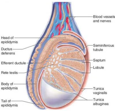
Figure 1 - Schematic representation of the mammalian testis and epididymis. 3
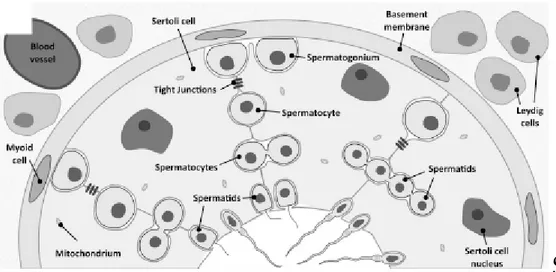
Figure 2 - Schematic illustration of seminiferous tubule, blood-testis barrier (BTB),
spermatogenesis and interstitial tissue. 5
Figure 3. mRNA expression of Aquaporin 0 in rat Sertoli cells. 19
Figure 4. mRNA expression of Aquaporin 1 in rat Sertoli cells. 20
Figure 5. Protein expression of Aquaporin 1 in rat Sertoli cells. 20
Figure 6. mRNA expression of Aquaporin 2 in rat Sertoli cells. 20
Figure 7. Protein expression of Aquaporin 2 in rat Sertoli cells. 21
Figure 8. mRNA expression of Aquaporin 3 in rat Sertoli cells. 21
Figure 9. Protein expression of Aquaporin 3 in rat Sertoli cells. 21
Figure 10. mRNA expression of transcript variants of Aquaporin 4 in rat Sertoli
cells. 22
Figure 11. Protein expression of Aquaporin 4 in rat Sertoli cells. 22
Figure 12. mRNA expression of Aquaporin 5 in rat Sertoli cells. 23
Figure 13. Protein expression of Aquaporin 5 in rat Sertoli cells. 23
Figure 14. mRNA expression of Aquaporin 6 in rat Sertoli cells. 24
Figure 15. Protein expression of Aquaporin 6 in rat Sertoli cells. 24
Figure 16. mRNA expression of Aquaporin 7 in rat Sertoli cells. 24
Figure 17. Protein expression of Aquaporin 7 in rat Sertoli cells. 25
Figure 18. mRNA expression of Aquaporin 8 in rat Sertoli cells. 25
Figure 19. Protein expression of Aquaporin 8 in rat Sertoli cells. 25
Figure 20. mRNA expression of Aquaporin 9 in rat Sertoli cells. 26
Figure 21. Protein expression of Aquaporin 9 in rat Sertoli cells. 26
Figure 22. mRNA expression of CFTR in rat Sertoli cells. 27
Figure 23. Protein expression of CFTR in rat Sertoli cells. 27
List of tables
Table 1 - AQPs in testis germ cells 11
Abbreviations
ABC - ATP-binding cassette AQPs - Aquaporins
ATP – Adenosine-5-triphosphate BSA - Bovine Serum Albumin BTB - Blood-Testis Barrier
cAMP - cyclic Adenosine Monophosphate cDNA - complementary Deoxyribonucleic acid CF - Cystic Fibrosis
CFTR – Cystic Fibrosis Transmembrane Regulator CREB - cAMP Response Element-Binding protein
DMEM: Ham’s F12 - Dulbecco’s Modified Eagle Medium Ham’s Nutrient Mixture F12 DNA - Deoxyribonucleic acid
EB - Elution Buffer EDs - Efferent Ducts
EDTA – Ethylene Diamine Tetra Acetic acid ENaC - Epithelial Na+ Channels
FBS – Fetal Bovine Serum
FSH - Follicle-Stimulating Hormone HBSS - Hank’s Balanced Salts Solution Isc - short-circuit current
LH - Luteinizing Hormone LWB - Last Wash Buffer
mAC - membrane-bound Adenylyl Cyclase
M-MLV RT - Moloney Murine Leukemia Virus Reverse Transcriptase mRNA - messenger Ribonucleic Acid
NCBes - Electrogenic Na+-coupled HCO
3- Transporters
NDCBEs - Na+-driven Cl-/HCO
3- exchangers
PBS – Phosphate Buffered Saline PCR – Polymerase Chain Reaction pHi - intracellular pH
PKA - Protein Kinase A
RIPA - Radio-ImmunoPrecipitation Assay RNA - Ribonucleic acid
RNAt - total RNA
RT-PCR - Reverse Transcription Polymerase Chain Reaction sAC - soluble Adenyly Cyclase
SCs - Sertoli cells
STF - Seminiferous Tubular Fluid Taq - Taq polymerase
Introduction
Infertility affects millions of couples worldwide and almost one third of the cases results from a male only fertility defect (Hamilton and Ventura 2006; Lutz 2006). When infertility is attributed to the male factor, treatment is not usually directed at a specific identified cause because a clear diagnosis is not established for the observed alterations in the male reproductive health. Moreover, the male is sometimes apparently clinically normal although suffering from abnormalities in sperm quality or quantity (including decreased sperm counts, low sperm motility and poor sperm-oocyte interactions) (Hirsh 2003; Pacey 2009). In these cases, treatment is usually associated with assisted reproductive technologies, which is frequently applied in an empirical manner to maximize the chances to conceive. Thus, it is imperative a deeper commitment upon basic and clinical research on the causes of male infertility.
The male reproductive tract is composed of highly heterogeneous tissues, which include the testis, the efferent ducts, the epididymis, and the vas deferens (Figure 1). Within the testis, spermatozoa are produced in a process termed spermatogenesis. However, spermatozoa maturation occurs along the excurrent ducts and epididymis. In fact, the formation of a competent spermatozoon is a complex process that involves the production of a large number of germ cells in the testis and several maturation steps that occur in the subsequent ducts. During the entire process, the establishment of the adequate environments, with the secretion of the numerous components of the different fluids throughout the male reproductive tract, is vital for the production of spermatozoa and for providing a means of transport for spermatozoon during its development (Rato, Socorro et al. 2010). A proper ionic content in the luminal milieus is important for the maintenance of membrane potentials, osmotic balances and fluid movements. Among the several ions, bicarbonate and H+ are key players in these processes. Bicarbonate ion (HCO
3-) together with
H+, result from the dissociation of the carbonic acid (H
2CO3), which is the product of the
reversible hydration of CO2, catalyzed by the enzyme carbonic anhydrase (Boron 2004). HCO3
-is essential to the ionic homeostas-is and a mobile physiological buffer that protects cells from fast and local changes in pH (Casey, Grinstein et al. 2009). Besides, intracellular processes only occur over a narrow pH range, so the adjustment of body fluids pH is important for the normal functions of the cells and tissues. Changes in the intracellular pH (pHi) affect the ionization state of weak acids and weak bases which corresponds to a wide array of cellular molecules, including all peptides and proteins, exerting a tight control on many biological processes (Boron 2004).
A central feature of the luminal fluids along the reproductive tract is its pH. In mammals, the pH of the luminal fluids of the reproductive system has significant effects on the male reproductive potential (Pastor-Soler, Piétrement et al. 2005). Moreover, disturbances in acid-base balance on the reproductive tract can be a cause for subfertility or infertility (Breton, Smith et al. 1996). During the spermatozoa transit along the male reproductive tract, the pH of the luminal fluids has to be properly controlled (Acott and Carr 1984; Jones and Murdoch 1996) and failure to maintain the pH homeostasis in the male reproductive tract impairs the production and/or maturation of spermatozoa causing infertility or subfertility (Acott and Carr 1984; Pastor-Soler, Piétrement et al. 2005).
Sertoli Cells
The mammalian testis is a complex organ. It is divided in compartments, called testicular lobules that are separated by fibrous inward extensions of the tunica albuginea (septum) (Figure 1) (Saladin 2003). Each lobule encloses coiled seminiferous tubules (Shubhada, Glinz et al. 1993; Walker and Cheng 2005), which contain the Sertoli cells and the germinal cells in the different development stages (Figure 2). These lobules are avascular and no nerves penetrate through their walls (Setchell 1986). The interstitial space of the testis comprises all the spaces between the seminiferous tubules, containing all the blood and lymphatic vessels, which are essential for the movement of hormones and nutrients into, and out of the testis (O'Donnell, Robertson et al. 2001). In this space we can also find the nerves, the Leydig cells (which are the primary sites of steroidogenesis in the testis) (Figure 2) and a significant population of macrophages (Setchell 1986).
The testis performs two major tasks: the production of steroid hormones and the formation of haploid germ cells. These functions are primarily regulated by pituitary gonadotropins, with luteinizing hormone (LH) acting on the testosterone-producing Leydig cells located in the interstitium, and follicle-stimulating hormone (FSH) acting on Sertoli cells in the seminiferous tubules (Griswold 1998; Walker and Cheng 2005). A chain of complex local interactions involving the various testicular cell types, such as germ, Sertoli, peritubular myoid and Leydig cells, are responsible for the control of spermatogenesis (Shubhada, Glinz et al. 1993; Walker and Cheng 2005). Spermatogenesis is the process by which immature germ cells undergo division, differentiation and meiosis to give rise to haploid elongated spermatids. This process takes place within seminiferous tubules, the functional unities of the testis, through close association of germ cells with epithelial somatic cells, the Sertoli cells.
Figure 1 - Schematic representation of the mammalian testis and epididymis. The testis is encapsulated by two layers: tunica vaginalis (the most outer tunic) and tunica albuginea. Extensions from tunica albuginea (septum) divide testis in lobules where the seminiferous tubules are located. Seminiferous tubules converge to the rete testis that is connected to the efferent ducts (EDs). The head of the epididymis is linked to the testis by various EDs. Adapted from (Saladin 2003).
Sertoli cells (SCs) are highly polarized epithelial cells that extend upwards from the basement membrane of the seminiferous tubule to its open lumen, and directly interact with the developing germ cells (Figure 2) (Mruk and Cheng 2004). From Enrico Sertoli works, in 1865, came out the concept that SCs function as “nurse cells” (Foley 2001) since they provide nutrients and regulatory factors for the sustenance of germ cells (Griswold 1995). They have important functions not only for the development of the testicular function, but also to the expression of the male phenotype (Sharpe, McKinnell et al. 2003; Mruk and Cheng 2004). The SCs are irregularly shaped, columnar cells with large dimensions and an enormous surface area, which allow each one of them to support a vast number of developing germ cells (Mruk and Cheng 2004). This is a crucial feature for spermatogenesis and germ cell movement (Mruk and Cheng 2004).
In mammals, between adjacent SCs, there is the blood-testis barrier (BTB), which is composed by tight junctions, basal ectoplasmic specialization, basal tubulobulbar complex and desmosome-like junctions (Figure 2) (Alves, Oliveira et al. 2012). The tight junctions between SCs are formed such that nothing larger than 1 kDa pass from the outside to the inside of the tubule, exerting a strict control over the passage of substances (Mruk and Cheng 2004; Walker and Cheng 2005; Walker 2010; Alves, Rato et al. 2013). The BTB, in rats, is
three components: (1) an anatomical/physical barrier, that restricts entry of molecules into the adluminal compartment; (2) an immunological barrier, that limits the movement of immune cells and regulates the level of cytokines in the seminiferous epithelium and (3) a physiological barrier, that is highly dynamic to encounter the needs of germ cells (Sikka and Wang 2008; Mital, Hinton et al. 2011). The BTB divides the seminiferous epithelium into the basal compartment, where spermatogonia and spermatocytes are found, and the adluminal (apical) compartment, containing different stages of meiotic spermatocytes, round spermatids, elongated spermatids, and spermatozoa (Figure 2) (Mruk and Cheng 2004; Su, Mruk et al. 2011). Thus, the developing germ cells that are present above BTB (in adluminal compartment) become effectively protected from direct access to plasma constituents and are highly dependent of SCs supply of nutrients and growth factors (Sharpe 1994; Sharpe, McKinnell et al. 2003; Mruk and Cheng 2004; Walker 2010; Rato, Alves et al. 2012). This highlights the essential role of SCs differentiation and of the formation of a competent BTB to the establishment of a normal spermatogenesis (Sikka and Wang 2008).
As discussed above, SCs face the lumen of the seminiferous tubule, provide structural support and create an immunologically protected space for developing germ cells from harmful agents and from the host’s own immune system (Petersen and Soder 2006). Moreover, they facilitate the progression of spermatogenesis and are responsible for the phagocytosis of the residual bodies and degenerating germ cells (Griswold 1995; Griswold 1998; Buzzard, Wreford et al. 2002; Mruk and Cheng 2004). SCs also regulate, among other things, the passage of ions and the selective flow of water, steroids and carbohydrates into the tubular lumen (Setchell and Waites 1975; Abraham 1991). They provide the germ cells with the appropriate nutrients, hormones and growth factors in a stage dependent manner, reflecting the ability of SCs to adapt to the changing needs of the germ cells (Mruk and Cheng 2004). Besides, SCs also control the composition of the seminiferous tubular fluid (STF), the physiochemical milieu where spermatogenesis occurs (Griswold 1995; Mruk and Cheng 2004; Rato, Socorro et al. 2010).
The secretion of STF commences during sexual maturation, after the formation of the BTB. It is dependent on FSH (Jegou, Le Gac et al. 1982; Jegou, Le Gac et al. 1983) and the pH of this fluid is known to be maintained slightly acidic (Levine and Marsh 1971; Caflisch and DuBose 1990).
Figure 2 - Schematic illustration of seminiferous tubule, blood-testis barrier (BTB), spermatogenesis and interstitial tissue. The Sertoli cells (SCs) reside on a basement membrane, under which are the lymphatic endothelium and the peritubular myoid cells. At the interstitial space are located the blood vessels and the Leydig cells. Between adjacent SCs, tight junctions are formed, representing the BTB that limits intercellular transport. Outside the BTB is the basal compartment where spermatogonia and spermatocytes are found, and inside the BTB is the adluminal compartment, containing different stages of meiotic spermatocytes, round spermatids, elongated spermatids, and spermatozoa. Adapted from (Alves, Oliveira et al. 2012).
The failure to maintain pH homeostasis in the male reproductive tract may impair the production and/or maturation of spermatozoa and therefore cause infertility or subfertility (Liu, Wang et al. 2012). Thus, the pH control of the STF is crucial for the male fertility. The regulation of SCs intracellular pH (pHi) plays a major role in this process (Tuck, Setchell et al. 1970; Mruk and Cheng 2004; Oliveira, Sousa et al. 2009; Oliveira, Sousa et al. 2009). pHi is mainly kept through the net balance between production and elimination of protons and by intracellular buffers (Roos and Boron 1981). The composition of the STF is influenced by net movements of water, K+ secretion, Na+, Cl- and HCO
3- reabsorption, and luminal acidification
(Levine and Marsh 1971; Au and Wong 1980; Turner 1984). Most of the knowledge of the ionic composition of this luminal fluid comes from the work of independent groups (Tuck, Setchell et al. 1970; Henning and Young 1971; Setchell 1978; Waites and Gladwell 1982; Fisher 2002; Clulow and Jones 2004), describing substantial differences in the ionic composition of STF, especially in the concentration of K+ but also in that of Na+, Cl- and HCO
3-, depending to the
methods of analysis used (Rato, Socorro et al. 2010). But all groups agreed that transepithelial ion and water transport through the BTB is mainly achieved by the intervention of SCs (Rato, Socorro et al. 2010).
SCs control the seminiferous fluid pH and electrolytes composition (Oliveira, Sousa et al. 2009; Oliveira, Sousa et al. 2009; Rato, Socorro et al. 2010). In these cells, distinct types of transport proteins have been identified, such as membrane pumps (Na+/K+-ATPase and
membrane transporters (NDCBEs, NBCes and Na+/H+ exchangers) (Boron 2001; Oliveira, Sousa
et al. 2009; Oliveira, Sousa et al. 2009), ion channels (voltage-dependent Cl- channels activated by acidic extracellular pH, CFTR Cl- channels, K+ channels and L- T- and N-type Ca2+
channels) (Taranta, Morena et al. 1997; Boockfor, Morris et al. 1998; Von Ledebur, Almeida et al. 2002; Auzanneau, Thoreau et al. 2003; Loss, Jacobsen et al. 2004), ion co-transporters (Na+–K+–2Cl- co-transporter and Na+/Ca2+ exchanger) (Grasso, Joseph et al. 1991; Pace, Lee et
al. 2000) and water channels (AQPs 0 and 8) (Tani, Koyama et al. 2001; Badran and Hermo 2002; Hermo, Krzeczunowicz et al. 2004). Although the involvement of such transporters in the establishment of the STF is not yet completely disclosed, it is certain that they have a key role in the cellular mechanisms responsible for determining ion composition, osmolarity and pH of the fluid (Rato, Socorro et al. 2010).
Cystic Fibrosis Transmembrane regulator (CFTR)
The CFTR is a cAMP-regulated membrane transporter that belongs to the ATP-binding cassette (ABC) superfamily (Liu, Wang et al. 2012). It is expressed in the apical membrane of secretory epithelial cells (Park and Lee 2012) and may conduct HCO3− directly as an anion
channel with measured bicarbonate permeability or indirectly as a Cl− channel, interacting
with other Cl−/ HCO
3− exchangers to provide a recycling pathway (Anderson, Gregory et al.
1991; Poulsen JH 1994; Shcheynikov, Yang et al. 2008) across those cells in many organs, including lungs, intestine, pancreas (Riordan 1993; Hug, Tamada et al. 2003) and skin (Reddy and Quinton 2003).
CFTR plays an important role in transepithelial salt absorption (Reddy and Quinton 2003), as well as in the HCO3- secretion and regulation of fluid volume in epithelial cells (Liu,
Wang et al. 2012). The permeability of CFTR to HCO3- was demonstrated in a study by Smith
and Welsh (1992) in human and canine airway epithelial cells that exhibited cAMP-regulated ionic currents, absent in cells isolated from individuals with a pathology called cystic fibrosis (CF), caused by mutations of CFTR gene. Those authors have also described a contribution of CFTR to the luminal fluid pH regulation. They reported that human cultured airway epithelia secreted an acidic fluid and that cAMP caused a reduction in the acid secretions making the luminal fluid more alkaline (Smith 1992; Smith 1993). It has also been reported that CFTR is responsible for the establishment of a fluid that contains a very high concentration of HCO3
-in the pancreas (Park and Lee 2012). Furthermore, -in sweat ducts, the two more abundantly expressed ion channels in the apical membrane are CFTR and epithelial Na+ channels (ENaC)
activity as well (Reddy and Quinton 2003). In fact, one way to diagnose CF is to measure the sweat chloride concentration (Gonska, Ip et al. 2009).
Furthermore, it has also been described that CFTR enhances osmotic water permeability in various cells or tissues (Schreiber, Nitschke et al. 1999; Schreiber, Pavenstadt et al. 2000; Pietrement, Da Silva et al. 2008). This interaction was initially described in human airway epithelial cells and in Xenopus oocytes (Schreiber, Nitschke et al. 1999; Schreiber, Pavenstadt et al. 2000) and more recently in rat epididymis (Cheung, Leung et al. 2003), however, the mechanisms underlying this cooperation and its physiological relevance remain obscure. It has been suggested that these results are consistent with a role for CFTR in controlling water permeability in vivo, and that this additional role of CFTR in controlling water permeability may have an impact on the development of the CF, in which men with a mutated CFTR gene are known have abnormal epididymal function and are infertile (Wong 1998).
CFTR in the male reproductive tract
In the male reproductive tract, high CFTR expression levels were detected in the epididymal and vas deferens epithelia and lower expression levels were observed in the mature testis (Tizzano, Silver et al. 1994).
As discussed above, in the seminiferous tubules, SCs are responsible for the secretion of intratubular fluid and play a major role on the establishment of its ionic composition (Rato, Socorro et al. 2010). Ko et al. (1998) studied the role of CFTR in SC ion and fluid secretion measuring the short-circuit current (Isc) of rat primary cultures grown on permeable supports. In those experiments, the Isc generated by the SCs was stimulated with ATP and cAMP agonists and inhibited by CFTR blockers (Ko, Chan et al. 1998), suggesting that CFTR does not play a major role in seminiferous ion and fluid secretion, but it is involved in HCO3- transport
(Xu, Chen et al. 2011). This was substantiated by directly measuring SCs pHi in the presence
of a specific CFTR inhibitor that caused a slight acidification of the cells that was reverted by the application of forskolin, a known CFTR activator (Xu, Chen et al. 2011). Therefore, even if the CFTR present in SCs does not have a major role in seminiferous fluid secretion, it might be crucial for testicular function. It is known that SCs nurture the germ cells by secreting various metabolites, proteins and growth factors in hormonally-regulated processes (Rato, Alves et al. 2012; Alves, Rato et al. 2013), particularly by FSH, known to be a key regulator of spermatogenesis (Sofikitis, Giotitsas et al. 2008). When FSH bounds to their receptors on SC, it activates the membrane-bound adenylyl cyclase (mAC), which in turn activates a cAMP/PKA signal transduction and the downstream transcription factors, such as CREB (Don and Stelzer 2002). More recently, it has been demonstrated that HCO3- entry in SCs causes the activation
phosphorylation could be potentiated by CFTR/HCO3-/sAC system. They described that lower
CFTR expression was accompanied by reduced phosphorylated CREB and total CREB expression in SCs from non-obstructive azoospermia patients (Xu, Chen et al. 2011). Based on these findings, we can assume that there is an involvement of CFTR in the FSH-mediated effect on spermatogenesis (Chen, Ruan et al. 2012). Indeed, mutations of CFTR gene have been associated with abnormal germ cell production and a reduction in germ cell quality and number (Boockfor, Morris et al. 1998).
CFTR expression has also been reported in germ cells of the various developmental stages (Gong, Li et al. 2001; Hihnala, Kujala et al. 2006; Teixeira, Sá et al. 2012). The presence of this ion transporter was observed in the cytoplasm and plasma membrane of primary spermatocytes (Hihnala, Kujala et al. 2006), in round spermatids (Trezise and Buchwald 1991; Trezise, Linder et al. 1993; Gong, Li et al. 2001) and in elongating spermatids (Gong, Li et al. 2001; Hihnala, Kujala et al. 2006). Its expression was also detected in the residual bodies resultant from cytoplasmatic mass sloughed off when germ cells develop into spermatozoa (Gong, Li et al. 2001). This distinct CFTR expression on spermatids differentiation into spermatozoa suggests a possible role for CFTR in spermiogenesis. In this process round spermatids change from a spherical form into an elongated and, ultimately, into a flagellar shape by reduction of volume, mainly due to a loss of cytoplasmic mass. These facts suggest the involvement of CFTR in volume reduction causing water efflux (Gong, Li et al. 2001), being that Aquaporin 7 (AQP7) and Aquaporin 8 (AQP8) are expressed in rat testis with a distribution remarkably similar to CFTR (Suzuki-Toyota, Ishibashi et al. 1999; Huang, He et al. 2006).
Aquaporins
The transmembrane water flow is a fundamental property of life. It was long assumed that the transport of water is due to simple diffusion through the lipid bilayer, but observations from experimental systems with high membrane water permeabilities, such as amphibian bladder and mammalian erythrocytes, suggested that the diffusion is not the only pathway for water to cross the membrane (Agre, King et al. 2002). While various explanations were proposed, no molecular water-specific transport protein was known until the discovery of aquaporin 1 (AQP1) (Preston, Carroll et al. 1992). With this, it is now well agreed that diffusion and channel-mediated water movements, named aquaporins (AQPs), both exists (Preston and Agre 1991; Verkman and Mitra 2000; Agre, King et al. 2002). Since diffusion of water occurs through all biological membranes at relatively low velocity, the AQPs water channels are found in a subset of epithelia with 10- to 100- fold higher capacity for water permeation (Agre, King et al. 2002). So AQPs are essential for the regulation of water
more of these members is ubiquitous in mammalian cells (Ishikawa, Cho et al. 2006; Jeyaseelan, Sepramaniam et al. 2006; Ishibashi, Hara et al. 2009).
AQPs are a family of small (25–34 kDa), hydrophobic, integral membrane channel proteins that facilitate rapid, passive movement of massive amounts of water (Zhang, Tan et al. 2012). Based on their sequence homology data, phylogenetic comparisons, and transport capabilities, AQPs comprise three major subtypes: the classical aquaporins, aquaglyceroporins and unorthodox aquaporins (Agre and Kozono 2003; Zardoya 2005; Nozaki, Ishii et al. 2008). Classical AQPs, includes AQP0, 1, 2, 4, and 5, are largely water-selective. Aquaglyceroporins, includes AQP3, 7, 9, and 10, are non-selective water channels, permeable to glycerol, urea and other small non-electrolytes, as well as water. Unorthodox aquaporins includes AQP6, 8, the superaquaporins (AQP11 and 12) being their functions under investigation. AQP1 and -11 may also localize in intracellular organelles, such as endoplasmic reticulum and mitochondria, to mediate intracellular water distribution (Morishita, Sakube et al. 2004; Nozaki, Ishii et al. 2008).
Some subtypes of aquaporins have been shown to facilitate gas transport (Herrera and Garvin 2011) including AQP1-dependent carbon dioxide (Cooper and Boron 1998; Nakhoul, Davis et al. 1998; Uehlein, Lovisolo et al. 2003), nitric oxide (Herrera, Hong et al. 2006; Herrera and Garvin 2007), and ammonia (Holm, Jahn et al. 2005; Musa-Aziz, Chen et al. 2009). AQP4 facilitates nitric oxide and oxygen transport (Wang and Tajkhorshid 2010) and AQP8 can also conduct other molecules, such as hydrogen peroxide, urea and ammonia (Wu and Beitz 2007). This could be explained by the analysis of AQP structures, where four single water channel molecules form a tetramer with the central pore of the tetramer being a passage for gas and ions (Yu, Yool et al. 2006; Wang and Tajkhorshid 2010).
So it is known that fluid secretion and absorption are vital processes in the physiology of male reproduction (Levine and Marsh 1971; Russell, Bartke et al. 1989). Most of the secretion of STF is done by SCs, but part of STF is also generated by differentiating germ cells (Sprando and Russell 1987). In fact, during the morphological differentiation of spermatids into sperm, a substantial amount of water is osmotically eliminated from the cytoplasm of the spermatids, with a reduction of cell volume of 70% (Cho, Svelto et al. 2003), suggesting that APQs can play a major role in the regulation and functions of reproduction events.
Aquaporins in testis and germ cells
Spermatogenesis, including the maturation of spermatozoa and their concentration and storage in seminiferous vessels, is associated with considerable fluid secretion and absorption in the male reproductive tract (Yeung, Barfield et al. 2005; Yeung, Barfield et al. 2005; Yeung, Callies et al. 2009). Such movements of fluid are consistent with the presence
Svelto et al. 2003). During spermatogenesis, and especially in the metamorphosis of round spermatids into elongated spermatids, one of the most distinct morphological changes is a striking reduction of germ cell volume, largely because of the osmotically driven fluid efflux (Huang, He et al. 2006), being expectable the intervention of the AQPs in this process.
So far, only a few isoforms have been described, although not always in a consistent manner, in the various testicular cells. For instance, in rat SCs, a specific expression pattern was described for AQP0. These cells express a semicircular pattern of AQP0 that varies with different stages of spermatogenesis. AQP0 expression in the seminiferous epithelium seems to be restricted to Sertoli and to Leydig cells, present in the interstitial space (Hermo, Krzeczunowicz et al. 2004).
In earlier studies, AQP1 was not detected in spermatozoa (Brown, Verbavatz et al. 1993; Curry, Millar et al. 1994; Liu, Gao et al. 1995) and AQP2 (Fushimi, Uchida et al. 1993), AQP3 and AQP4 were all absent from testis (Frigeri, Gropper et al. 1995). AQP1 antibody labels microvessel endothelial cells, though not endotubular and interstitial cell lines, which indicates the involvement of AQP1 in the regulation of fluid transport across endothelial cell membranes of testicular microvessels (Nicotina, Romeo et al. 2004). AQP5 localizes to the interstitial tissue of the testis including Leydig cells (Skowronski, Kwon et al. 2009). Confocal microscopy reveals co-expression of AQP5 with capillary AQP1 in testis by observation of a double-labeling (Skowronski, Kwon et al. 2009).
The AQP7 and AQP8 have been identified in rat testis and, by in situ hybridization, and they have been shown to be abundantly expressed in germ cells (Ishibashi, Kuwahara et al. 1997). This suggests that AQP7 and AQP8 could play a role in spermatogenesis. Despite sharing a similar transmembrane structure, AQP7 and AQP8 are different in function and distribution pattern in testis. AQP7 mRNA is transiently expressed at a late phase of spermatogenesis and cellular and subcellular localization of AQP7 protein varies with different stages of spermatogenesis (Suzuki-Toyota, Ishibashi et al. 1999; Calamita, Mazzone et al. 2001; Calamita, Ehringer et al. 2001). AQP7 is expressed in the round and elongated spermatids (Yeung, Callies et al. 2010) and is also localized to the rat sperm tail (Ishibashi, Kuwahara et al. 1997; Suzuki-Toyota, Ishibashi et al. 1999; Kageyama, Ishibashi et al. 2001). The role of AQP7 in male reproductive physiology is unclear. Infertile men lacking AQP7 expression in ejaculated sperm have lower sperm motility, compared to fertile men with AQP7-positive sperm. AQP7 may be involved in the maintenance of sperm motility, and lack of AQP7 in sperm may be an underlying mechanism of male infertility (Saito, Kageyama et al. 2004). However, male AQP7-knockout mice are fertile, with sperm not showing any morphologic and functional abnormalities, or any differences in pregnancy outcomes (Sohara,
faster in AQP7-knockout sperm than wild-type, with this faster water movement in knockout mouse spermatozoa possibly being mediated by AQP8 (Yeung, Callies et al. 2009). In situ hybridization of testis demonstrated expression of AQP8 mRNA in germ cells at all stages of spermatogenesis, from primary spermatocytes to spermatids (Ishibashi, Kuwahara et al. 1997), and also in SCs (Tani, Koyama et al. 2001; Badran and Hermo 2002). Although there are many studies on AQP8 in the testis, reports on the cellular localization are inconsistent, ranging from spermatogenic cells in general (Elkjaer, Nejsum et al. 2001; Kageyama, Ishibashi et al. 2001; Yang, Song et al. 2005), in elongated spermatids (Yeung, Callies et al. 2009), residual bodies and primary spermatocytes (Calamita, Mazzone et al. 2001) and in SCs (Badran and Hermo 2002). A phenotypic analysis of AQP8 null mice (-/-) showed few and only mild phenotype differences between wild-type and AQP8-deficient (-/+) mice. The testis weight and size in AQP8-null mice were increased, but no impaired fertility or abnormalities in sperm count or morphology were found (Yang, Song et al. 2005). Thus, it is expectable that AQP7 and AQP8 may both contribute to the generation of seminiferous tubule fluid, due both to SC secretion and water efflux from germ cells (Russell, Bartke et al. 1989).
Table 1 – Aquaporin expression and putative role in testis and testicular cells
Subtypes Distribution Major functions in reproduction
AQP0 Sertoli and Leydig cells A role in spermatogenesis
AQP 1 Epithelial cells of the testis Regulation of fluid transport across endothelial cell membranes of testicular microvessels; reduction of abnormal fluid retention in endotubular cells and extracellular matrix in Leydig cells
AQP 5 Testis Related to water and small uncharged solute molecule transport at critical sites
AQP 7 Testis Sperm maturation and storage; sperm motility; generation of seminiferous tubule fluid, from both SC secretion and water efflux of germ cells
AQP 8 Sertoli cells and all stages of spermatogenesis
Generation of seminiferous tubule fluid; physiological sperm volume regulation
AQP 9 Leydig cells Transport of water and non-charged solutes in Leydig cells
AQP 11 Elongated spermatids Maintaining cytoplasmic components of elongated spermatids; sustaining Sertoli cell capacity
AQP9 mRNA expression has been detected in interstitial Leydig cells (Tsukaguchi, Shayakul et al. 1998), while AQP9 protein is conspicuously expressed at the plasma and intracellular membrane of these cells (Elkjaer, Vajda et al. 2000; Badran and Hermo 2002), indicating that AQP9 plays a specific role in the transport of water and non-charged solutes in
It has also been described, using immunohistochemical techniques, that in the rat adult testis AQP11 localizes in elongated spermatids and no other cell types, except for residual bodies inside SCs (Yeung and Cooper, 2010), suggesting that AQP11 plays a role in recycling surplus cytoplasmic components of elongated spermatids, sustaining SC phagocytic capacity (Yeung and Cooper, 2010).
Aims of the Project
Sertoli cells perform important roles that go from providing structural and nutritional support of the developing germ cells, to the dislocation of the germ cells and the formation of the blood-testis barrier. Sertoli cells also regulate, among other things, the passage of ions and the selective flow of water into the tubular lumen.
The bicarbonate is an essential element for maintenance of intracellular and extracellular pH in all cells and tissues. Herein we focused on the identification and role aquaporins and CFTR in rat SCs and the possibility that they could act as molecular partners.
It has been reported that SCs express the CFTR protein, which is permeable to bicarbonate. The bicarbonate is an essential element for the maintenance of intracellular and extracellular pH in all cells and tissues. Mutations in the gene that encodes the CFTR protein are associated to a variety of abnormalities on the male reproductive system, which in most of the cases results in infertility. So, the identification of specific CFTR-binding partners will provide new pathways in the explanation of how CFTR functions as an ion channel and as a regulator of other membrane transporters and channels. Ultimately, it will not only allow the identification of new therapeutics for cystic fibrosis and other diseases resulting from CFTR dysfunction, but also offer insights in the cause of many diseases that are linked to these membrane transporters. Possible candidates to act as molecular partners of CFTR, are the water channels involved in water balance and ion homeostasis, known as aquaporins (AQPs).
Thus, the first objective of this work was to evaluate protein and mRNA expression of CFTR by slot blot and RT-PCR, respectively, in SCs using primary cultures obtained from 20-day old rats. The second was to detect the protein and mRNA expression of aquaporins isoforms (AQP0-9), with the same methodology. And the third objective, using the co-immunoprecipitation technique, was to investigate the possibility of the physical interaction between CFTR and the AQPs isoforms previously detected in SCs.
Materials and methods
1. Chemicals
Hank’s Balanced Salts Solution (HBSS), Dulbecco’s Modified Eagle Medium Ham’s Nutrient Mixture F12 (DMEM: Ham’s F12), Ethylene Diamine Tetra Acetic acid (EDTA), Soybean Trypsin Inhibitor, DNAse, Collagenase type I, Bovine Serum Albumin (BSA), trypsin-EDTA and other chemicals were obtained from Sigma-Aldrich (St. Louis, MO, USA). Fetal Bovine Serum (FBS) was obtained from Biochrom AG (Germany). NZY M-MuLV Reverse Transcriptase (M-MLV RT), random hexamer primers, dNTPs and NZTaq 2x Green Master Mix, agarose and Greensafe were obtained from NZYTech (Lisboa, Portugal). Primers were obtained from STABVIDA (Oeiras, Portugal). Tween 20 was obtained from Applichem (Darmstadt, Germany). Dried milk was obtained from Regilait (Saint-Martin-Belle-Roche, France). Regilait, France. Polyclonal antibodies were obtained from Gentaur (Gdansk, Poland) and Santa Cruz Biotechnology (Heidelberg, Germany).
2. Primary Cultures of Rat SCs
Male Wistar rats (20-day-old) were sacrificed by cervical dislocation. The testes were immediately excised in aseptic conditions and washed two times in a conical tube in 30 mL of ice cold HBSS (potassium chloride 0,4 g/L, potassium phosphate monobasic anhydrous 0,06 g/L, sodium chloride 8 g/L, sodium phosphate dibasic 0,045 g/L, D-glucose 1 g/L, sodium bicarbonate 0,35 g/L) containing 10000 U/mL of penicillin, 10 mg/mL streptomycin and 25 μg/ml amphotericin B (pH 7.4). After removal of the adherent epididymis and vas deferens, the testis were decapsulated in HBSS, and the loosen tissue was washed three times in the same solution. SCs were isolated by a method described by Oliveira and collaborators (2012) with slight modifications. Briefly, to remove contaminating peritubular cells, the tissue from decapsulated testes was washed in a conical tube and dispersed in glycine solution (HBSS plus 1 M glycine, 2 mM EDTA, 0,002% (w/v) Soybean Trypsin Inhibitor; pH 7.2). To further remove residual peritubular cells, the tubules were placed and dispersed in a Petri dish containing a glycine solution with 0,5 mg/ml DNAse I during 10 minutes at room temperature. To uncoil the tubules and further release the interstitial tissue/cells, the dispersed tubules were forced through a large-pore Pasteur pipette. The tubular pellet was then digested for 15-20 minutes (at room temperature) in a HBSS plus 0,225 mg/ml collagenase type I and 0,05 mg/mL DNAse I. After digestion, the disaggregated seminiferous tubules were washed three times in HBSS by centrifuging the cell suspension 3 minutes at 300.g. The SC suspension was collected and
disaggregate large SC clusters, the cellular suspension was forced though a 20G needle. For cell culture, the concentration of the clusters on the cellular suspension obtained was adjusted to 5000 clusters/ml, plated on 25 cm2 culture flasks (Cell+; Sarstedt), and incubated
at 33ºC in an atmosphere of 5% CO2: 95% O2. The day of plating was considered day 0 of
culture and the cultures were left undisturbed until day 2.
3. RNA Extraction
The extraction of total RNA (RNAt) was performed using the E.Z.N.A. Total RNA Kit (Omega bio-tek, Norcross USA) as indicated by the manufacturer. Total RNA was isolated from lung, liver, brain, kidney and eye using 15 mg of tissue. Total RNA from SCs was isolated after detaching cells from the culture flasks using a trypsin-EDTA solution. To eliminate residual trypsin, detached cells were washed with 3 mL of phosphate buffered saline (PBS), by centrifugation at 3000.g during 5 minutes. RNA concentration and absorbance ratios (A260/A280) were determined by spectrophotometry (NanophotometerTM, Implen, Germany).
4. RT-PCR
The RNAt obtained was reversely transcribed in a mixture containing 0.5 mM of each
dNTP, 250 ng of random hexamer primers, 1 μg of RNAt and sterile H2O. The mixture was
incubated 5 minutes to 65ºC. Then, M-MLV RT and Reaction Buffer were added and incubated sequentially at 25ºC for 10 minutes, 37ºC for 50 minutes and 70ºC for 15 minutes. The resulting complementary deoxyribonucleic acid (cDNA) was used with exon-exon spanning primer sets designed to amplify CFTR, AQP1, AQP2, AQP3, AQP4_13, AQP4_24, AQP5, AQP6,
AQP7, AQP8, AQP9 and AQP0 cDNA fragments (Table 2). Polymerase chain reactions (PCR)
were carried out using 0.5 μg of cDNA in 12.5 μL of total volume of a mixture containing 6 μL of NZYTaq Green Master Mix 2x, 0.1 μL of 50 μM of each primer and sterile H2O. Primer’s
sequences, optimal annealing temperature, the number of cycles required for exponential amplification phase of fragments, the fragments size and the positive control are indicated in Table 2. cDNA-free sample was used as negative control. At the end of the experiments, samples were run in 1% agarose gel electrophoresis with 25 μL of Greensafe in 100 mL, during 30 minutes at 120 V. The agarose gel was visualiszd using software Molecular Imager FX Pro Plus MultiImager (Biorad, Hercules, USA) coupled to an image acquisition system (Vilber Lourmat, Marne-la-Vallée, France). The size of the expected products was compared to a DNA ladder (NZYDNA Ladder VI, Nzytech).
Table 2 - Oligonucleotides and Cycling Conditions for PCR Amplification of Cystic fibrosis transmembrane conductance regulator (CFTR), aquaporin-0 (AQP0), aquaporin-1 (AQP1), aquaporin-2 (AQP2), aquaporin-3 (AQP3), aquaporin-4 transcript variants 1 and 3 (AQP4_1.3), aquaporin-4 transcript variants 2 and 4 (AQP4_2.4) aquaporin-5 (AQP5), aquaporin-6 (AQP6), aquaporin-7 (AQP7), aquaporin-8 (AQP8), aquaporin-9 (AQP9).
Gene Sequence (5'- 3') (ºC) AT Amplificon Size (bp) Cicles C+
CFTR AN: NM_031506.1 Forward: GTTGGGAATCAGCGATGGAG 65 156 40c. Lung Reverse: TGACTGTGTAGGGAAGCACA AQP1 AN: NM_012778.1 Sense: CCTGCTCGAGAACTCACTTG 56 186 40c. Kidney Antisense: TGTCCAAGAGCCACAGACAA AQP2 AN: NM_012909.2 Sense: CAGTAGAAATCCGTGGGGAC 56 189 35c. Kidney Antisense: GGCCCAGGGTAACAGAGAAA AQP3 AN: NM_031703.1 Sense: GAGATGCTCCACATCCGCTA 57 156 35c. Kidney Antisense: GCCAAAAGCCAAGTTGATGG AQP4_1.3 AN: NM_012825.3 or NM_001270558.1 Sense: CAGGGAAGGCATGAGTGACG 56 198 40c. Kidney Antisense: TCTGAGCCACCCCAGTTAAT AQP4_2.4 AN: NM_001142366.1 or NM_001270559.1 Sense: GAAGACAGCACCTGTGATAGC 58 234 40c. Brain Antisense: CACAGGTAGGGGGTTCTCTG AQP5 AN: NM_012779.1 Sense: GCCATTACTCTGGCCCTCTT 55 175 30c. Lung Antisense: CAGGCGTTGTGTTGTTGTTC AQP6 AN: NM_022181.1 Sense: GGTGTCCGAGAGACCCTTG 58 354 35c. Kidney Antisense: TACGGTCTTGGTGTCAGGAAA AQP7 AN: NM_019157.2 Sense: GCGGAGTTCCTGAGTACCTA 54 126 35c. Kidney Antisense: CATGGTCACTCCGAAACCAA AQP8 AN: NM_019158.2 Sense: AGTCCAAATACTGGGCTCCT 54 196 35c. Liver Antisense: CTCCGATCATTCCCCCAAAC AQP9 AN: NM_022960.2 Sense: CCCAGTTTTTGGGAGCCTTT 65 97 45c. Liver Antisense: CCTACGACGAGCAGTTTTCC AQP0 AN: NM_001105719.1 Sense: TCCTTTGCTCCTGCTATCCT 59 131 40c. Brain Antisense: GAAACACTCTTGAGCCGTGG
Abbreviations: AN – Genbank Accession Number; AT - annealing temperature; c. - Number of cycles during exponential phase of amplification; C+ - Positive Control
tissues/cells were allowed to stand 15 minutes on ice and the suspension was centrifuged at 14000.g for 20 minutes at 4ºC. The resulting pellet was discarded. The total protein concentration was measured using the Bradford assay.
6. Slot Blot
Total protein extracted from the tissues and SCs were transferred on activated polyvinylidenedifluoride (PVDF) membranes using a Hybri-slot manifold system (Biometra, Göttingen, Germany) by means of a vacuum pump. The membranes were then blocked by incubating during 90 min in Tris-buffered saline solution (TBS) with 0.05% Tween 20 containing 5% skimmed dried milk.
Afterwards, the blocked membranes were incubated at 4ºC overnight with rabbit anti-AQP1 antibody (1:1000; Ref. anti-AQP11-A; Gentaur, Gdansk, Poland) or rabbit anti-AQP2 antibody (1:5000; Ref. AQP21-A; Gentaur, Gdansk, Poland) or rabbit anti-AQP3 antibody (1:5000; Ref. AQP31-A; Gentaur, Gdansk, Poland) or rabbit anti-AQP4 antibody (1:5000; Ref. AQP41-A; Gentaur, Gdansk, Poland) or rabbit anti-AQP5 antibody (1:5000; Ref. AQP51-A; Gentaur, Gdansk, Poland) or rabbit anti-AQP6 antibody (1:5000; Ref. AQP61-A; Gentaur, Gdansk, Poland) or rabbit anti-AQP7 antibody (1:5000; Ref. AQP71-A; Gentaur, Gdansk, Poland) or rabbit AQP8 antibody (1:1000; Ref. AQP81-A; Gentaur, Gdansk, Poland) or rabbit anti-AQP9 antibody (1:5000; Ref. anti-AQP91-A; Gentaur, Gdansk, Poland) or goat anti-CFTR antibody (1:500; Santa Cruz Biotechnology Heidelberg, Germany, SC-8909). The immune-reactive proteins were detected separately with goat anti-rabbit IgG-AP (1:5000, Santa Cruz Biotechnology Heidelberg, Germany, Sc-2004) or donkey anti-goat IgG-AP (1:5000, Santa Cruz Biotechnology Heidelberg, Germany, Sc 2020). Membranes were then reacted with ECF (GE Healthcare, Buckinghamshire, UK) and read using a BioRad FX-Pro-plus (Bio-Rad, Hemel Hempstead, UK). The densities from each band were obtained using the Quantity One Software (Bio-Rad, Hemel Hempstead, UK), according to standard methods.
7. Co-Immunoprecipitation
Immunoprecipitation of CFTR was performed using the Dynabeads® Co-Immunoprecipitation Kit (Life Technologies, Calsbad, USA). Anti-CFTR goat antibody was conjugated to magnetic beads according to the manufacturer’s protocol. Briefly, harvested SCs (50 mg) were lysed in 450 μL of extraction buffer (1x IP Buffer, NaCl 100 mM, Protease Inhibitors 1:400) and centrifuged, at 2600 x g for 5 minutes, to remove large cell debris and nuclei. The final supernatant contained the cell lysate ready to be used immediately for co-immunoprecipitation. For the co-immunoprecipitation, 1.5 mg of the antibody-coupled beads was first washed in 900 uL of extraction buffer using a DynaMag™-2 magnet (Life
in the cell lysate. This suspension was incubated at 4ºC on a rotator for 30 minutes. The supernatant was then removed using the DynaMag™-2 magnet and the beads were washed three times with 200 uL of extraction buffer. Subsequently, the beads were washed in 200 uL of Last Wash Buffer (LWB) (1x LWB, Tween 20 0,02%), with subsequent removal of the supernatant by using the DynaMag™-2 magnet. Finally, the beads were resuspended in 60 μL of Elution Buffer (EB) and gently mixed. The supernatant obtained by using the DynaMag™-2 magnet contained the purified protein complexes. 2 μL of the solution containing purified protein complexes were transferred to activated PVDF membranes using a Hybri-slot manifold system (Biometra, Göttingen, Germany) by means of a vacuum pump. The membranes were then blocked by incubating during 90 min in TBS with 0.05% Tween 20 containing 5 % skimmed dried milk. Afterwards, the blocked membranes were incubated at 4ºC overnight with goat anti-CFTR antibody (1:500; Santa Cruz Biotechnology Heidelberg, Germany, SC-8909) or rabbit anti-AQP4 antibody (1:5000; Ref. AQP41-A; Gentaur, Gdansk, Poland). The immune-reactive proteins were detected separately with goat anti-rabbit IgG-AP (1:5000, Santa Cruz Biotechnology Heidelberg, Germany, Sc-2004) or donkey anti-goat IgG-AP (1:5000, Santa Cruz Biotechnology Heidelberg, Germany, Sc 2020). Membranes were then reacted with ECF (GE Healthcare, Buckinghamshire, UK) and read using a BioRad FX-Pro-plus (Bio-Rad, Hemel Hempstead, UK). The densities from each band were obtained using the Quantity One Software (Bio-Rad, Hemel Hempstead, UK), according to standard methods.
Results
1. Expression of the Aquaporin isoforms in rat Sertoli cells
The expression of the different Aquaporin isoforms in cultured rat SCs was evaluated by identifying the mRNA transcripts, using the RT-PCR technique, and the corresponding protein, using the slot-blot technique.
1.1. Aquaporin 0
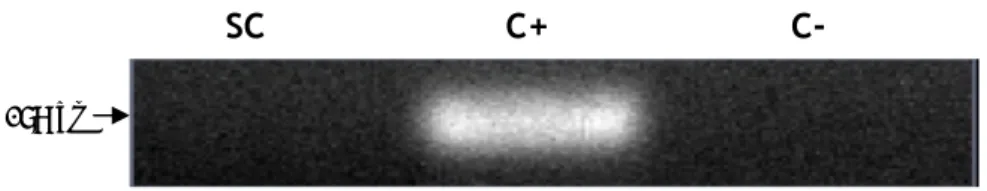
The Aquaporin 0 (AQP0) is also known by the name of major intrinsic protein (MIP) and initially was thought to be a gap junction protein (Ebihara, Beyer et al. 1989). After discovery of the function of AQP1, expression of MIP in oocytes was shown to induce a relatively small increase in water permeability that was not inhibited by mercurials – thus the symbol AQP0 (Mulders, Preston et al. 1995). In our experiments, using total RNA from rat SCs, we were able to identify the expression of mRNA transcripts of AQP0 (Figure 3).
Figure 3. mRNA expression of Aquaporin 0 in rat Sertoli cells. SC: Sertoli cells; C+: positive control (rat brain total lysate) for Aquaporin 0; C-: control without reverse transcriptase.
1.2. Aquaporin 1
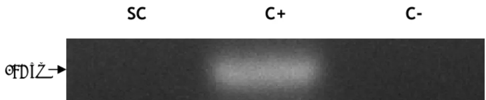
Aquaporin 1 (AQP1) is expressed in various organs and cells, as for instance kidney, central nervous system, eye, gastrointestinal tract, female and male reproductive system and skin (Benga 2012). Besides, it has been associated with a huge capacity for water transport. Using specific primers for this aquaporin, we were able to detect the expression of mRNA transcripts of AQP1 on rat SCs (Figure 4).
SC C-
C+
131 bpFigure 4. mRNA expression of Aquaporin 1 in rat Sertoli cells. SC: Sertoli cells; C+: positive control (rat kidney total lysate) for Aquaporin 1; C-: control without reverse transcriptase.
On the other hand, the presence of the Aquaporin 1 protein was not confirmed in the cultured rat SCs. When using a specific anti-AQP1 antibody, we were not able to detect any specific staining correspondent to this aquaporin isoform (Figure 5).
Figure 5. Protein expression of Aquaporin 1 in rat Sertoli cells. SC: Sertoli cells; C+: positive control (rat kidney total lysate) for Aquaporin 1.
1.3. Aquaporin 2
The Aquaporin 2 (AQP2) has been described as the vasopressin-regulated water channel and is widely expressed in renal collecting ducts (Nielsen, Smith et al. 1993). It has been associated with the movements of water in those ducts (Nielsen, Smith et al. 1993). In rat SCs we were not able to observe the expression of mRNA transcripts of AQP2 by means of RT-PCR (Figure 6)
Figure 6. mRNA expression of Aquaporin 2 in rat Sertoli cells. SC: Sertoli cells; C+: positive control (rat kidney total lysate) for Aquaporin 2; C-: control without reverse transcriptase.
SC C-
C+
SC C+
SC C-
C+
186 bpFigure 7. Protein expression of Aquaporin 2 in rat Sertoli cells. SC: Sertoli cells; C+: positive control (rat kidney total lysate) for Aquaporin 2.
1.4. Aquaporin 3
Aquaporin 3 (AQP3) expression is distributed in multiple organs including kidney (Ecelbarger, Terris et al. 1995), airways (Nielsen, King et al. 1997), skin, and eye (Hamann, Zeuthen et al. 1998). Besides the transport of water, AQP 3 is permeable to glycerol (Zeuthen and Klaerke 1999). In our experimental work, we were not able to detect the expression of mRNA transcripts of AQP3 in rat SCs (Figure 8).
Figure 8. mRNA expression of Aquaporin 3 in rat Sertoli cells. SC: Sertoli cells; C+: positive control (rat kidney total lysate) for Aquaporin 3; C-: control without reverse transcriptase.
Furthermore, the presence of the AQP 3 protein was also not detected in the cultured rat SCs. When using a specific anti-AQP3 antibody we were not able to observe any specific staining correspondent to this aquaporin isoform (Figure 9).
Figure 9. Protein expression of Aquaporin 3 in rat Sertoli cells. SC: Sertoli cells; C+: positive control (rat kidney total lysate) for Aquaporin 3.
1.5. Aquaporin 4
The Aquaporin 4 (AQP4) is constitutively expressed in the basolateral cell
SC C+
SC C+
SC C-
C+
exit these cells (Agre and Nielsen 1996). AQP4 is also expressed in astrocytes (Nagelhus, Mathiisen et al. 2004). It has been described that this aquaporin can present four alternative transcript variants (Aquaporin 4 variants 1 to 4). When we analyzed the expression of the mRNA transcript variants 1 and 3 of AQP 4 in rat SCs, we were not able to detect any expression of these variants (Figure 10). However, when we evaluated the expression of mRNA transcript variants 2 and 4 of AQP 4 we could observe a distinctive staining in SC (Figure 10), correspondent to the presence of the mRNA of these transcript variants.
Figure 10. mRNA expression of transcript variants of Aquaporin 4 in rat Sertoli cells. AQP4 1.3: Aquaporin 4 transcript variants 1 and 3; AQP4 2.4: Aquaporin 4 transcript variants 2 and 4; SC: Sertoli cells; C+: positive control, rat kidney total lysate for AQP4 1.3 and rat brain total lysate for AQP4 2.4; C-: control without reverse transcriptase.
The presence of the protein of AQP 4 was identified in the cultured rat SCs. Using a specific anti-AQP4 antibody we were able to detect specific staining correspondent to this aquaporin isoform (Figure 11).
Figure 11. Protein expression of Aquaporin 4 in rat Sertoli cells. SC: Sertoli cells; C+: positive control (rat kidney total lysate) for Aquaporin 4.
SC
C+
SC C-
C+
AQP4 2.4
AQP4 1.3
198 bp1.6. Aquaporin 5
Aquaporin 5 (AQP5) has been localized to the apical membrane of multiple secretory glands, including lacrimal glands, salivary glands and submucosal glands of airways (Nielsen, King et al. 1997). Due to its localization it has been postulated that AQP5 activity may be a rate limiting step for glandular fluid release, as was confirmed in salivary glands and airways from mice bearing targeted disruption of the gene encoding AQP5 (Ma, Song et al. 1999; Song and Verkman 2001). In rat SCs we were not able to perceive the expression of mRNA transcripts of AQP 5 as can be seen in Figure 12.
Figure 12. mRNA expression of Aquaporin 5 in rat Sertoli cells. SC: Sertoli cells; C+: positive control (rat lung total lysate) for Aquaporin 5; C-: control without reverse transcriptase.
Furthermore, the presence of the AQP 5 protein was equally not confirmed in the cultured rat SCs. When using a specific anti-AQP5 antibody, we were not able to detect the specific staining correspondent to this aquaporin isoform (Figure 13).
Figure 13. Protein expression of Aquaporin 5 in rat Sertoli cells. SC: Sertoli cells; C+: positive control (rat lung total lysate) for Aquaporin 5.
1.7. Aquaporin 6
The presence of Aquaporin 6 (AQP6) has been distinctively described in acid secreting α-intercalated cells from renal collecting duct, where this protein has been restricted to intracellular sites (Yasui, Kwon et al. 1999). Further analysis showed that AQP6 is co-localized alongside H+-ATPase in intracellular vesicles and not in the plasma membrane (Yasui, Hazama
et al. 1999).
SC
C+
SC C-
C+
175 bpFigure 14. mRNA expression of Aquaporin 6 in rat Sertoli cells. SC: Sertoli cells; C+: positive control (rat kidney total lysate) for Aquaporin 6; C-: control without reverse transcriptase.
In our experimental work, the expression of mRNA transcripts of AQP 6 was not detected in SCs (Figure 14). Interestingly, when using a specific anti-AQP6 antibody we were able to confirm the presence of the AQP 6 protein in cultured rat SCs, as a specific staining correspondent to this aquaporin isoform was clearly detected (Figure 15).
Figure 15. Protein expression of Aquaporin 6 in rat Sertoli cells. SC: Sertoli cells; C+: positive control (rat kidney total lysate) for Aquaporin 6.
1.8. Aquaporin 7
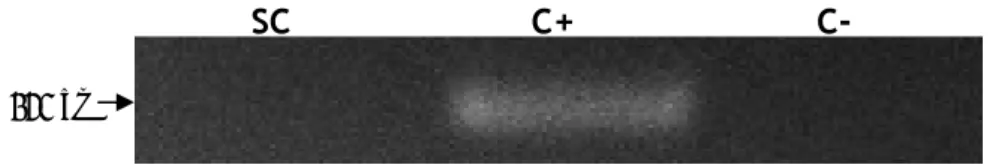
Aquaporin 7 (AQP7) expression was described in a small distal segment of the proximal tubule, though its deletion in mice is not associated with significant impairment in urinary concentrating ability, but rather with an impairment of glycerol clearance (Sohara, Rai et al. 2005). AQP7 was also identified in adipose tissue where was shown to be permeated by water plus glycerol (Kishida, Kuriyama et al. 2000) suggesting its participation on adipocyte metabolism. Similar to the results we obtained for AQP6, we were not able detect the expression of mRNA transcripts of AQP7 in rat SCs (Figure 16).
Figure 16. mRNA expression of Aquaporin 7 in rat Sertoli cells. SC: Sertoli cells; C+: positive control (rat kidney total lysate) for Aquaporin 7; C-: control without reverse transcriptase.