Ana Rita Machado dos Santos
Maio de 2013
Impact of Astrocytes and Gliogenesis on
the Pathophysiology of Depression
UMinho|20
13
Ana Rita Mac
hado dos Sant
os
Impact of Astrocytes and Gliogenesis on t
he P
at
hoph
ysiology of Depression
Universidade do Minho
Trabalho realizado sob a orientação da
Professora Doutora Luísa Pinto
e co-orientação do
Professor Doutor João Filipe Oliveira
Ana Rita Machado dos Santos
Maio de 2013
Dissertação de Mestrado
Mestrado em Ciências da Saúde
Impact of Astrocytes and Gliogenesis on
the Pathophysiology of Depression
Universidade do Minho
iii
Patience, persistence and perspiration make
an unbeatable combination for success.
v Acknowledgements/Agradecimentos
À Professora Doutora Luísa Pinto, agradeço do fundo do coração pela sábia orientação, pela constante presença durante toda esta etapa, pela amizade, entusiasmo e pela leitura sempre crítica do trabalho, que tornou a execução desta tese um verdadeiro prazer. Ambiciono, um dia, chegar ao nível de excelência com que nos presenteias diariamente.
Ao mestre Dinis Alves, agradeço todo o empenho prestado ao trabalho, todos os serões passados no biotério a planear as tarefas seguintes, toda a organização que tanto me chocava mas que foi tão necessária, a amizade e o bom-humor que foi tão bem-vindo durante este ano.
À mestre Patrícia Patrício, agradeço o apoio prestado, a valiosa e constante ajuda no laboratório, a partilha de conhecimentos, a amizade e a boa-disposição sempre presente no dia-a-dia.
Ao mestre António Pinheiro, agradeço o apoio prestado, a fomentação da discussão dos resultados, a amizade e a partilha de conhecimentos que tanto me ajudaram durante este último ano.
À mestre Mónica Morais, agradeço toda a disponibilidade, ajuda e amizade.
Ao Professor Doutor João Bessa e ao Professor Doutor Nuno Sousa, agradeço a oportunidade de integrar uma equipa fantástica e de desenvolver um trabalho que tanto gozo me deu.
Com uma equipa assim, tudo se torna mais fácil!
Ao Professor Doutor João Filipe Oliveira, agradeço o entusiasmo, disponibilidade, todos os desafios propostos e pela visão crítica sobre o trabalho.
Aos NeRDs, agradeço pelo carinho, pelas sugestões e pela visão crítica sobre o meu trabalho.
Às minhas colegas de laboratório e mestrado Ana Maria Silva, Márcia Santos, Vanessa Sardinha, Magda Reis, Joana Silva e Sofia Lopes, agradeço a boa-disposição, a constante partilha de bons momentos, os sorrisos e as caminhadas. Vocês tornaram-se verdadeiras amigas durante este último ano!
Às minhas colegas de casa Cláudia Miranda, Carla Calçada e Margarida Araújo, agradeço o bom-ambiente que me proporcionaram em Braga, agradeço a ajuda quando tinha menos tempo e, acima de tudo, agradeço a amizade. Braga tornou-se uma cidade mais acolhedora por vossa causa!
vi
Às minhas fiéis amigas Mariana Urbano, Rita Sousa, Filipa Santana e Sofia Brito, agradeço a constante amizade que, apesar de durar tantos anos, continua a alegrar os meus dias e a tornar-me uma pessoa mais feliz e grata por vos ter no meu caminho.
Ao Afonso Costa, tenho a agradecer a amizade, a boa-disposição e a ajuda na formatação desta tese. À minha família, agradeço a compreensão pelo pouco tempo dispensado e a boa-disposição aos fins-de-semana, que me fizeram sempre querer voltar a casa. À minha mãe agradeço por ser um grande exemplo de força e por nunca me deixar desistir dos meus sonhos.
Ao Amílcar Cordeiro, agradeço com todo o meu coração o apoio constante em todas as etapas da minha vida pessoal e profissional, o amor e carinho dispensados mesmo quando tinha muito trabalho e pouco tempo, a dedicação e a ajuda. Obrigada por me dizeres sempre que sou capaz! Contigo a meu lado, o meu caminho torna-se francamente mais fácil e muito mais feliz!
vii Abstract
Major depression is a highly prevalent disorder that poses a significant social burden in society nowadays. The pathophysiology of this disease is still poorly understood but growing evidence suggests that impaired neuroplasticity may be a key underlying mechanism for the precipitation of the disorder. This theory is substantiated by the fact that depression leads to a decrease in neurogenesis and several antidepressants (ADs) stimulate hippocampal neurogenesis, but it is still unclear if these pro-neurogenic effects are responsible for their mood-, emotional- and cognitive-improving actions. Recent studies also showed an important role for astrocytes in the pathophysiology of this disorder, which are crucial for neurotransmission and neurovascular coupling, evidenced by astrocytes loss in major depressive disorder (MDD). However, the importance of astrocytes in the precipitation of and recovery from MDD is still largely unknown.
Therefore, we proposed to study the role of astrocytes and adult gliogenesis, more precisely the generation of new astrocytes, in the precipitation of and recovery from depressive-like cognitive behavior in rats both untreated and treated with ADs in a longitudinal manner - at short-term, long-term and recurrence perspective -, using a pre-validated model of depression - the unpredictable chronic mild stress (uCMS).
Regarding the cognitive behavior assessment, although short-term memory seems to be impaired through the course of the disease, at short-term, long-term and recurrence, administration of fluoxetine and imipramine was effective in reverting the cognitive impairments induced by depression. The long-term memory was also highly affected by this disorder in all analyzed time-points, but the treatment with the ADs was not effective in reverting the cognitive impairments observed. Regarding the hippocampal dentate gyrus (DG) astrocytic population, at the long-term perspective of the disease, imipramine, but not fluoxetine, was able to elicit a strong pro-gliogenic effect. The same does not happen at recurrence for both ADs, suggesting that an adaptation to the stressful environment by these specific type of cells might be happening throughout time. Thereby, we provide some consistent evidences for the causative implication of gliogenesis and astrocytes in the pathophysiology of depression, having significant impacts in the long-term development and maintenance of cognitive deficits, as well as in the long-term recovery of those impairments by ADs. Our results endorse the view of the adult hippocampal DG gliogenesis process as a promising therapeutical target in future therapies in the neuropsychiatric field. Moreover, this study shows for the first time that alterations in the hippocampal DG resident astrocytes can be further analyzed as a predictive target for depression.
ix Resumo
A depressão é uma doença bastante prevalente que, atualmente, representa um peso importante na sociedade. A fisiopatologia desta doença é ainda pouco compreendida, mas evidências crescentes sugerem que o comprometimento da neuroplasticidade pode ser um mecanismo fundamental subjacente à precipitação da doença. Esta teoria é apoiada pelo facto de a depressão induzir uma diminuição da neurogénese e, também, ao facto de vários antidepressivos levarem à estimulação da neurogénese. No entanto, ainda não é claro se esses efeitos pró-neurogénicos são responsáveis pelas melhorias a nível de humor, emocionais e/ou cognitivas, nos pacientes. Estudos recentes demonstraram a importância dos astrócitos na fisiopatologia desta doença, mostrando ser cruciais para a neurotransmissão e acoplamento neurovascular, evidenciada por uma perda de astrócitos em episódios depressivos. Contudo, a importância dos astrócitos na precipitação e recuperação desta doença ainda é, em grande parte, desconhecida.
Por isso, propusemo-nos a estudar o papel dos astrócitos e da gliogénese adulta, mais precisamente da geração de novos astrócitos, na precipitação e recuperação de défices cognitivos associados à depressão. Este estudo foi elaborado com ratos tratados e não tratados com antidepressivos e seguiu uma linha longitudinal no episódio depressivo - a curto prazo, a longo prazo e na recorrência da doença -, usando um modelo pré-validado de depressão – um protocolo de exposição crónica a stress. Quanto à avaliação comportamental a nível cognitivo, embora a memória a curto prazo ser afetada com o decorrer da doença, a administração de fluoxetina e imipramina foi eficaz na reversão dos danos cognitivos induzidos pela depressão. A memória a longo prazo foi igualmente afetada por esta doença em todos os tempos experimentais analisados, mas o tratamento com os antidepressivos não se mostrou eficaz em reverter os défices cognitivos observados. Quanto a alterações da população astrocítica no girus denteado do hipocampo, o tratamento com imipramina, contrariamente ao tratamento com fluoxetina, foi capaz de induzir um efeito pró-gliogénico, a longo termo. O mesmo não acontece na recorrência para ambos os antidepressivos, sugerindo que uma adaptação ao novo ambiente hostil por parte das novas células que estão continuamente a ser geradas, esteja a ocorrer ao longo do tempo. Neste trabalho, fornecemos algumas evidências consistentes do envolvimento ativo da gliogénese e dos astrócitos na fisiopatologia da depressão, tendo impactos significativos no desenvolvimento e manutenção de défices cognitivos a longo termo, bem como na recuperação desses mesmos défices após o tratamento com antidepressivos. Os resultados apresentados apontam para a gliogénese no girus denteado do hipocampo como um alvo terapêutico promissor neste contexto patológico. Mais ainda, este estudo demonstra, pela primeira vez, que alterações a nível da população astrocítica residente no girus denteado do hipocampo poderão ser futuramente analisadas como um alvo preditivo para a depressão.
xi
T
ABLE
O
F
C
ONTENTS
1. INTRODUCTION 3
1.1. DEPRESSION:CURRENT STATUS,TREATMENT AND COGNITIVE RELATED ASPECTS 3 1.2. NEUROGENESIS IN THE ADULT BRAIN: RELEVANCE FOR THE PATHOLOGY OF DEPRESSION 5 1.3. ADULT NEUROGENESIS AND GLIAL BIOLOGY: THE MISSING LINK 6 1.4. GLIAL CELL PATHOLOGY IN THE DISEASE CONTEXT 10
1.5. GLIOGENESIS IN DEPRESSION 12
1.6. IMPACT OF ASTROCYTES IN DEPRESSION 14
1.7. RESEARCH OBJECTIVES 17
2. MATERIALS AND METHODS 21
2.1. ANIMALS 21
2.2. CHRONIC MILD STRESS PROTOCOL 21
2.3. DRUGS 22
2.4. BRDU INJECTIONS 22
2.5. BEHAVIORAL TESTS 23
2.5.1. Novel Object Recognition 23
2.5.2. Sucrose Preference Test 24
2.5.3. Forced Swimming Test 24
2.5.4. Open Field Test 25
2.6. MEASUREMENT OF PLASMA CORTICOSTERONE LEVELS 25
2.7. TISSUE PREPARATION AND SECTIONING 25
2.8. IMMUNOSTAININGS 25
2.8.1. Immunofluorescence 25
2.8.2. DAB (3,3`diaminobenzidine tetrahydrochoride) immunohistochemistry 26
2.9. NEUROSPHERES CULTURE SET UP 27
2.10. DATA ANALYSIS 28
3. RESULTS 31
3.1. STUDY OF THE COGNITIVE BEHAVIORAL DIMENSION OF DEPRESSIVE-LIKE RATS TREATED WITH THE ADS FLUOXETINE AND IMIPRAMINE AT SHORT-TERM, LONG-TERM AND RECURRENCE 31
3.1.1. Establishment and validation of the animal model of depression 31
3.1.2. Cognitive evaluation of depressive-like behavior in animals treated with ADs in a longitudinal manner – at short-term, long-term and recurrence 35 3.2. MORPHOLOGICAL ALTERATIONS AND TOTAL NUMBER OF ASTROCYTES ANALYSES IN THE HIPPOCAMPAL DG IN
DEPRESSION AND AFTER ADS TREATMENT 37
3.2.1. Total number of astrocytes analyses in depression and after ADs treatment 37
3.2.2. Morphological alterations of astrocytes by depression and after ADs treatment 39 3.3. IMPACT OF ADULT GLIOGENESIS IN DEPRESSION AND AFTER ADS TREATMENT 42
3.3.1. Cell-fate and density of newborn glial cells analysis on hippocampal DG 42
3.3.2. Morphological alterations of newly born astrocytes by depression and after ADs treatment, at long-term 45
3.3.3. Study of glial proliferation and differentiation in vitro, using neurosphere cultures 47
4. DISCUSSION 53
5. CONCLUDING REMARKS AND FUTURE PERSPECTIVES 65
xiii
Abbreviations
% Percentage
µm Micrometre
ADs Antidepressants
ANPs Transiently amplifying neural progenitors BLPB Brain lipid binding-protein
BPD Bipolar disease
BrdU Bromodeoxyuridine
CNS Central nervous system
CTRL Control
DAPI 4´-6´- diamidino-2-phenylindole
DG Dentate gyrus
FLX Fluoxetine
FST Forced swimming test
GCL Granular cell layer
GFAP Glial fibrillary acidic protein GLAST Glutamate aspartate transporter HPA Hypothalamic pituitary adrenal
IMIP Imipramine
LIF Leukemia inhibitory factor
LT Long-term
MAM Methylazoximethanol
MDD Major depressive disorder
MIN Minutes
NOR Novel object recognition
NSC Neural stem cells
OB Olfactory bulb
OF Open field
PDFG Platelet derived growth factor
PFA Paraformaldehyde
PFC Prefrontal córtex
REC Recurrence
RMS Rostral migratory stream
RT Room temperature
SAL Saline
SEM Standard error of the mean
xiv
SGZ Subgranular zone
SPT Sucrose preference test
SSRI Selective serotonin reuptake inhibitors
ST Short-term
TBS Tris-buffered saline
1
3
1. Introduction
“Canst thou not minister to a mind diseased, Pluck from the memory a rooted sorrow, Raze out the written troubles of the brain, And with some sweet oblivious antidote Cleanse the stuffed bosom of that perilous stuff Which weighs upon the heart?”William Shakespeare, Macbeth
1.1. Depression: Current Status, Treatment and Cognitive related aspects
Depression is a complex mood disorder that poses a massive burden in current society and is foreseen as the leading cause for disability during an individual’s most productive years. This disorder affects several behavioral domains in patients, such as mood, anxiety and cognition (Bessa, Mesquita, et al., 2009; Clelland et al., 2009) and is characterized by emotion dysregulation – the hallmark of depression - and sustained negative effect.Although impairments in cognitive processes, such as attention and memory, can be correlated with depressive episodes, they can also increase individual´s susceptibility for a first hit and recurrence of this disorder. In fact, more than 75% of the depressed individuals relapse within two years of recovery from the first depressive episode (Gotlib & Joormann, 2010), leading us to believe that there are some specific factors that are empowered to increase patients´ risk for a new depressed episode. Even though there is no knowledge of a real cause for the precipitation of Major depressive disorder (MDD), it is accepted that there is a familial basis for susceptibility to this disorder; however, on the majority of the population only the interplay between a genetic predisposition and some environmental factors (e.g. stress-related factors) are sufficient to cause depression (Nestler et al., 2002). Furthermore, this disorder is characterized by several pathophysiological alterations in the brain such as differences in size of specific brain regions, changes in neuronal morphology, neurochemical and signaling alterations and also changes in genetic and epigenetic regulation. Taking this into account, it is imperative to understand the neural mechanisms behind the onset, maintenance and recurrence of this gradually decadent disorder.
4
There are currently several leading hypotheses that attempt to elucidate the neural and molecular mechanisms of depression. The monoamine hypothesis of depression has been the most prevalent. In fact, the current treatments in clinics were developed based on the classical monoamine hypothesis of depression (Charney, 1998) as most classic antidepressants (ADs) operate through increasing the levels of serotonin and noradrenaline. It is noticeable that this neurophysiologic theory of depression stands from the drugs that are used to treat it. The most widely ADs used in the clinics are the tricyclic agents (e.g. imipramine), the serotonine-selective reuptake inhibitors (SSRIs; e.g.fluoxetine) and norepinephrine-selective reuptake inhibitors. Despite these drugs have provided the possibility to develop a high range of behavioral tests to allow the validation of phenotypes of depressed-like animals, the number of patients that present a total remission after treatment with these ADs is still far from the desired one – around 50% (Nestler et al., 2002). Furthermore, the monoamine hypothesis theory states that depression is the result of underactivity of monoamines, being almost all ADs monoamine agonists. However, several other hypotheses on the etiology of depression have been put forward - the neurotrophin hypothesis, the cytokine hypothesis, the hypothalamic pituitary adrenal (HPA) axis modulation hypothesis and the neurogenic hypothesis. Importantly, none of these hypotheses are mutually exclusive.
Regarding the several dimensions commonly affected in this neuropsychiatric disease, cognition is, indeed, one of the less marked but still relevant behavioral dimension affected in depressed individuals, being attention and memory the most strongly impaired. In 1976, a cognitive model have emerged and highly contributed to our current knowledge regarding the neuropathophysiology of depression. This model states that early adverse events combined with other intrinsic factors, such as genetic factors, can lead to the self creation of depressive schemas, powered enough to interfere with attention, memory and cognition. By continuously interpreting their experiences in a negative way, these individuals will be susceptible to depression. Actually, the same author claimed that changes in cognition can lead to an amelioration of others symptoms related with this disorder, such as sustained negative affect and anhedonia (Beck, 1976). Anhedonia, commonly defined as the loss of interest or pleasure in all or almost all activities, is an important symptom experienced by depressed patients (Der-Avakian & Markou, 2012). Hereupon, the idea of an interplay and continuity between emotional changes and cognitive impairments has emerged (Swaab et al., 2005; Sotiropoulos et al., 2008). So, tackling this major dimension – cognition – we can possibly revert some of the other symptoms that usually come along with MDD.
5
It is, though, of major importance to elucidate the cognitive and neurobiological factors that are involved in the onset, resilience and recurrence of this disorder in order to prevent and treat it by developing targeted strategies.
1.2. Neurogenesis in the adult brain: relevance for the pathology of depression
One of the most surprising findings in the context of depression was the involvement of adult neurogenesis imbalances, along with dendritic arborization impairments, in the pathophysiology of MDD, as in the action of ADs, leading to the substantiation of the so called “neurogenic hypothesis of depression” (Warner-Schmidt & Duman, 2006). In a simplified version, this hypothesis states that new neurons in the adult brain are needed for both proper mood control and AD efficacy (Petrik et al., 2012).
Although Cajal´s had claimed that the central nervous system (CNS) was immutable, a great deal of evidences has proved the opposite: CNS features regenerative and plasticity potentials. Moreover, neurogenesis, a process that comprises the generation, differentiation and integration of new neurons in the preexisting brain neuronal networks, occurs in the adult brain and persists throughout life in specific brain locus (Doetsch et al., 1999; Gage, 2002). Nowadays, adult neurogenesis is known to occur mostly in two defined mammalian brain regions: the subependymal zone of the lateral ventricles (SEZ) and the subgranular zone (SGZ) of the hippocampal dentate gyrus (DG). Within these regions, there are located resident progenitor cells, also known as neural stem cells (NSCs). These particular subtype of cells have both morphological and antigenic glial properties, being constantly described as stem cells with glial properties and radial-glia like appearance (Filippov et al., 2003; Rakic, 2003). NSCs can give rise to intermediate progenitor cells, designated as transit amplifying neural progenitors (tANPs, or Type-2 cells) that are mitotically active and divide to give rise to neuroblasts (also known as Type-3 cells). These latter cells will then fully maturate into granule neurons, elongating their axons and making the appropriate axonal connections. The neuroblasts are born in the SEZ and migrate along the rostral migratory stream (RMS) becoming mostly mature GABAergic granule and periglomerular interneurons in the olfactory bulb (OB), whereas those which are born in the adult SGZ migrate into the granular cell layer (GCL) of the DG and differentiate into glutamatergic granule cells. The adult-born neurons become integrated in the pre-existing neuronal network 5 to 7 weeks after their birth (Van Praag et al., 2002; Ambrogini et al., 2004; Espósito et al., 2005; Zhao et al., 2006). Shortly, this complex phenomenon is nothing more than a synchronized orchestra that culminates in the formation of new neurons.
6
Interestingly, the initial proposal saying that the neurogenic modulatory effects of ADs were responsible for all behavioral improvements observed after chronic treatment with these drugs was an oversimplification, as demonstrated by many studies (Holick et al., 2008; Boldrini et al., 2009; Jayatissa et al., 2009). In fact, it was observed that the short-term mood-improving actions of ADs depend on neuronal remodeling, rather than on neurogenesis (Bessa, Ferreira, et al., 2009). This observation makes all sense since a large number of studies were successful in showing that the time window required by a newborn cell to be fully mature and integrated in the pre-existing neural network is around 5 (Espósito et al., 2005; Zhao et al., 2006) to 7 (Van Praag et al., 2002; Ambrogini et al., 2004) weeks, like mentioned above. Thus, to unravel the importance of neurogenesis in the recovery from depression after treatment with ADs, we need to consider the long-term perspective of this disease, taking in consideration the time that new neurons need to integrate the network and become functional. Indeed, recent data from our group shows, for the first time, that despite triggering an immediate pro-neurogenic response, the neurobiological importance of this ADs effect becomes of particular relevance later on the course of the disease (4 weeks after the treatment with ADs). At this time point, the suppression of cytogenesis (with methylazoximethanol (MAM) administration) significantly compromises behavioral long-term recovery, an effect that is counteracted by ADs treatment (Mateus-Pinheiro et al., 2013). This particular study suggests that slower neuroplastic changes, regarding neurogenesis and remodeling of the neuro-glial networks, are apparently necessary to determine the extent of recovery from depressive symptoms.
1.3. Adult Neurogenesis and Glial Biology: the missing link
Along the entire scientific history, there have always been some secondary actors, forgotten towards the brightest ones. Glial cells are the perfect example of those secondary actors, always obfuscated by neurons. In fact, glia´s central role in cortical and neuronal function was always underestimated and glial cells were frequently seen only as neuron partners, supporting them (Coyle & Schwarz, 2000). However, this particular view has changed dramatically when it was found that NSCs in the developing brain and in the adult neurogenic zones exhibit astroglial properties (Morrens et al., 2012). Hereupon, glial cells immediately jumped to the front line and a vast number of studies were conducted in order to elucidate the role of these cells. Glia was found to interact closely with neuronal cells, participating in brain metabolism, synaptic neurotransmission and in interneuronal communication (Volterra &
7
Meldolesi, 2005). Moreover, glial cells occupy about half the volume of the brain (Jessen, 2004) and are responsible for actively maintaining the tissue homeostasis (Devinsky et al., 2013). These neuron-partners, the glial cells, are subdivided into distinct classes, possessing different characteristics: Astrocytes, Oligodendrocytes, Microglia and NG2-positive cells.
Starting by describing oligodendrocytes, these mature glial cells are mostly involved in the production of myelin in the brain and spinal cord (Bradl & Lassmann, 2010). Axons are unsheathed with this lipoprotein – Myelin - and, periodically, some gaps are formed, named as Nodes of Ranvier (Morrens et al., 2012), enabling axons to accelerate the conduction of the action potential, by a saltatory mode (Hartline & Colman, 2007). The absence of myelin (demyelination) severely compromises the conduction of the action potential trough the axon, resulting in several neurological deficits (Patel & Balabanov,2012). Due to the absence of myelin, axons can be even more predisposed to severe injury because of the deprivation of the trophic effects of the oligodendrocytes (Trapp et al., 1998; Waxman, 2001).
A great number of oligodendrocyte progenitor cells are generated in the SEZ (Morrens et al., 2012) being, just like adult oligodendrocytes, very vulnerable to certain conditions such as oxidative stress and inflammation. Thus, several pathologies, such as spinal cord injury, Parkinson´s and Alzheimer´s diseases, lead to oligodendrocytes dysfunction or even dead. However, some data reports spontaneous replacement of these cells, under specific conditions (McTigue & Tripathi, 2008).
Regarding Microglia, these parenchymal tissue macrophages constitute about 10% of all cells in the CNS (Aguzzi et al., 2013). Microglia can have a ramified appearance, ordinarily found in the brain parenchyma, or be attached to the vasculature and within the perivascular extracellular matrix (ECM), named as perivascular microglia. Although microglia activation seems not to be pro- or anti-neurogenic per se, the molecules secreted by them are the ones responsible for the cellular net outcome (Morrens et al., 2012). These cells can be activated by neurons and are responsible for eliminating and maintaining synapses, thus leading to a normal function of the neural circuit (Aguzzi et al., 2013).
Microglia functions are closely related with phagocytosis, being these cells the ones responsible for eliminating the neural precursor cells and thus regulating adult neurogenesis (Sierra et al., 2010). Several studies regarding the contribution of microglia to disease states have shown beneficial, adverse and dispensable functions of these cells, putting forward an angel/devil perspective of microglia (Morrens et al., 2012). However, more studies are needed to really address the microglial function on both health and disease states.
8
Mentioning now the NG2 chondroitin Sulfate Proteoglycan expressing cells, the discovery of these cells lead to the existence of a fifth major cell population in the CNS, having as colleagues neurons, astrocytes, mature oligodendrocytes and microglia (Xu et al., 2011). Although NG2 expressing cells are known as oligodendrocyte progenitor cells because of their property to differentiate into oligodendrocytes (Nishiyama et al., 1997; Lu et al., 2002; Zhou & Anderson, 2002; Kitada & Rowitch, 2006; Ligon et al., 2006; Zhu et al., 2008; Komitova et al., 2009), recent data also showed that these specific cells, which are expressed on immature myelinating glia in the CNS, can also give rise to subpopulations of astrocytes during normal development. Besides acting as a plastic progenitor pool for more differentiated cells, this cell population may constitute a unique glial network, constantly interacting with neurons (Jabs et al., 2005; Bergles et al., 2010). NG2 expressing cells represent the most numerous population of proliferating cells in the adult brain (Dawson et al., 2003) and can be found in both neurogenic zones, although in a fewer percentage in the SGZ. Some reports argue about the multipotency of these cells but they seem to be controversial (Nishiyama et al., 2009; Richardson et al., 2011), being the most solid aspect the fact that NG2 positive cells receive synaptic input from neurons and may be involved in glutamate signaling modulation (Bergles et al., 2010; Mangin & Gallo, 2011).
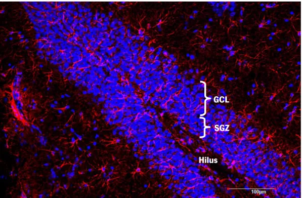
Lastly, regarding astrocytes, the main glial subtype, these cells interact closely with neurons, participating in the regulation of synaptic neurotransmission by releasing chemical transmitters: the so called “tripartide synapse” (Araque et al., 1999) (see Figure 1). These star-shaped cells have functional receptors for neurotransmitters and respond to their stimulation by releasing gliotransmitters, including glutamate. Astrocytes can increase the intracellular calcium ([Ca2+]i) upon an elevation of synaptically
released neurotransmitters, resulting in the release of glutamate via regulated exocytosis (Rossi & Volterra, 2009). Data reports that this increase in [Ca2+]i is extremely important, in a functional view,
for astrocyte-astrocyte and also astrocyte-neuron intercellular communication (Sofroniew & Vinters, 2010; Cornell-Bell et al., 1990; Charles et al., 1991).
9
Figure 1. The tripartide synapse. Astrocytes express many of the same receptors as neurons. When
neurotransmitters are released from the presynaptic terminal of a neuron, astrocytic receptors are thought to be activated, leading to a rise in calcium ions in the astrocyte and the release of various active substances, such as ATP, which act back on neurons to either inhibit or enhance neuronal activity. Astrocytes also release proteins, which control synapse formation, regulate presynaptic function and modulate the response of the postsynaptic neuron to neurotransmitters (Allen & Barres, 2009).
Astrocytes can also couple to neighboring astrocytes through gap junctions and, putting this in a multicellular network perspective, they can play a role in both normal function and CNS disorders (Nedergaard et al., 2003; Seifert et al., 2006). These findings led astrocytes to the spotlight, bringing a new concept of neuron–glia intercommunication where astrocytes play an active role by integrating neuronal inputs and modulating synaptic activity (Rossi & Volterra, 2009). It is also noteworthy that astrocytes are able to synthesize glutamate de novo and to store glucose in the form of glycogen, unlike neurons (Hertz & Zielke, 2004), thus contributing to brain metabolism (Hertz et al., 2007). This phenomenon is only possible due to astrocytes´ high oxidative metabolism.
As mentioned above, NSCs express several radial glia and astrocytic markers, including brain lipid binding-protein (BLPB) and the glutamate aspartate transporter - GLAST (Steiner et al., 2006). Although they express the common glial fibrillary acidic protein isoform alpha (GFAP)-a, the progenitor cells also specifically express the GFAP isoform delta and can be isolated based on its specific expression (Van Den Berge et al., 2010). The fact that NSCs in both neurogenic zones of the adult brain (SGZ and SEZ)
10
have plenty of astroglial properties makes it possible to link adult neurogenesis and glial cells (Morrens et al., 2012). In fact, genetic ablation of GFAP-expressing cells showed to be capable of eliminating adult neurogenesis (A. D. R. Garcia et al., 2004; Imura et al., 2003; Morshead et al., 2003). Moreover, astrocytes from the SEZ and SGZ were shown to promote the proliferation of progenitor cells and their neuronal differentiation ex vivo (Lim & Alvarez-Buylla, 1999; Song et al., 2002). Although astroglia seems enough to support synaptic integration and functional maturation of newly born neurons (Hong-jun Song et al., 2002), the same cells derived from a non-neurogenic region fosters glial over neurogenic fate (Lie et al., 2002). In vivo, astrocytes seem to provide highly physical support to progenitor cells and newly born neurons (Shapiro et al., 2005; Plümpe et al., 2006), thus also playing a possible role in adult neurogenesis regulation in vivo (Morrens et al., 2012).
Citing Ben Barres, an expert in neuron-glial interactions: “Quite possibly saving astrocytes from dying in neurological disease would be a far more effective strategy than trying to save neurons (glia already know how to save neurons, whereas neuroscientists still have no clue)” (Barres, 2008).
1.4. Glial Cell Pathology in the disease context
Astrocytes dysfunction has been related with neural impairments, mostly because of the tripartide synapse disturbance. There is now a growing body of evidences showing that either loss of normal astrocytic functions or gain of abnormal effects can contribute to the progress of several diseases, with these cells playing numerous roles in clinical and pathological mechanisms (Sofroniew, 2005; Seifert et al., 2006; Barres, 2008; De Keyser et al., 2008; Takano et al., 2009).
Focusing in genetic diseases with cognitive delays, astrocytes seem to have a preponderant role, being the alteration of the astrogliogenesis timing more associated with mental impairments, in animal models (Gauthier et al., 2007). Moreover, a recent and specific study focused in Down Syndrome, was successful in showing a gliogenic shift from neural progenitors from Down syndrome patients, with a concomitant decrease in neurogenesis (Lu et al., 2011).
Astrocytes were also found to be related with epilepsy due to their effects both on glutamate transport and release as in buffering potassium and interstitial volume control (Wetherington et al., 2008; De Lanerolle et al., 2010). Astrocytic dysfunction has been shown to be related with abnormal neuronal excitability, regarding adult model systems (Gómez-Gonzalo et al., 2010); additionally, data showed that inducing reactive astrocytosis can lead to the formation of hippocampal epileptic foci
11
(Ortinski et al., 2010). All these observations have raised several questions, mostly regarding the possibility of increased susceptibility to epileptogenesis resulting from an abnormal astrocyte development (which could result in an alteration of excitatory-inhibitory balance of the developing brain) (Molofsky et al., 2012).
Astrocytes can also see themselves involved in Alzheimer´s disease, being the reactive astrogliosis a well-known feature of this neurodegenerative disease. This specific process seems to be focal in this disease, such that reactive astrocytes are closely associated with amyloid plaques, surrounding them with a high density of processes and acting like neuroprotective barriers (Sofroniew & Vinters, 2010). Some reports state that reactive astrocytes have the capability of taking up and degrade extracellular deposits of a specific form of amyloid beta (Aβ42), leading to the belief on a role for astrocytes in the progression of the disease (Wyss-Coray et al., 2003). Some other studies reported the decrease of astrocyte glutamate transporters in Alzheimer´s disease, suggesting a resulting increased vulnerability of local neurons to excitotoxicity (Simpson et al., 2010).
Giving us a new insight about astrocytes´s role in cognitive functions, an amazing study came out this year, claiming that the engraftment of human glia progenitor cells in mice was enough to differentially enhance both activity-dependent plasticity and learning of the animal (Han et al., 2013). With this study, the authors were able to show that human astrocytes generated within the mouse brain were able to maintain their complex phenotype in a cell-autonomous fashion, suggesting that the specific aspects of human cognition could reflect the course of astrocytic evolution (Oberheim et al., 2006).
To further analyze astrocytes´ specific functions, transgenic mice models have been used along the scientific route, in which some astrocytic functions are blocked or attenuated. An example of this approach is the transgenic mouse model in which the expression of an inositol 1,4,5-trisphosphate absorbent is capable to attenuate astrocytic Ca2+ signaling. With this specific model, researchers showed
that the attenuated activity of Ca2+ was correlated with reduced astrocytic coverage of asymmetric
synapses in a specific hippocampal region, resulting in behavioral impairments in reference memory and remote contextual fear memory (Tanaka et al., 2013).
Although controversial, these studies gave as a first clue about the possible role of glial cells and gliogenesis in the disease context, leaving an open window to be further explored.
12 1.5. Gliogenesis in Depression
Unlike neurons, glial cells retain their ability to proliferate in most brain areas of postnatal and adult subjects (Kraus-Ruppert et al., 1975; Gensert & Goldman, 2001; Kornack & Rakic, 2001). The generation of astrocytes is detectable in the neocortex and the hippocampus of adult human brain and, although the majority of newly generated cells in the adult rat hippocampal DG are neurons (about 75%), there is still around 15% of new cells that are positive for the astrocytic marker GFAP and might be astrocytes. This neuron to glia ratio does not change with AD treatment, indicating that these treatments increase the number of newly generated glial cells in the adult brain (Rajkowska & Miguel-Hidalgo, 2007).
It is well known that there are multiple extrinsic and intrinsic mechanisms acting in concert to repress gliogenesis during the neurogenic period, and further induce gliogenesis when an appropriate number of neurons have been reached (Miller & Gauthier, 2007). However, in contrast to neurogenesis, the function of gliogenesis in the healthy adult brain has so far not been elucidated.
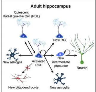
During development, several molecules act together to further determine the fate of multipotent precursor cell, later generating either neurons or glial cells (see Figure 2 for a schematic representation of the gliogenic process).
Figure 2. The Gliogenic Process in the Young Adult Mouse Hippocampus under basal conditions. There are at
least three critical choice points: (1) a radia glia (RGL) cell decides to remain in quiescence or to become activated and enter the cell cycle; (2) an activated RGL can undergo one of three models of self-renewal: (i) symmetric self-renewal to expand the RGL pool, (ii) neurogenic, or (iii) astrogliogenic asymmetric self-renewal to generate a differentiated progeny while maintaining the RGL pool; and (3) The RGL makes a choice between returning to quiescence and maintaining the stemness or differentiating into an astrocyte via transition to astroglia. It is also possible that a quiescent RGL can directly differentiate into an astrocyte without cell division. (Bonaguidi et al., 2011)
13
After several years of controversy, it is now well accepted that radial glial cells in the developing CNS are multipotent cells that have the capacity to give rise to separate precursors for neurons and mature glial cells (Campbell & Götz, 2002; Malatesta et al., 2003). These glial precursors can differentiate into astrocytes or oligodendrocytes, due to specific factors in the microenvironment of the cell: when exposed to platelet derived growth factor (PDGF) the differentiation culminates in the generation of oligodendrocytes, whereas when exposed to leukemia inhibitory factor (LIF), they produce astrocytes (Bonni et al., 1997; Rajan & McKay, 1998). Moreover, basic FGF (bFGF) has been proved to be relevant in both early neuronal development, maintaining the multipotent precursors, and postnatally, being produced by astrocytes and some neurons and inducing the oligodendrocyte lineage by glial precursors (Rogister et al., 1999; Rajkowska & Miguel-Hidalgo, 2007).
Similarly to the effects on the production of glial cells that occur during development, the factors mentioned above can also act in the proliferative zones of the adult brain and may, according to some reports, participate in the pathophysiology of depression (Horner & Palmer, 2003). Moreover, those factors can be manipulated to further achieve an effective AD action. Regarding this, bFGF, a stimulator of astrocyte and oligodendrocyte proliferation, can be an important factor in the depression field (Skaper & Varon, 1987; Hunter et al., 1993). Depressed patients showed a reduction in the mRNA level of bFGF in the dorsolateral prefrontal cortex, contrarily to ADs treatment that increased bFGF expression in the hippocampus and neocortex in an animal model (Mallei et al., 2002; Evans et al., 2004; Maragnoli et al., 2004).
Besides all these observations, there are still several gliogenic windows that must be further studied and explored in the context of this disease.
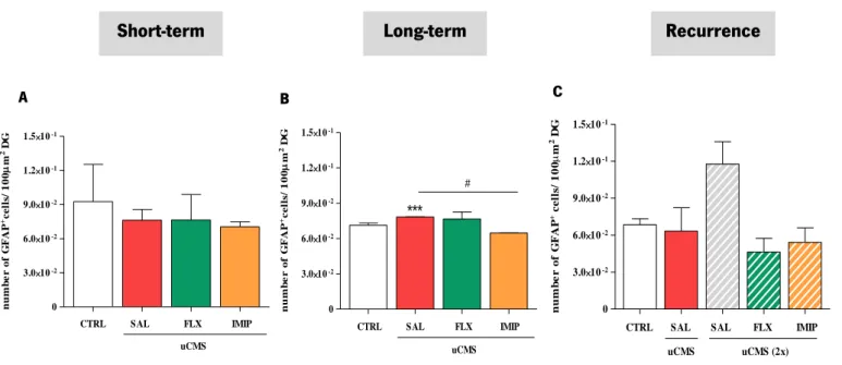
Regarding in vivo results, data from our lab showed that impairments induced by unpredictable chronic mild stress (uCMS) exposure, a validated animal model of depression (Bessa, Mesquita, et al., 2009), were reversed by both imipramine and fluoxetine ADs. Interestingly, whereas fluoxetine failed to restore working memory when neurogenesis was blocked, the cognitive-improving efficacy of imipramine did not depend on active neurogenesis. Fluoxetine treatment, as previously reported (Boldrini et al., 2009), was more effective at promoting differentiation of newly-born cells into neurons rather than astrocytes, contrarily to imipramine treatment, that showed to elicit a strong pro-gliogenic effect (Mateus-Pinheiro et al., 2013). These striking preliminary results suggest that the efficacy of imipramine in the recovery from depression depends directly on active gliogenesis and not neurogenesis.
14
However, until now little is known about the molecular changes regulating the decreased adult gliogenesis in depression, as well as about the counteracting mechanisms triggered by ADs. With growing evidence supporting the possible role of glial cells in the ethiopathogenesis of depression and the pro-gliogenic action of ADs, such as imipramine, adult gliogenesis becomes a promising area to study that may help to unravel novel therapeutic options for this pathology.
Since ADs treatment have showed to reverse the reduction in astroglial density in animal models of depression, and being the glial alterations more pronounced than those experienced by neurons, glial cells may represent a good target to give an anticipated and better prognosis of this deleterious disorder.
1.6. Impact of Astrocytes in Depression
During the last years, some studies came up showing the involvement of astrocytes in the pathophysiology of MDD (Hercher et al., 2009). Indeed, as mentioned above, astrocytes have been pointed out as an important player in brain function (Wang & Bordey, 2008; Perea et al., 2009), cross-talking with neurons, complementing and modulating neurotransmission (Araque et al., 1999); and to possess unique phenotypic features that allow them to monitor their neighbourhood, dynamically responding to neurovascular changes (Wang & Bordey, 2008).
In depression, a loss of astrocytes in specific regions of the brain was observed and this phenomenon lead us to an open window that was ill explored (Gosselin et al., 2009). Several studies, conducted in postmortem brain tissue of subjects diagnosed with MDD and/or bipolar disease (BPD), reported prominent decreases in the packing density and number of glial cells in several different frontolimbic areas, including prefrontal and medial prefrontal cortex, the dorsolateral and orbitofrontal cortex, the amygdala and also the hippocampus (Cotter et al., 2001; Harrison, 2002; Rajkowska & Miguel-Hidalgo, 2007; Drevets et al., 2008; Hercher et al., 2009).
However, the opposite pattern - increased glial cell density - was also seen in the GCL of the DG in depressed patients. Actually, this phenomenon could be explained by a reduction on glial processes, rather than a loss of glial cells, which could induce a decrease in hippocampal volume, currently seen in neuroimaging studies in the depression field (Stockmeier et al., 2004). Importantly, the study of glial pathology in mood disorders has not been extensive enough in subcortical structures to draw valid conclusions. It is also crucial to mention that several findings indicate that lower density of astrocytes
15
and decreased GFAP expression are associated with younger depressed subjects who had early onset of depression. Indeed, some studies indicate that GFAP expression levels are reduced in younger but not older depressed subjects. Thus, an increase in GFAP expression might not simply be related to biological aging, it may also be associated with the progression of cellular changes of depressive illness. This last observation implies that the involvement of GFAP expression is different in early versus late life depression. Increasing clinical evidence confirms that late onset depression (first depressive episode when older than 50 years) differs from early-onset depression by its etiology, phenomenology and cerebrovascular pathology (Rajkowska, 2000; Rajkowska et al., 2005).
Besides cell density, it seems that glial cell size and shape also suffer alterations in mood disorders. Some studies reported the increase of the glial cell bodies (Rajkowska et al., 1999, 2001; Chana et al., 2003) in depressed individuals and, regarding this observation, Rajkowska et al. proposed the existence of a compensatory mechanism capable of responding to the metabolic needs of the surrounding neurons. Since reduction in glial density was followed by increased glial nuclei, the authors claimed that the functional glial cells (the ones not affected by stress - related mechanisms) would be forced to work, due to the shrinkage of the damaged ones (Rajkowska & Miguel-Hidalgo, 2007). Afterwards, their nuclei would be bigger and with a different conformation. Fascinatingly, this adaptation – more specifically the increased size of glial nuclei - seems to be targeted to depressive disorders, since the glial size was not found to be altered in other disorders, such as schizophrenia (Rajkowska et al., 1998; Selemon et al., 1998).
Although astrocytes represent the most numerous type of glial cell, we cannot forget about the other glial cells, which also suffer alterations in this disease context. Furthermore, specific decreased number of oligodendrocytes was seen in the amygdala in MDD (Hamidi et al., 2004), as additional alterations in the microglial population in BPD (Manji et al., 2000).
With these observations, we can conclude that those alterations currently seen in depression models are not astrocytes – directed, affecting also the other glial populations (despite the astrocytic alteration are the most reported ones).
Lately, an exciting study came out showing that the specific ablation of astroglial cells in the prefrontal cortex (PFC) of adult rats (with L-alpha-aminoadipic acid) was enough to induce a depressive-like behavior on the animals. They presented a phenotype quite similar to those animals that are submitted to uCMS, an animal model of depression (Banasr & Duman, 2008). In order to show that it was specific for astrocytes, the researchers also injected neurotoxic ibotenate but it showed to be harmless to the animals.
16
Regarding the ADs administration, some studies also suggest that the treatment not only affects neurons, but also activates astrocytes. This activation can lead them to carry out specific functions that result in the reactivation of cortical plasticity and may cause the readjustment of neuronal networks, thus helping depressed individuals to recover (Czéh & Di Benedetto, 2012). Indeed, in a study conducted in Tupaia belangerie, a specie phylogenetically close to primates (Martin, 1993), animals were subjected to a chronic stress paradigm (Fuchs & Flügge, 2002) and the reduced number of astroglia found in response to stress was prevented by concomitant fluoxetine treatment (Czéh et al., 2006). Besides treatment with fluoxetine, chronic administration of lithium and antipsychotic medication have led to an increased glial number both in hippocampus and PFC of rats and nonhuman primates (Rocha et al., 1998; Selemon et al., 1999). As seen in other studies, chronic treatment with lithium induced an upregulation of GFAP expression and an alteration of astrocytes original morphology, more specifically in astrocytic orientation.
Taken together, further studies are needed to address the importance of all these glial alterations at the onset, maintenance and recurrence of a depressive episode. It is extremely relevant to determine whether therapies based on gliogenic factors will attenuate all the depressive symptoms. Moreover, it is of major importance to establish a state marker related with glial alterations, during episodes of depression.
17 1.7. Research Objectives
This project aims to correlate cognitive behavior with alterations in astrocytic cells and gliogenesis in the hippocampal DG of adult rats exposed to uCMS and treated with ADs, in a longitudinal manner: at short-term, long-term and recurrence time-points. Since treatment with the AD imipramine, but not fluoxetine, has been shown to elicit a strong pro-gliogenic effect, we also aim to establish a hippocampal derived-neurospheres culture to further study the differentiation of astrocytes in vitro, conditioning the culture with norepinephrine or serotonine (the neurotransmitters that mediates the effect of imipramine and fluoxetine, respectively).
In order to do that, our main objectives are:
To study the impact of the uCMS model of depression in astrocytic cells;
To explore how exposition to uCMS and treatment with distinct ADs (fluoxetine and imipramine) modulate hippocampal gliogenesis, more precisely the generation of new astrocytes, at different time-points: immediately after ADs treatment (short-term); 4 weeks after the end of the uCMS protocol and ADs treatment (long-term) and after exposition to a second period of uCMS (recurrence);
To correlate the cognitive performance of depressive-like rats (rats exposed to uCMS) and AD-treated rats, with astrocytes morphological alterations at short-term, long-term and recurrence;
To establish a hippocampal-derived neurosphere culture to further assess the proliferation and differentiation of astrocytes versus neuronal cells induced by the administration of distinct ADs in vitro, conditioning the neurospheres with serotonin or norepinephrine.
19
21
2. Materials and Methods
2.1. Animals
Male Wistar rats (Charles-River Laboratories), with 2 months of age and weighing 200-300g were group-housed (three per cage) under 12h light: 12h dark cycles, at 22°C, relative humidity of 55% and with food and water ad libitum.
Forty animals were randomly assigned to five main experimental groups (n=8): one control group (CTRL) not exposed to stress and treated with saline; three groups exposed to uCMS and treated with saline (CMS), fluoxetine (FLX) or imipramine (IMIP); and one group with the same treatment as the ones before but exposed to an extra stress period (DOUBLE).
All procedures were carried out in accordance with EU Directive 2010/63/EU and NIH guidelines on animal care and experimentation.
2.2. Chronic mild stress protocol
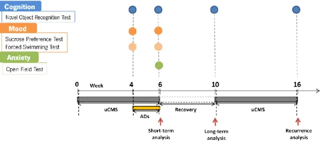
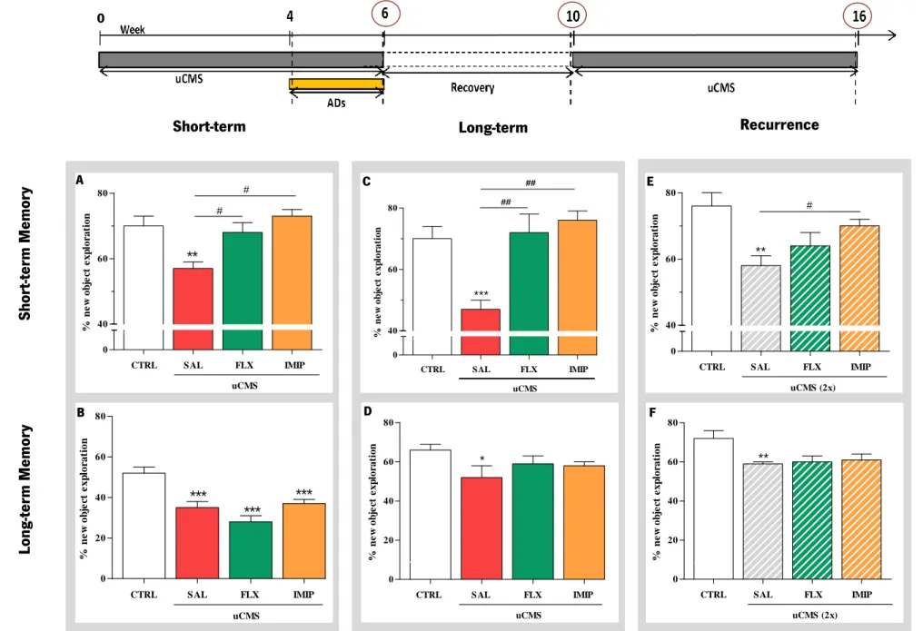
A protocol of uCMS was applied for 6 weeks as previously validated and described (Willner, 2005; Bessa, Ferreira, et al., 2009; Bessa, Mesquita, et al., 2009). Briefly, the uCMS protocol comprises several mild stressors (confinement to a restricted space for 1h; overnight food deprivation followed by 1h of exposure to inaccessible food; overnight water deprivation followed by 1h of exposure to an empty bottle; overnight damp bedding; inverted light/dark cycles; exposure to stroboscopic lights during 4h and noise exposure during 4h) to which animals were random- and uninterruptedly exposed during 6 weeks. In the last 2 weeks of the uCMS protocol, subsets of animals were administered daily with the ADs imipramine or fluoxetine and all other animals were injected daily with saline. On the following 4 weeks, all the animals were allowed to recover due to the stress protocol´s cessation, being kept unexposed to any kind of stressor. After 4 weeks resting, three of the five groups (FLX, IMIP and DOUBLE) were exposed to a second period of uCMS during 6 weeks. The behavioral analysis were performed during the stress protocol (week 4), after the cessation of the stress period (week 6) – short term analysis - ; after the recovery period (week 10) – long term analysis - ; and after the second exposure to the stress protocol (week 16) – recurrence analysis (Figure 3).
For the molecular and cellular analyses, the subsets of rats exposed to only one period of uCMS during 6 weeks and the ones exposed to one period of uCMS that were allowed to recover for additional 4
22
weeks, were already performed by other researchers and were available for this project for molecular and cellular analyses.
Figure 3. Schematic representation of the behavior analysis. The cognitive dimension was evaluated by the
novel object recognition test (NOR) in animals exposed to uCMS; the assessment was done at weeks 4, 6, 10 and 16 (after the exposition to the second period of uCMS). The mood dimension was assessed by both Sucrose Preference Test (SPT), giving us an anhedonic information regarding the animals profile, and Forced Swimming Test (FST) at weeks 4 and 6. Anxiety dimension was evaluated trough the Open Field Test (OF) at week 6.
2.3. Drugs
The ADs fluoxetine (10mg.kg-1; Kemprotec, Middlesborough, UK) and imipramine (10mg.kg-1;
Sigma-Aldrich, St.Louis, USA) were administered intraperitoneally (i.p.: 1 ml.kg-1). These drugs were dissolved
in DMSO (5%; Sigma-Aldrich) and saline (0,9%; B.Braun) and administered daily during the last two weeks of the uCMS protocol (Figure 3).
2.4. BrdU injections
Short-term analysis. BrdU (100mg.Kg-1; i.p.) was administered for one day at the end of the uCMS
period.
Long-term analysis. In order to assess cell proliferation, BrdU (50mg.Kg-1; i.p.) was administered daily
for five consecutive days at the end of the uCMS period, including the last two days of uCMS protocol and the first three days of the recovery period.
23
Recurrence analysis. At this time-point, BrdU (50mg.Kg-1; i.p.) was administered daily for seven
consecutive days at the end of the first uCMS period, including the last three days of uCMS protocol and the next four days of the recovery period.
2.5. Behavioral Tests
2.5.1. Novel Object Recognition
The cognitive function was assessed longitudinally by the novel object recognition test (NOR) and was performed at weeks 4, 6, 10 and 16 (always with objects different from each other in terms of shape, colour and texture), as schematized on Figure 3 by the blue dots. For this purpose, a black acrylic box (50x50x150cm) with an open field space (51x51x39,5cm) and illuminated with a white lamp (100-140 lux) was used. This test is phased in 4 days and was already tested and described in the literature (Bevins & Besheer, 2006; Dere et al., 2007; Winters et al., 2008; Ennaceur, 2010):
Exploration. On the first day, the animals were allowed to explore the test apparatus without any object during 10 minutes (min).
Sample Phase. On the second day, two identical objects were placed in the back left and right corners of the apparatus and the animal was able to explore them during 10 min. Within an interval of approximately one hour, a second sample object exposure (3 min) was performed. This time, one of the sample objects (left object) was repositioned in a new corner of the apparatus (middle of the left wall). This second trial gives an insight on hippocampus function and it works as a memory “reinforcement” of the sample object.
Choice Phase – Long-term memory. On the third day, the long-term memory was assessed 24 hours after the memory reinforcement (done on the second day of the test) by a 3 min trial with the replacement of one of the sample objects for a novel object.
Choice Phase – Short-term memory. Lastly, on the fourth day, animals were tested for their short-term memory condition. The previous objects were switched for two identical new sample objects and the animal was left for exploration in the apparatus, during 10 min. Within an interval of approximately one hour, a choice phase test was performed. To do this, one of the sample objects was replaced for a
24
completely new object and, for 3 min, the exploration of the animal was determined in order to test its memory.
Trials were video-recorded and the discrimination index (D) was calculated by the following formula: D = (N-F)/(N+F); being N the time spent exploring the Novel object and F the time spent exploring the Familiar object. For this test, it is crucial to define what we considered as the object exploration by the animal; for that, we assumed exploration of an object as directing the nose to the object at a distance of less than 2 cm or touching it with the nose.
2.5.2. Sucrose Preference Test
To assess anhedonia, the sucrose preference test (SPT) was conducted at week 4 and 6 of the uCMS period (Figure 3). Animals were allowed to habituate to the sucrose solution (2% m/v) 1 week prior to the uCMS protocol, in order to establish the baseline values for sucrose preference. For both assays, animals were food- and water-deprived for 12h during the non-active period. The room was cleaned with ethanol 96% and the test was performed under dimly illumination. Animals were placed in each cage, further covered with both the grid and the lid. Two pre-weighted bottles containing the sucrose solution and tap water were placed simultaneously in the cage and consumption was measured for 1h.
Sucrose preference was calculated by the following formula: sucrose preference=[(sucrose consumption / Total consumption) x 100] like previous described (Bessa, Mesquita, et al., 2009). Anhedonia was defined as a reduction in sucrose preference in relation to the baseline levels.
2.5.3. Forced Swimming Test
Learned-helplessness was assessed through the forced swimming test (FST) and was conducted at week 6 of the uCMS protocol (Figure 3). The test was performed 24h after the 5 min pre-test session, consisting in placing the animals in cylinders filled with water (25°C; 50cm of depth) during 10 min. Trials were video-recorded and both the immobility time and the latency to immobility were measured through the Etholog (vs.2.2) software. Learned-helplessness was defined as an increase in the immobility time and a decrease in the latency to immobility.
25 2.5.4. Open Field Test
Anxiety-like signs were assessed through the open field (OF) test like some authors have reported (Prut & Belzung, 2003), in the week 6 of the uCMS protocol (Figure 3). The test apparatus consisted of a brightly illuminated square arena of 43.2 x 43.2 cm closed by a wall of 30.5 cm high. Animals were placed individually in the center of the arena and their movement was traced during 5 min, using a two 16-beam infrared system. The resulting data was analysed using the Activity Monitor software (Med Associates,Inc.), considering two previously defined areas: a central and an outer area. Time spent in each of the zones was recorded and analysed, further.
2.6. Measurement of Plasma Corticosterone Levels
Blood samples were collected from the rat’s tails in different time-points: 8.00 am and 8.00 pm, at weeks 4 and 6 of the uCMS protocol.
Blood plasma was separated by centrifugation (2500 rpm, 30 min) and corticosterone levels were determined using the Correlate(tm)-EIA ELISA Kits (Assay Design Inc., Ann Arbor, MI, USA).
2.7. Tissue Preparation and Sectioning
Animals were deeply anaesthetized with sodium pentobarbital (20%; Eutasil, Sanofi) and were transcardially perfused through the ascending aorta with saline (0.9% NaCl; B Braun). Brains were dissected from the skull, embedded in Neg-50 Frozen Section Medium (Thermo Scientific) and further frozen in liquid nitrogen. The tissue was processed in series of 20µm cryosections, extending over the entire length of the hippocampus formation. The slides where then maintained at -20°C for immunostaining procedures.
2.8. Immunostainings
2.8.1. Immunofluorescence
Cryosections were immersed in 4% paraformaldeyde (PFA; Sigma-Aldrich) for 30 min at room temperature (RT) and then rinsed in Tris-Buffered Saline (TBS). Eventual pretreatment of the sections is mentioned in the list of used antibodies in Table 1. After 10 min of permeabilization in TBS-Triton (T; Thermo Scientific) 0,2%, sections were incubated with the primary antibody in solution (TBS/10% Fetal Bovine Serum (FBS)) overnight, at 4°C. On the second day, sections were washed 3 times in TBS and incubated with secondary antibody for 2h, at RT. The used secondary antibodies are listed in the Table
26
2. Sections were rinsed with TBS and further stained with DAPI (1:1000; Invitrogen) and mounted with immune-mounting medium (Immumount, Thermo Scientific) for following analysis by confocal microscopy. Solutions composition is described in Table 3.
For each animal, GFAP-positive cells and GFAP co-localized with BrdU-positive cells within the DG were analyzed in the confocal microscope Olympus FluoViewTM FV1000 (Hamburg, Germany).
Estimation of cell density in the DG was obtained by crossing the GFAP+ cell number values within the
corresponding DG areas, determined using the same confocal microscope. Moreover, in order to analyze the density of newborn astrocytes within the area of the DG (from the short-term, long-term and recurrence), cells that co-localized GFAP+ and BrdU+ were selected and analyzed.
For morphologic analysis of the new astrocytes (BrdU+GFAP+), we used the Neurolucida software, with
the AutoNeuron extension module (MBF Bioscience).
2.8.2. DAB (3,3`diaminobenzidine tetrahydrochoride) immunohistochemistry
For the purpose of analyzing the astrocytic morphology, this specific technique was used. Basically, the first day of the protocol is similar to the one explained on the section before, except the blockage of endogenous peroxidases, performed immediately before the primary antibody incubation. This blocking solution consists of TBS with 10% H2O2 and it was added for 10 min, shaking. On the second day, the
slides were washed with TBS buffer and then incubated with biotinylated secondary antibody (ThermoScientific) for 30 min. After that, streptavidin peroxidase (ThermoScientific) was added for 30 min. A washing step took place with TBS buffer and, before the develop step, the tissue was washed with a Tris-HCl solution for 5 min. The develop solution (0,025 g DAB on 100 ml Tris-HCl solution and 500 µl H2O2) was added to the tissue and the reaction was stopped around 5 min after with TBS. The
sections were then dehydrated manually, staying 2 minutes in alcohol 96° and 2 min more in alcohol 96° for total tissue dehydration, 2 min in alcohol 100° and 2 final min in xylol.
For each selected astrocyte (localized on the DG of the hippocampus), all processes were reconstructed at 100x (oil) magnification using a motorized microscope (Axioplan2; Carl Zeiss) and Neurolucida software. A three-dimensional analysis of the reconstructed astrocytes was performed using NeuroExplorer software (Microbrightfield).
27 Table 1. Primary Antibodies.
Prim. Antibody /
Specie Working Dilution Pretreatment Company
BrdU Mouse 1:50 30 min. in HCL (2 M) + 20 min. in pre-heated
Cytrate buff. (80ºC) Dako
GFAP Rabbit 1:200 None Milipore
Table 2. Secondary Antibodies.
Sec. Antibody / Antigenicity Working
Dilution Pretreatment Company
Alexa Fluor 488 anti-mouse 1:1000 None Invitrogen
Alexa Fluor 568 anti-rabbit 1:1000 None Invitrogen
Table 3. Solutions Composition.
Solution Composition
TBS 50 mM Tris base, 150 mM NaCl, pH 7.6 TBS-T TBS, 0.2% TritonX – 100
PFA 4% TBS, 4% PFA, pH 7.4 Cytrate Buffer 0.1 M Sodium citrate, pH 6.0
2.9. Neurospheres Culture Set Up
For establishing the neurospheres culture, 10 rats, 6 days old, were sacrificed and their brains removed. The hippocampal DGs were isolated and minced using a bisturi and the dissociated tissue was digested in 0.1% trypsin (Sigma-Aldrich) for 15 min at 37°C. After that, the tissue was triturated with a 5 mL pipette (10 times up and down), and the resulting solution was filtered through a 70 µm strainer (BD Biosciences), followed by a centrifugation at 1300 rpm for 5 min. The resulting pellet was resuspended in 1 ml of neurosphere medium (supplemented with growth factors), and a single cell suspension was achieved by gentle trituration. The growth factors added were 10 ng/ml epidermal growth factor (EGF; Invitrogen) and 10 ng/ml basic fibroblast growth factor (bFGF; Invitrogen). The cells were then plated in a T25 flask at a clonal density of 8-10 cells per µL and the growth factors were added every 2 days.
28 2.10. Data Analysis
All the statistical analysis was performed with GraphPad Prism 5.01. The unpaired Student´s t-test was used to examine whether data sets differed significantly, when the experimental setup was composed by only two experimental groups, and also to compare the control group with the uCMS group. One-way ANOVA was used in multiple statistical comparisons between groups and with only one level of analysis, with Tukey’s multiple comparison test post hoc analysis. Statistical significance was accepted for p<0.05.
29
31 0 50 100 150 200 250 CTRL CMS *** Im m o b il it y t im e ( s)
3. Results
3.1. Study of the cognitive behavioral dimension of depressive-like rats treated with the ADs fluoxetine and imipramine at short-term, long-term and recurrence
3.1.1. Establishment and validation of the animal model of depression
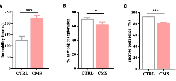
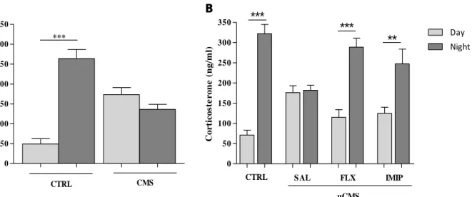
As it was reported by our group, the appliance of the uCMS paradigm, the same used in this study, induces typical depressive-like signs in all three behavioral dimensions that are commonly affected by depression – anxiety, mood and cognition (Bessa, Mesquita, et al., 2009). In order to proceed to further tasks, it was imperative to first assess and characterize both the behavioral profile of the animals and the levels of corticosterone as a powerful tool to measure stress induction, since the increased levels of plasma corticosterone can be related with a stressful episode (Kant et al., 1987).
Four weeks after starting the uCMS protocol (described in the 2.2 section of materials and methods), animals were tested in order to evaluate the extension of the psychological damages, which can lead to several affected behavioral dimensions. Therefore, learned helplessness was assessed through the FST, anhedonia was assessed by the SPT and the cognitive domain was evaluated through the NOR test (Figure 4).
Figure 4. Behavioral characterization of the animals on the fourth week of the uCMS protocol. (A) Learned-helplessness was assessed through the FST; (B) Cognition integrity was assessed through the NOR test; and (C) Anhedonia was evaluated with the SPT. Data is represented as mean ± sem. *p<0.05, **<0.01, ***<0.001. Abbreviations used: CTRL – Control Group; CMS – uCMS group.
A B C 0 20 40 60 80 + CTRL CMS * % n e w o b je ct e x p lo ra ti o n 0 20 40 60 80 100 CTRL CMS *** su cr o se p re fe re n ce ( % ) 0 20 40 60 80 + CTRL CMS * % n e w o b je ct e x p lo ra ti o n 0 50 100 150 200 250 CTRL CMS *** Im m o b il it y t im e ( s) 0 20 40 60 80 100 CTRL CMS *** su cr o se p re fe re n ce ( % )