Gabriela Teixeira Carrasqueiro
Dissertation in the Master's Degree in
Oncobiology
Molecular Mechanisms of Cancer
Epigenetic Alterations in Sepsis
Departamento de Ciências Biomédicas e Medicina
2017
Gabriela Teixeira Carrasqueiro
Epigenetic Alterations in Sepsis
Master's Degree in Oncobiology
Molecular Mechanisms of Cancer
Work carried out under the supervision of:
Prof. Dr. Pedro Castelo-Branco e Drª Alexandra Binnie
Departamento de Ciências Biomédicas e Medicina
2015/2017
Epigenetic Alterations in Sepsis
Declaração de autoria do trabalho
Declaro ser o(a) autor(a) deste trabalho, que é original e inédito.
Autores e trabalhos consultados estão devidamente citados no texto e
constam da listagem de referências incluída.
A Universidade do Algarve reserva para si o direito, em conformidade com o disposto no Código do Direito de Autor e dos Direitos Conexos, de arquivar, reproduzir e publicar a obra, independentemente do meio utilizado, bem como de a divulgar através de repositórios científicos e de admitir a sua cópia e distribuição para fins meramente educacionais ou de investigação e não comerciais, conquanto seja dado o devido crédito ao autor e editor respetivos
I
Agradecimentos
Em primeiro lugar gostaria de agradecer ao meu orientador, o professor Dr. Pedro Castelo-Branco e a minha coorientadoraDr.ª Alexandra Binnie pela grande oportunidade que me deram para fazer esta tese de mestrado sob a sua orientação. Similarmente, gostaria de referir a importância destes na elaboração desta tese, uma vez que estiveram sempre presentes e, sempre que foi oportuno, contribuíram com o seu conhecimento e experiência, contribuindo de forma decisiva para a minha evolução como estudante e investigador durante a elaboração deste trabalho.
Uma menção especial para a Dr.ª Alexandra Binnie que sempre me acompanhou de perto durante a realização deste trabalho, uma vez que este foi feito no âmbito de várias temáticas que tiveram como base o trabalho realizado por ela e os seus colegas. Queria agradecer à Dr.ª Cláudia Santos e ao Dr. Chris Walsh pela ajuda prestada durante algumas etapas essenciais deste trabalho.
Não posso esquecer também de agradecer a todos os meus colegas no laboratório de
epigenético do grupo de epigenética e doenças humanas (Epigenetics and
HumanDiseaseGroup), pois ajudaram-me sempre necessitei e com os quais partilhei muitas horas.
Queria agradecer ao meu namorado, Tiago, pelo apoio, a paciência e a força que sempre me deu durante a realização deste trabalho. E um agradecimento muito especial à minha família, especialmente, minha querida mãe, Sónia, o meu querido irmão, Luís, e a minha grande amiga de infância Jéssica, que desde sempre me deram apoio e também viveram comigo todas as etapas desta tese.
II
Resumo
A sepsis representa a resposta inflamatória sistémica do hospedeiro a umainfecção, (Iskanderet al., 2013) e é a 15ª causa mais significativa de morbidade e mortalidade em todo o mundo. A sepsis provoca 20 a 30 milhões de casos anuais no mundo inteiro causando uma morte a cada 3-4 segundos (WSD et al., 2013). Os pacientes com sepsis requerem custos substanciais de cuidados de saúde durante a sua hospitalização. Para além disso, os sobreviventes incorrem em custos adicionais devido ao aumento dos requisitos de cuidados de saúde nos anos que se seguem ao episódio da sepsis (Iskanderet al., 2013)
A sepsis é um processo complexo que pode ser iniciado por uma ampla gama de infeções, bacterianas, virais ou fúngicas, numa ampla gama de locais corporais. Muitos são os fatores de risco que despoletam um choque séptico ou o desenvolvimento da sepsis severa. Estes incluem, idade (75-85 anos), etnia e sexo (Alexander Melamed et al., 2009), bem como comorbidades como o cancro (linfomas ou leucemias), ou outras doenças imunossupressivas como HIV ou diabetes(Martin GS et al., 2006). As infeções nosocomiais, resultantes da exposição ao ambiente hospitalar, também são uma causa importante de sepsis (Farinas Alvarez et al., 2001).
Na fisiopatologia desta doença heterogénea, uma resposta inflamatória intensa é desencadeada, e seguida por uma cascata de eventos secundários, incluindo disfunção endotelial, coagulopatia, apoptose celular e imunossupressão (Stearns-Kurosawaet al., 2011). Uma infeção localizada torna-se assim uma síndrome sistémica, mediada por fatores solúveis como citoquinas e células imunes circulantes, como neutrófilos e leucócitos (Stearns-Kurosawaet al., 2011). Estes eventos contribuem para a disfunção progressiva dos órgãos e, posteriormente, a morte.
O diagnóstico precoce da sepsis pode ser desafiador devido à existência de outras doenças com apresentações similares. Os “imitadores” de sepsis incluem insuficiência cardíaca, insuficiência renal, pancreatite, anafilaxia, embolia pulmonar, isquemia intestinal, cetoacidose diabética e vasculite, entre muitos outros. Os marcadores de proteínas da sepsis, incluindo a procalcitonina e a proteína C-reativa, não são confiáveis (José Raimundo Araújo prognóstico.
Este projeto visou estudar esta doença complexa de uma forma inovadora. Com colaboração com um grupo de investigadores no Canadá, criou-se pela primeira vez uma coorte de pacientes sépticos (n=68) e pacientes com terapia intensiva não-séptica (n=66). Um
III
estudo chamado EPSIS (EpigeneticProfiling in Severe Sepsis) visou compreender a importância da regulação epigenética na patogénese, progressão, resposta ao tratamento e resultados de pacientes sépticos críticos.
Esta tese focou-se na hipótese que pacientes com e sem sepsis apresentavam diferentes padrões de metilação de DNA nos genes implicados na patogénese de sepsis. Como primeiro objetivo e fase neste projeto, identificou-se genes-chave na regulação epigenética na sepsis. Identificou-se genes que são diferencialmente regulados na sepsis, e que se correlacionam com diferentes vias e consequências metabólicas importantes, como a coagulopatia, disfunção endotelial, imunossupressão eimunoactivação e apoptose. Assim, após esta revisão bibliográfica extensa criou-se um cluster inicial de 128 genes.
De seguida, com os dados de metilação provenientes do coorteda EPSIS, analisou-se as diferenças de metilação de DNA entre as várias sondas dos 128 genes, potenciado, desta forma, a criação do segundo cluster de genes que se mostraram significativos na distinção de um paciente com sem sepsis e sem sepsis.
Na terceira fase, e como o nosso foco assentava na análise desta doença de uma maneira robusta, analisámos trêsconjunto de dados, provenientes do GeoDatasets, que avaliaram expressão de genes em pacientes com e sem sepsis. Assim, apesar de não podermos fazer uma correlação direta entre os nossos dados de metilação e os dados da transcriptómica, conseguimos entender quais os genes que estão diferencialmente regulados na patogénese desta doença.
Com as análises de metilação e de transcrição criou-se, pela primeira vez, um grupo de 10 genes que se mostraram estar diferencialmente metilados e expressos na sepsis. Entre esses incluem-se HLA-A, HLA-C, HLA-DOB, HLA-DQB1, FADD, APOL3, ITGB2, NLRP12, VARS2 e C3AR1. Este grupo de genes desempenha diferentes funções como inflamação, apoptose, imunidade inata e adaptativa. As análises mostraram que metade destes genes apresentaram um elevado grau de metilação, nomeadamente HLA-DOB, APOL3, FADD, NLRP12 e VARS2, enquanto os outros cinco (HLA-A, HLA-C, HLA-DQB1, C3AR1, ITGB2) se encontravam hipometilados. Analisando as diferenças ao nível de expressão genica a maioria destes genes encontravam-se subregulados.
A última fase deste projeto focou-se na correlação de parâmetros clínicos relevantes com a metilação dos 10 genes que anteriormente se revelaram significativos. Diferentes parâmetros foram analisados para a coortede pacientes, incluindoos dias de permanência da
IV
positiva ou gram-negativa); administração de vasopressores, esteróides, ventilação mecânica; e presença de doenças crónicas incluindoinsuficiência cardíaca e doença cardíaca isquémica. Em cada um destes parâmetros analisou-se varias sondas de CpG que apresentaram significância estatística e pudemos estabelecer uma relação entre metilação destes genes eo parâmetro clínico.
Este projeto centrou-se no estudo da sepsis, uma doença altamente complexa, heterogénea, e frequentementefatal. Nós reunimos, para a primeira vez, perfis de metilação de DNA de 66pacientes sépticos e 68pacientes com terapia intensiva não-séptica e compáramos esses dados com informações de transcriptómica de 3coortes diferentes de pacientes com sepsis. Adotamos uma abordagem de bioinformática para identificar e estabelecer assinaturas de metilação de DNA da sepsis em um conjunto focado de genes relacionados a sepsis. Dez desses genes, incluindo HLA-A, ITGB2, FADD, e APOL3, mostraram mudanças significantes de metilação e transcrição, sugerindo regulação epigenética. Além disso, identificamos correlações entre várias sondas CpG e parâmetros clínicos, incluindo infecção gram-positiva versus gram-negativa, sobrevivência global, e necessidade de vasopressores.
Palavras-chave:
V
Abstract
Sepsis is a life-threatening complication of infection. Typically, a localized infection triggers a systemic inflammatory cascade resulting in widespread organ damage, organ failure, and often death. Of the 31.5 million cases of sepsis worldwide annually, it is estimated that there are 5.3 million deaths (Fleischmann et al., 2016). The pathophysiology of sepsis involves widespread reprogramming of gene expression. Bioinformatic approaches have revealed multiple gene pathways that are activated or inhibited in sepsis (DC Angus and Tom Van der Poll et al., 2013). Epigenetic mechanisms, including histone modifications, DNA methylation, and non-coding RNAs (such as microRNAs, siRNAs, andribosomal RNAs) are master regulators of gene expression in both normal and pathological states. Although there is limited data on epigenetic regulation of sepsis, localized epigenetic changes have been identified in individual genes.However no genome-wide data has yet been published. In this studywe define a number of sepsis-related DNA methylation changes in sepsis-associated genes. We correlate these changes with gene expression and with clinically-relevant outcomes. Finally, we will investigate the mechanisms by which DNA methylation regulates individual gene expression in sepsis.
Keywords:
Epigenetic, Sepsis, DNA Methylation, Biomarker
A sepsis é uma complicação potencialmente fatal derivada de uma infecção. Normalmente, uma infecção localizada desencadeia uma cascata inflamatória sistémica, resultando em danos generalizados nos órgãos, falência de órgãos e, muitas vezes, a morte. Dos 31,5 milhões de casos de sepsis reportados em todo o mundo anualmente, estima-se que haja cerca de 5,3 milhões de mortes (Fleischmann et al., 2016). A fisiopatologia da sepsis envolve a reprogramação generalizada da expressão génica. As abordagens bioinformáticas revelaram múltiplas vias genéticas que são ativadas ou inibidas na sepsis (DC Angus e Tom Van der Poll et al., 2013). Os mecanismos epigenéticos, incluindo modificações nas histonas, metilação do DNA e RNAs não codificante (como microRNAs, siRNAs ou RNAs ribossómicos) são os principais reguladores da expressão genica, em estados normais e patológicos. Embora haja dados limitados sobre a regulação epigenética da sepsis, ainda não foram publicados dados genome-wide. Neste projecto, definiremos as mudanças de metilação
VI
correlacionar essas mudanças com a expressão génica e com parâmetros clínicos relevantes. Finalmente, investigaremos os mecanismos pelos quais a metilação do DNA está a regular individualmente cada gene envolvido na sepsis.
VII
Abbreviations
3
3´UTR - 3′ untranslated region
5
5´UTR - 5′ untranslated region
A
A2AP - Alpha 2-antiplasmin
Alpha – 1B - Adrenergicreceptor (α1B adrenoreceptor) ANOVA- Analysis of Variance
APAF-1 - Apoptotic protease activating factor 1 APC – Activated protein C
APCs – Antigen- presenting cells APOL3 - Apolipoprotein L3 ATP – Adenosine Triphosphate
B
BATF - Basic Leucine Zipper ATF-Like Transcription Factor BAX - Bcl-2-like protein 4
BCL-2 - B-cell lymphoma 2
BET - Bromodomain and extra-terminal BID - BH3 Interacting Domain Death Agonist BIM - BCL-2-like protein 11
BIP - Immunoglobulin protein
BIRC1 - Baculoviral IAP Repeat Containing 1 BIRC2 - Baculoviral IAP Repeat Containing 2 BIRC5 - Baculoviral IAP Repeat Containing 5
VIII
BIRC8 – Baculoviral IAP Repeat Containing 8 BOK-L - Bcl-2 related ovarian killer
BTN3A2 - Butyrophilin Subfamily 3 Member A2
C
C21ORF56 - Spermatogenesis And Centriole Associated 1 Like C3AR1 - Complement C3a Receptor 1
C6ORF15 - Chromosome 6 Open Reading Frame 15 C9ORF95 - Nicotinamide Riboside Kinase 1
CARS- Compensatory Anti-inflammatory response syndrome CCL-4 - Chemokine (C-C motif) ligand 4
CD14 - Cluster of differentiation 14 CD2 - Cluster of differentiation 2 CD28 - Cluster of differentiation 28 CD4 - Cluster of differentiation 4 CD80 - Cluster of differentiation 80 CD86 - Cluster of differentiation 86
CEACAM1 - Carcinoembryonic Antigen Related Cell Adhesion Molecule 1 CHN2 - Chimerin 2 CLDN1 - Claudin 1 CNX – Calnexin CRT - Calreticulin C-SMAC - Central-SMAC
D
DEFB1 - Defensin Beta 1
DIC - Disseminated intravascular coagulation DNA - DesoxiriboNucleicAcid
DNMT - DNA methyltransferaseenzymes DNMT 1 - DNA Methyltransferase 1 DNMT 3a - DNA Methyltransferase 3a
IX
DNMT 3b - DNA Methyltransferase 3b DNMT b - DNA Methyltransferase b DPP4 - Dipeptidyl Peptidase 4 D-SMAC - Distal-SMAC dsRNA - Double stranded RNA
DYNAMICS - DNA as a Prognostic Marker in ICU patients
E
EPSIS - Epigenetic Profiling in Severe Sepsis study EWAS – Epigenome-wide association study
F
FADD - Fas-associated protein with death domain FDR - False Discovery Rate
FGA - Fibrinogen Alpha Chain FGB - Fibrinogen Beta Chain FGG - Fibrinogen Gamma Chain
G
G-CSF - Granulocyte colony-stimulating factor
GM-CSF - Granulocyte macrophage colony-stimulating factor GNA12 -G Protein Subunit Alpha 12
GRP79 - Glucose related protein 78
GSTM3C - Glutathione S-Transferase Mu 3
H
H3K9me2 - Dimethylation of histone H3 at lysine 9 HAS-2 - Hyaluronan Synthase 2
HLA - Human leukocyte antigen
HLA-DQA2 - HLA Class II Histocompatibility Antigen, DQ(6) Alpha Chain HMWK - High-molecular-weight kininogen
X
Hsp100 - 100 Kilodalton heat shock protein Hsp60 - 60 Kilodalton heat shock protein Hsp70 - 70 Kilodalton heat shock protein Hsp90 - 90 Kilodalton heat shock protein HSPG2 - Heparan Sulfate Proteoglycan 2
I
ICAM1 - Intercellular Adhesion Molecule 1 ICAM2 - Intercellular Adhesion Molecule 2 ICU – Intensive care unit
IL-1 – Interleukin-1
IL-1 RA - IL-1 receptor antagonist IL-10 – Interleukin-10
IL-12 – Interleukin-12 IL-4 – Interleukin-4 IL-6 – Interleukin-6 IL-8 – Interleukin-8
INO80 - INO80 Complex Subunit ITGB2 - Integrin Subunit Beta 2
K
KDM6b - Lysine Demethylase 6BL
LPB - Lipopolysaccharide-binding protein LPS – LipopolysaccharidesM
MHC-I - Major histocompatibility complex type I MHC-II - Major histocompatibility complex type II miRNA – MicroRNA
XI
MODS – Multiple organ dysfunction syndrome MTCH1 - Mitochondrial carrier homolog 1
MyD88 - Myeloid differentiation primary response 88
N
ncRNA – Non-coding RNA NF-κB - nuclear factor kappa B
NK – Natural killer cell
NLRP12 - NLR Family Pyrin Domain Containing 12 NMP - Nuclear matrix protein
NOD-1 - Nucleotide Binding Oligomerization Domain Containing protein 1
O
OCLN – Occludin
P
PAMPs - Pathogen-associated molecular patterns PaO2 -Arterial partial pressure of oxygen
PCLN - Paracellin-1
PDI - Protein disulfide isomerase PF4 - Platelet Factor 4
piRNA - Piwi-interacting RNAs
POL1RD -RNA Polymerase I Subunit D PRRs - Pattern recognition receptors P-SMAC - Peripheral SMAC
R
RAGE - Receptor for advanced glycation end products RFX1 - Regulatory Factor X1
RNA - Ribonucleic acid
XII
S
SESN-2 - Sestrin 2
sFas - Soluble Fas receptor siRNA - Short interfering RNAs
SIRS – Systemic inflammatory response syndrome SLE - Systemic lupus erythematosus
SNPs - Single nucleotide polymorphisms SPAKs - Stress-activatedprotein kinases SRNX1 - Sulfiredoxin 1
SSBP1 - Single Stranded DNA Binding Protein 1 ssRNA - Single Stranded RNA
T
TET- Ten eleven translocation TLR-10 – Toll-like receptor 10 TLR-2 – Toll-like receptor 2 TLR-4 – Toll-like receptor 4 TLR-6 – Toll-like receptor 6 TLRs - Toll-like receptors TNF-ɑ - Tumor necrosis factor ɑ TPA -Tissue plasminogen activator TSS – Transcription start site
TSS1500 - Transcription start site to 1500 nucleotides upstream of TSS TSS200 - Transcription start site to 200 nucleotides upstream of TSS
V
VARS2 - Valyl-TRNA Synthetase 2 VWF - Von Willebrand factor
W
XIII
Y
Índice
AGRADECIMENTOS ... I RESUMO ...II ABSTRACT ... V ABBREVIATIONS ... VII 1. INTRODUCTION ... 1 1.1- Sepsis ... 11.2- Infection and Immunity ... 1
1.2.1- Infection ... 1
1.2.2- Innate Immunity ... 2
1.2.3- Adaptive Immunity ... 3
1.3- Pathophysiology of Sepsis... 4
1.3.1- Immunoactivation and Immunosuppression ... 4
1.3.2- Apoptosis ... 4 1.3.3- Endothelial Integrity ... 4 1.3.4- Coagulopathy... 5 1.4- Epigenetics ... 6 1.4.1- DNA Methylation ... 7 1.4.2- Histone Modifications ... 7 1.4.3- Non-coding RNAs ... 8
1.4.4- Epigenetic changes in sepsis... 9
1.5- Summary ... 9
2- MATERIALS AND METHODS ...10
2.1- DYNAMICS ... 10
2.2- Data Extraction and Normalization ... 11
2.3- Phase 1: Literature Search ... 12
2.4- Phase 2: Methylation Arrays ... 12
2.5- Characterization of the differentially-methylated CpG sits ... 12
2.6- Phase 3: Transcriptomic data analysis ... 13
2.7- Phase 4: Correlating DNA methylation changes with clinical parameters ... 13
2.8- Statistical analysis ... 13
3- RESULTS ...16
3.1- EPSIS Study ... 16
3.2- Phase 1: Literature Search ... 19
3.3- Phase 2: Methylation Arrays ... 24
3.4- Heatmap analysis ... 25
3.5- CpG Localization ... 30
3.6- Phase 3: Transcriptomic data analysis ... 34
3.7- Phase 4: Correlating DNA methylation changes with clinical parameters ... 41
4- DISCUSSION ...49
4.1- Phase 1: Literature screen ... 49
4.2- Phase 2: Methylation arrays ... 50
4.3- Phase 3: Transcriptomic data ... 52
4.4- Phase 4: Correlating DNA methylation changes with clinical parameters ... 53
4.6- Comparison with EWAS study ... 55
4.7- Future work ... 55
5- CONCLUSION ...56
1. Introduction
1.1- Sepsis
Sepsis represents the host´s systemic inflammatory response to a severe infection (Iskanderet al., 2013). It is the 15th most significant cause of morbidity and mortality worldwide. In 2016 it was estimated there were 31.5 million cases of sepsis and 19.4 million cases of severe sepsis worldwide, with over 5 million deaths (Fleischamann et al., 2016). Sepsis patients incur substantial health care costs during hospitalization and survivors incurs additional costs due to increased health care requirements in the years following their sepsis episode (Iskander et al., 2013). Although sepsis episodes are short, lasting only days to weeks (as opposed to cancer or other chronic diseases), the associated costs are very high accounting for more than 5% of total US hospital costs ((KN Iskanderet al., 2013, Torio CM et al., 2011). Mortality from severe sepsis is also extremely high, with over 40% of patients admitted to the ICU ultimately dying, despite receiving all necessary care.
1.2- Infection and Immunity
Sepsis is a complex process that can be initiated by a wide range of infections, including bacterial, viral and fungal, in almost any body site including the lungs, kidney, abdomen, brain, soft tissues, and blood stream. Risk factors for sepsis include age, ethnicity and sex (Alexander Melamed et al., 2009) as well as comorbidities such as cancer, HIV, and diabetes (Martin GS et al., 2006; Kang et al., 2011). Nosocomial infections, resulting from exposure to the hospital environment, are also an important cause of sepsis (Farinas Alvarez et al., 2001). In US studies the groups with the worst outcomes from sepsis are African-Americans, elderly males between age 75 and 84, and those with nosocomial infections (Alexander Melamed et al., 2009).
1.2.1- Infection
The immune system consists of a network of organs, cells and molecules thataims to maintain homeostasis of the body during an infection (Cruvinelet al., 2010). The non-specific response is immediate and maximum, independent of the antigen and does not result in
can recognize structures shared by microorganisms, named pathogen-associated molecular patterns (PAMPs). Among them are nucleic acids (e.g. single stranded RNAor double stranded RNA present in viruses), proteins, cell wall lipids (LPS in gram negative bacteria) and carbohydrates (e.g. beta-glucan, present in fungi). Innate immune cells, namely macrophages, defend against pathogensby means of surface receptors that recognize and bind to microbial components (https://www.ncbi.nlm.nih.gov/books/NBK27090/). The receptors are known as pattern recognition receptors (PRRs), among which the family of toll-like receptors (TLRs) is the most studied. TLRs play a central role in binding to pathogens and triggering the initiation of aninflammatory response (Takeda et al., 2005). TLR-4, for examplebinds to the gram-negative cell wall component LPS, while TLR-2 recognizes specific structures on the membrane of gram-positive bacteria (Cohen et al., 2002; Opal et al., 2002).
1.2.2- Innate Immunity
This first line of defence involves physicial barriers, including the epitheliumand endothelium at entry points such as the skin,respiratory and gastrointestinal systems. These are also the sites where the main effector cells of innate immunity act, including macrophages, neutrophils, dendritic cells and Natural Killer (NK) cells. Phagocytosis the release of inflammatory mediators, and activation of the complement system constitute the main defence mechanisms of innate immunity. The innate immune response also triggers the release of cytokines and chemokines, secreted proteins involved in the immune response and management of immune cell trafficking. These proteins have growth, differentiation and activation functions (Borishet. al., 2003) and their production is induced by an immune insult. Cytokines may have both pro and anti-inflammatory potential; their ultimateactivity dependingon the immune cells present and their state of responsiveness to the cytokine (Mark D. Turner et. al., 2014). Early recognition of bacterial substances is important for the immune response and for overall survival. The role of the innate immune system is to detect microbialproducts in the tissues and to trigger an effector function to the site of infection (Martin et al., 2000).
1.2.3- Adaptive Immunity
Unlike theinnate response, the adaptive immune response depends on the activation of specialized cells, lymphocytes and soluble molecules. Itis more targeted and specialized to fight an infection. The adaptive immune response is characterized by specificity and diversity of pathogen recognition, response expertise, tolerance to the body's own components, and the creation of immunological memory. In addition to lymphocytes, antigen presenting cells (APCs) play a key role in the activation of the adaptive response by presenting antigens to T lymphocytes.
This secondphase of the immune response begins when a pathogen is ingested by an immature dendritic cell in the infected tissue. These immune cells are derived from the same bone marrow precursors as macrophages.After ingestion of a potential pathogen, the dendritic cell migrates to the lymph nodes where it “presents” the microbial antigens to circulating lymphocytes. Lymphocytes are white blood cells that provide the effector function of the adaptive immune system. They are divided into two classes - B cells, that mediate antibody responses, and T cells, that provide cell-mediated immune responses.
Illustration 1 provides a brief overview of immune system activation in response to a viral infection:
1.3- Pathophysiology of Sepsis
1.3.1- Immunoactivation and Immunosuppression
The pathophysiology of sepsis is complex and only partially understood. In response to infection, an intense inflammatory response is mounted. A set of inflammatory mediators are produced and released into the bloodstream, including TNF-α, IL-1, IL-6, and IL-8. These mediators have the capacity to bind to target cells and trigger signal transduction and activate secondary response pathways (Rubinstein et al., 1998).). The immune response is complex and includes simultaneous immune activation and immunosuppression(Hotchkiss et al., 2003).
As a result of increased osmotic pressure extravascularly and increased hydrostatic pressure intravascularly, fluid, proteins, red blood cells and white blood cells escape from the intravascular space. This facilitatesthe flow of chemical mediators and inflammatory cells towards the stimulus but also causes tissue edema and damage (Hunter et al., 2012). Other secondary events in sepsis includeendothelial dysfunction, coagulopathy, and cellular apoptosis, all of which contribute toorgan failure (Stearns-Kurosawa et al., 2011), A localized infection thus becomes a systemic syndrome (Stearns-Kurosawa et al., 2011). These events contribute to progressive dysfunction of organs and subsequently death.
1.3.2- Apoptosis
Apoptosis, or “programmed cell death”, is also dysfunctional in sepsis. Widespread cell death among immune cells (dendritic cells, monocytes, macrophages and lymphocytes) contributes to a state of secondary immunosuppression (Hotchkiss et al., 2003). This compromises the ability of the patient to eradicate the initial infection and predisposes them to secondary infections, such as nosocomial infections (Wesche et al., 2005).
1.3.3- Endothelial Integrity
In normal conditions, fibrinolysis is strongly regulated and kept in balance by the endothelium, such that it allows blood to flow freely without systemic bleeding or clotting (Levi M et al., 2013). However, in an hyperinflammation situation during severe sepsis
drives hemostasis toward a prothrombotic and antifibrinolytic state, which can lead to disseminated microvascular thrombosis, organ ischemia, and MODS (Levi M et al., 2013). Inflammatory meditors like IL-6, vWF, protein C, plasma-free hemoglobin, are some of the essential mediators for the disseminated platelet, activating the coagulation products or activating the fibrinolytic pathway (Can Ince et al., 2016). These mediators are directly correlated with coagulation and fibrinolysis. For example, protein C is one of the molecules produced by endothelial cells in cases of intact endothelium. These endothelial cells serve as a barrier between blood products and procoagulant molecules, such as heparin, residing in the extracellular matrix.
Under normal conditions, large numbers of peripheral blood neutrophils, induced by the colony stimulating factors granulocyte colony-stimulating factor (G-CSF) and granulocyte macrophage colony-stimulating factor (GM-CSF), enter sites of bacterial infection by first adhering to activated endothelial cells and then migrating along a gradient of chemotactic factors. These chemotactic factors are produced at the local site of infection. Neutrophils use toll-like receptors (TLR-2 or TLR-4) to interact with pathogen-associated molecular patterns (PAMPs) on bacteria to phagocytize and eliminate the pathogens. However, neutrophils from septic patients have increased expression of surface integrins, which promote firm adhesion to endothelial cells. As a consequence, the neutrophils remain bound more tightly to the endothelial cells and fail to migrate appropriately into the site of the bacterial infection, which result in a continuous bacterial infection and an accumulation of neutrophils on the endothelial wall.
This endothelial disruption comes begins briefly this way because of increased expression of adhesion molecules on the endothelial cells, resulting in attachment of white blood cells. Also become increasingly the cross talk exists between the coagulation system and the inflammation system in sepsis (Abraham et al., 2000).
Due to its importance in the pathophysiology of sepsis, endothelium function has been one of the routes that have been chosen to create new therapeutics, namely inducing the release of proinflammatory cytokines such as IL-6 (Cianferoni S et al., 2013).
1.3.4- Coagulopathy
anticoagulant mechanisms and the procoagulant response is altered in sepsis with the balance tipped towards coagulation. Activated protein C (APC), an endogenous vitamin K-dependent anticoagulant, plays a major role in the down-regulation of the procoagulant arm and was briefly used as a treatment for sepsis although it ultimately proved ineffective (Robert Satran et al., 2003).
In sepsis there is a generalized activation of the clottingcascade leading to the formation of clots in small vessels along with tissue hypoperfusion and a cytopathic hypoxia (Fink et al., 1997; Remick et al., 2007). Cytopathic hypoxia refers to conditions in which mitochondria have reduced ability to perform aerobic respiration and oxidative phosphorylation due to relative lack of oxygen.
1.4- Epigenetics
Epigenetic is the "science of change" (Bob Weinhold et al.,2006). The word epigenetic literally means "additional changes that occur in the genetic sequence". This term encompasses any process that alters gene expression without changing the DNA sequence. These changes are heritable and are therefore passed on to daughter cells. Many diseases have some level of evidence linking them with epigenetic mechanisms, including cancer of almost all types, cognitive and cardiorespiratory disorders, such as cardiac hypertrophy (Charbel Khalil et al., 2014); reproductive disorders, namely obesity and several cancers of the reproductive system, such as endometrial, ovarian, breast, testicular and prostate cancers (Crujeiras et al., 2015(, and also autoimmune diseases, including SLE (Systemic lupus erythematosus), autoimmune thyroid disease, inflammatory bowel disease, psoriasis (Judith Greer et al., 2012) and neuro-degenerative diseases, such asHuntington's disease (Luca Lovrečić et al., 2015,Weinhold et. al.,2006). Common epigenetic modifications include DNA methylation; histone modifications such as methylation, acetylation, phosphorylation and ubiquitination; and non-coding RNAs, including microRNAs.
Of all the epigeneticprocesses, the best-known and most studied is DNA methylation. This change occurs at cytosine bases, which are converted into 5-methylcytosine by DNA methyltransferase enzymes (DNMT). The modified cytosine residues are almost always associated with a guanine residue and are known as CpG sites.
1.4.1- DNA Methylation
DNA methylation is the most studied of the epigenetic changes. It involves addition of a methyl group to a cytosine residue in a cytosine-guanine pair. In mammals, there are 3 major DNA methyl transferases: DNMT1, DNMT3a and DNMT3b. DNMT1 is a maintenance methyltransferase, while DNMT3a and 3b are de novo methyltransferases (Phillips et al.,2008). DNMT1 is the most abundant DNMT in adult cells (Robertson et al.,1999). It binds to hemimethylated DNA (DNA with only one strand methylated), at CpG sites. After DNA replication, although the parent strand remains methylated the newly synthesized strand is not. DNMT1 binds to these hemimethylated CpG sites and methylate the cytosines on the newly synthesized strand. The de novo DNA methyltransferases DNMT3a and DNMT3b do not require hemimethylated DNA to bind: they show an equal affinity for hemimethylated and non-methylated DNA (Okano et al.,1998). Both DNMT3a and DMNTb are essential for early development, and the loss of either is lethal. It should be noted that the role of these enzymes is not restricted and absolute, DNMT1 can function as a de novo DNMT whileoverexpression of DNMT1 leads to de novo methylation of CpG islands (Vertino et al.,1996).
A new class of ezymes,ten eleven translocation (TET) enzymes oxidize 5-methylcytosines (5mCs) and promote locus-specific reversal of DNA methylation. The TET family comprises three main proteins, namely, TET1, TET2, and TET3—that catalyze the successive oxidation of methylcytosine (5mC) to hydroxymethylcytosine (5hmC), 5-formylcytosine (5fC), and 5-carboxylcytosine (5caC) (He et al., 2011). Disruption of epigenetic landscapes, including DNA methylation patterns, is a hallmark of different diseases, including cancer. When theses enzymes are muted a disruption of crucial proteins happens (Kasper Dindler Rasmussen et al., 2016).
1.4.2- Histone Modifications
Histone modifications are covalent post-translational modifications (PTM) of the histone proteins, which bind to DNA and give chromatin its structure. Histones can be post-translationally modified to restructure chromatin in many ways, including by phosphorylation, ubiquitination, acetylation, and methylation (Kouzarides et al.,2007).
recruiting histone modifiers. Among these different modifications, histone acetylation, at the ε-amino group of lysine residues in H3 and H4 tails, is the one most consistently associated with promoting transcription (Diane Handy et al.,2011). Histone proteins form histone cores, consisting of 8 histone proteins, and act to package DNA, which wraps around the histone cores into chromosomes. With respect to histone modification, histone acetylation seems to be important for the regulation of proinflammatory gene expression in lung epithelial cells (Mélanie Anne Hamon et al.,2008). Expression of H3-acetyl, H3K4me2 and H3K9me2 are associated with modulation of IL-8 transcriptional activity in response to LPS exposure (T.Angrisano et al.,2010). In monocytes, chromatin remodelling occurs at the promoters of pro-inflammatory and anti-inflammatory genes after LPS stimulation, increasinglevels of H3K9me2 in pro-inflammatory genes like IL-6, IL-12 and TNF-α. The expression of H3K9me2 in monocytes increases the capacity to produce anti-inflammatory cytokines, such as the IL-1 receptor antagonist (IL-1RA) and IL-10 (Cavaillonet al.,2006; Biswas et al.,2009).
1.4.3- Non-coding RNAs
A non-coding RNA (ncRNA) is a functional RNA molecule that is transcribed from DNA but not translated into protein. ncRNAs function to regulate gene expression at the transcriptional and post-transcriptional level. The three major classes of short non-coding RNAs are microRNAs (miRNAs), short interfering RNAs (siRNAs), and piwi-interacting RNAs (piRNAs). MicroRNAs (miRNAs) are endogenous 22 nt RNAs that play important regulatory roles in animals and plants by targeting mRNAs for cleavage or translational repression (Fazli Wahid et al.,2010). Both major groups – miRNAs and siRNAs- are shown to play a role in heterochromatin formation, histone modification, DNA methylation targeting, and gene silencing (David Bartel et al.,2004). miRNAsplay an important role in the maturation, development and regulation of B cells, as well the maturation of the antibody response (Danger et al.,2014). Expression of the miR-17-92 cluster (miR-155 and miR-181a) participates in B cell proliferation and apoptosis. This cluster functions by targeting BCL-2-like protein 11 (BIM) transcripts, which belong to a family of apoptosis regulators. Expression of another miRNA, miR-34a, blocks B cell development, which leads to aberrant antibody responses to foreign antigen (Danger et al.,2014; Rao et al., 2010).
1.4.4- Epigenetic changes in sepsis
Although prior studies of epigenetic changes in sepsis are limited, there are some data to suggest a role for epigenetics in sepsis pathophysiology. For instance, clinical studies show CD4+ T cell loss in septic patients accompanied by histone methylation and chromatin remodelling in the promoters of the T-bet and GATA-3 genes (Javier Cabrera-Perez et al.,2014). This contributes to anergy of the CD4+ T cells, which are crucial players in the humoral immune response andare essential for formation of functional CD8+ T cell memory (Green et al., 2013; Weinstein et al.,2012).
As mentioned above, LPS exposure affects histone methylation in a number of pro-inflammatory genes including IL-6, IL-8, IL-12 and TNF-α (T.Angrisano et al., 2010; Cavaillonet al., 2006; Biswas et al., 2009).
DNA methylation changes may also occur in direct response to the pathogen. The tumour suppressor gene, runx3, is methylated in gastric epithelial cells in response to Helicobacter pylori infection resulting in its downregulation (Y. Katayama et al.,2009). This may play a role in the connection between H.pylori infection and gastric cancer (Y. Katayama et al., 2009).
1.5- Summary
The present project aims to study sepsis, a highly complex disease, in an innovative way. We have assembled, for the first time, DNA methylation profiles from 66septic and 68non-septic intensive care patients. Using these samples, we will adopt a bioinformatics approach to identifying DNA methylation signatures associated withsepsis. We will also correlate epigenetic changes with important clinical outcomes, such as survival and organ failure and will look for markerswith prognostic and diagnostic potential. In this way, we hope to clarify the role of DNA methylation in the pathogenesis of sepsis and to identify new diagnostic tools that will improve our ability to diagnose and manage septic patients.
2- Materials and Methods
2.1- DYNAMICS
The data used in this study were derived from human DNA samples collected as part of the DYNAMICS trial (DNA as a Prognostic Marker in ICU patients study) (NCT01355042). DYNAMICS was a multi-center prospective observational clinical trial of the prognostic value of cell frees DNA (cfDNA) in sepsis. The aim of this proposal is to analyze DNA methylation patternsvalidate the prognostic utility of cell free DNA (cfDNA) in critically ill septic and non-septic patientsand to use this data to establish two different epigenetic signatures that correlate with the septic state as well as with severe disease and mortality.
The DYNAMICS cohort comprises 1000 patients (400 septic patients and 600 non-septic critically ill patients) enrolled at 9hospital sites in Canada (Hamilton General Hospital; St. Joseph’s Hospital, Hamilton; Ottawa Civic Hospital; and Ottawa General Hospital. Four of these hospital sites collected whole blood samples, resulting in a total of 481 available DNA samples. For the purposes of the DNA methylation analysis a cohort of 72 septic and 69 non-septic critically ill patients was selected from the patients enrolled in DYNAMICS.Four groups of patients were specifically created:
1. Septic patients with low severity of illness (MODS ≤ 8) – 34 patients 2. Septic patients with high severity of illness (MODS ≥ 9) – 32 patients 3. Non-septic patients with high MODS (≥ 9) at ICU admission – 32 patients 4. Non-septic patients with low MODS (< 9) at ICU admission – 36 patients
Groups were balanced in terms of male-female ratio and age. Relevant clinical parameters such as survival, presence of septic shock, days to ICU discharge, days to hospital discharge, and the presence of co-morbidities (including liver disease, diabetes, chronic heart failure, ischemic heart failure, chronic lung disease, AIDS, cancer, chronic renal insufficiency, and chronic dialysis) were also recordedfrom the DYNAMICS database. Details of the patient’s treatment plan were also available including the presence or absence of mechanical ventilation, vasopressors, and steroids and the results of any positive cultures.
On Day 1 of ICU admission, a whole blood sample was collected from each enrolled patient. Depending on the hospital site, the sample was collected in either a DNA PaxGene
tube or in a tube containing anti-coagulant and then centrifuged to obtain a buffy coat. DNA extraction was performed using the DNA QiaQuick kit (Qiagen) andthe samples were subjected to bisulfite conversion using the EZ-96 DNA Methylation Kit (Zymo Research, Irvine, CA), according to the manufacturer’s specifications. DNA methylation analysis was performed by hybridization to the Illumina HumanMethylation 450k beadchip at The Centre for Applie Genomics in Toronto, Canada.
2.2- Data Extraction and Normalization
The power calculation was based on a case-control design comparing septic patients (N=64) and non-septic critically ill patients (N=64). The log transformed DNA methylation level measure (beta value) was assumed to benormally distributed and the Bonferroni-corrected significance level was set at0.05. It was assumed thatapproximately 400K of the 450K CpG probes would pass quality control for the final analysis. A two tail T-test was used to evaluate the power given the available sample sizes under different effect sizes. The sample size was selected to achieve 80% power given an effect size equal to or larger than 1.15.
Data cleaning and normalization was performed by our collaborators in Canada. In brief, the ChAMP analysis pipeline, which integrates a number of Bioconductor packages, loaded raw data (IDAT files) and performed quality control, normalization, and batch correction as described below. Patient samples were excluded if 1) > 20% of sample probes had detection p-value > 0.01 or 2) median bisulfite conversion efficiency (BSCE) control probe signal < 4000 in the green channel. Seven patient samples were removed. A total of 134 high quality patient samples were included in the final analysis. Probes with the following criteria were excluded: 1) detection p-value < 0.01 in at least 10% of samples, 2) probes mapping to sex chromosomes, 3) probes with non-unique genomic mapping (cross-reactive probes), and 4) probes containing single nucleotide polymorphism (SNP)-introduced artifacts. A total of 414,826 probes passed quality control and were used for further analysis.
Three replicate samples from the same patient were processed on 3 separate batches. Correlation between replicates was 0.998 after batch correction. White blood cell-type proportions were estimated from the methylation data using the reference-based model proposed by Housemanusing the function champ.refbase.
2.3- Phase 1: Literature Search
Sepsis is a complex disease and many proteins are involved in its pathogenesis. To identify genes associated with sepsis we conducted an extensive literature search. Our focus was on identifying proteins involved in 5 key sepsis pathways – namely coagulopathy, apoptosis, immune activation, immunosuppression and endothelial dysfunction. Relevant citations were identified in Pubmed using the key word “sepsis” in combination with one of these pathways. In-depth review of papers published in the most recent 5 years resulted in a set of target genes. The genes of interest were subsequently converted to a set of CpG sites using the Illumina documentation provided with the HumanMethylation 450k BeadChip array.
2.4- Phase 2: Methylation Arrays
The methylation data obtained from the 450k arrays is represented as beta values. These values estimate the site-specific methylation level using the ratio between methylated and unmethylated alleles. β values therefore range from 0 and 1, with 0 being unmethylated and 1 fully methylated.
From the genome-wide data we extracted the beta-values for our CpG sites of interest from each of our 134patients. We averaged the beta values across the septic and non-septic groups and then used the T-test correction for multiple comparison testing, using Bonferroni comparisons to determine which CpG sites were significantly differentially-methylated. To identify the most significant methylation changes we then applied a Δbeta value cut-off of 0.02 (Δβ≥ 0.02) as very small methylation differences are unlikely to be of biological significance.
2.5- Characterization of the differentially-methylated CpG sits
Annotation of individual CpG sites is available from Illumina including gene location (1st exon, gene body, 5'UTR or 3'UTR, TSS1500, TSS200) and CpG Island location (open sea, S-shore, S-shelf, N-shore, N-shelf, island).
2.6- Phase 3: Transcriptomic data analysis
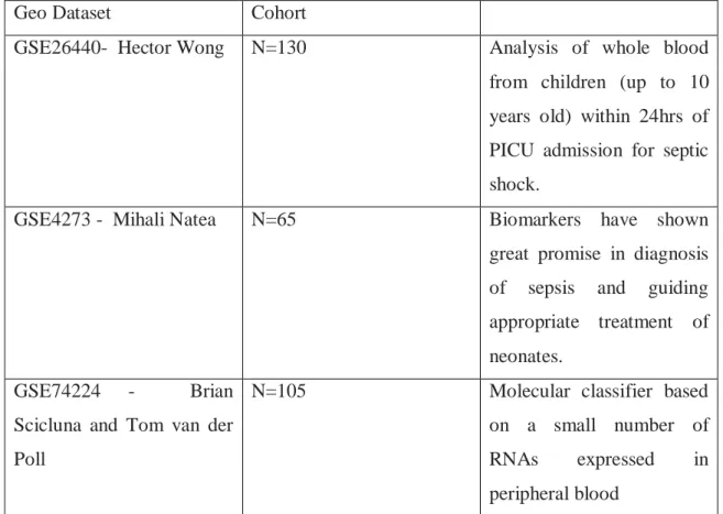
Three datasets relevant to sepsis were identified and analyzed. These include: 1. Hector Wong - pediatric sepsis (GSE26440)
2. Mihali Natea- neonatal sepsis (GSE4273)
3. Brian Scicluna and Tom Van der Poll- pneumonia versus noninfectious respiratory failure (GSE74224)
Analysis of expression data was performed in GEO2R by allocating the patients as either septic or non-septic and then using the built-in R script to identify differentially-expressed genes. We also imposed a minimum expression change ratio of 1.5 fold. This ratio was chosen as it suggests biological significance.
2.7- Phase 4: Correlating DNA methylation changes with clinical
parameters
Clinical data including survival, length of ICU and hospital stay, culture results, use of vasopressors, mechanical ventilation and steroids, was available for our cohort. Clinical criteria were used to divide septic patients into groups, which were then compared to methylation status at specific CpG sites. Correlations were assessed using a two tailed T-test with adjustment for multiple comparison testing, using FDR.
2.8- Statistical analysis
Statistical analyses wereperformed using the GraphPad Prism 5.0 software. Since the methylation values were normalized we were able to perform unpaired t-tests to determine statistical significance.
Correction for multiple comparisons was performed via adjusted p-values using the Bonferroni and FDR. Both these methods attempt to control the expected proportion of false discoveries. These corrections are based in an adjustment made to P- values when several dependent or independent statistical tests are being performed simultaneously on a single data
set. Do to these comparisons the p-value must be divide by the number of comparisons being made.
2.9- Study pipeline
Gene selection (literature): Genes related with sepsis and different metabolic consequences, including
coagulopathy, apoptosis, immune activation, immunosuppression and
endothelial dysfunction 128 genes
Pipeline: ≥0.2 and p-value <0.05 61 genes and 595 probes
Gene selection (datasets): GEO datasets from Hector Wong, Mihali Natea, Scicluna and Tom Van der Poll.
Pipeline: 1.5 fold-ratio and
p-value<0.05 28 genes
10 genes that are differentially methylated and expressed (HLA-A, HLA-C, HLA-DOB, HLA-DQB1, ITGB2, VARS2, NLRP12, C3AR1, APOL3, FADD)
Correlation between methylation levels and clinical features
Septic shock, days to ICU discharge, days to hospital discharge, core temperature, liver disease, diabetes,
chronic heart failure, ischemic heart failure, chronic lung disease, AIDS, cancer and the cancer type, chronic renal insufficiency, chronic dialysis.
3- Results
3.1- EPSIS Study
The current study is a nested case-control study of DNA methylation changes in sepsis. It utilizes genomic DNA samples obtained from a subset of patients in the DYNAMICS (DNA as a Prognostic Marker in ICU patients (clinicaltrials.gov ID NCT01355042) cohort to investigatethe importance of epigenetic regulation in the pathogenesis, progression, treatment response and outcomes of critically ill septic patients.
DYNAMICS was an observational multi-site study of critically patients in Canada for which 1000 ICU patients were enrolled, of whom 400 had severe sepsis and 600 had non-septic critical illness. Four of the nine sites involved in DYNAMICS (Hamilton General Hospital; St. Joseph’s Hospital, Hamilton; Ottawa Civic Hospital; and Ottawa General Hospital) also consented patients for whole blood DNA samples drawn on day 1 of study enrolment. A total of 260 septic and 221 non-septic patients consented to provide whole blood samples.
For the present study, a total of 141 patient samples from DYNAMICS were selected for genomic DNA methylation analysis. Samples were selected to provide equal numbers of patients in 4 distinct groups based on sepsis status and severity of illness, as indicated by the Multi Organ Dysfunction Score (MODS):
Group 1: Septic patients with high MODS (≥ 9) at ICU admission – 32 patients Group 2: Septic patients with low MODS (< 9) at ICU admission – 34 patients Group 3: Non-septic patients with high MODS (≥ 9) at ICU admission – 32 patients Group 4: Non-septic patients with low MODS (< 9) at ICU admission – 36 patients
Genomic DNA was extracted by our collaborators in Toronto and bisulfite-converted (see methods). Hybridization to the Infinium HumanMethylation450 BeadChip array, comprising over 450,000 individual CpG sites, was performed at The Centre for Applied Genomics in Toronto, Canada. The Human Methylation450 array interrogates methylation at >485,000 CpG sites, providing coverage to >99% of RefSeq genes, targeted across gene regions including the promoter, 5’UTR, first exon, gene body, and 3’UTR (Sandoval et al., 2011).
Bisulfite-converted gDNA (500 ng) was hybridized to Infinium Human Methylation 450K (HM450K) BeadChips (Illumina, San Diego, CA) and scanned with iScan (Illumina) in accordance with manufacturer’s protocol. From the initial cohort of 141 patients, only 134 high quality patient samples were included in the final analysis (samples were excluded if >20% of sample probes had detection p-value >0.01 or median bisulfite conversion efficiency (BSCE) control probe signal <4000 in the green channel).A total of 414,826 probes passed quality control and were used for further analysis.
Inclusion Criteria for Sepsis: Patient must have A or B and C
Inclusion Criteria for Non-Sepsis: Patient must have A or B or C
A. Severe Sepsis
1. 3 SIRS criteria (systemic inflammatory response syndrome)
2. Infection (suspected or confirmed) 3. 1 acute organ dysfunction
A.
1. Multiple trauma with episode of shock on presentation
2. Patient expected to remain in ICU for 72 hours
B. Septic Shock
1. All of the above in “A” and currently on vasopressors
B.
1. Critically Ill Severe Sepsis
2. three SIRS criteria (systemic inflammatory response syndrome)
3. Infection (suspected or confirmed) 4. 1 acute organ dysfunction
C. Patient expected to remain in ICU for 72
hours C.
i.e.: - Intracranial hemorrhage, subarachnoid hemorrhage, subdural hemorrhage
- No shock - No organ failure
C. 1. Non-Septic Shock i.e.: - Cardiogenic shock - Heat shock
- Burns - Hypovolemia - Pulmonary embolism
- Abdominal aortic aneurysm rupture
2. Patient expected to remain in ICU for 72 hours
Patient data collection for DYNAMICS study
A. Demographic data 1. Baseline
- Date of birth; gender, height and weight
- Admission diagnosis; admission type – medical vs surgical - Severity of illness measurement: APACHE II
2. Presence of SIRS criteria
- Fever/hypothermia, tachycardia, tachypnea, leukocytosis/leucopenia 3. Presence of organ dysfunction at the time of enrolment
- Cardiac – systolic blood pressure - Respiratory – PaO2/FiO2 ratio - Renal – creatinine
- Hematological – platelet 4. Chronic health variables
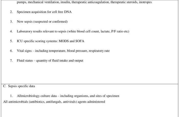
B. Daily data
pumps, mechanical ventilation, insulin, therapeutic anticoagulation, therapeutic steroids, inotropes 2. Specimen acquisition for cell free DNA
3. New sepsis (suspected or confirmed)
4. Laboratory results relevant to sepsis (white blood cell count, lactate, P/F ratio etc) 5. ICU specific scoring systems: MODS and SOFA
6. Vital signs – including temperature, blood pressure, respiratory rate 7. Fluid status – quantity of fluid intake and output
C. Sepsis specific data
1. Allmicrobiology culture data – including organisms, and sites of specimen All antimicrobials (antibiotics, antifungals, antivirals) agents administered
I r
The goal of the current study is to identify sepsis-related genes that are both epigenetically modified AND regulated in sepsis. Our collaborators in Canada have undertaken an epigenome-wide association study (EPSIS) looking for DNA methylation changes that correlate with the presence of sepsis anywhere in the genome. The disadvantage of this approach is that with a highly heterogeneous patient population and a large number of candidate CpG sites it may miss DNA methylation changes that are biologically relevant to sepsis. The objective of our study is therefore to look at a subset of genes that we believe to be important in the pathophysiology of sepsis. By limiting our analysis to this small group of genes we hoped to minimizefalse positives and maximize the chances of identifying methylation changes that are important for the pathogenesis and prognosis of sepsis.
3.2- Phase 1: Literature Search
The first step in our pipeline was to perform an extensive bibliographic investigation of the metabolic pathways and genes involved in the pathophysiology of sepsis. As such, dozens of articles and journals were read in order to understand which proteins are implicated in the pathogenesis and progression of this heterogeneous disease and its systemic effects.
Five specific pathways were identified as central to the pathogenesis of sepsis: immunoactivation, immunosuppression, coagulopathy, apoptosis and endothelial dysfunction. Our literature review resulted in a list of 128 genes that were highly relevant to the pathogenesis and systemic effects of sepsis (table 3). Genes are divided into 12 groups based on their association with the 5 pathways described above.
Genes relevant to immunoactivation include those involved in innate immunity, adaptive immunity, and inflammation. The innate immune system consists of cells and proteins that are always active and ready to mobilize and fight microbes at the site of infection. The main components of the innate immune system are 1) physical epithelial barriers, 2) phagocytic leukocytes, 3) dendritic cells, 4) natural killer (NK) cells, and 5) circulating plasma proteins (Alberts et al., 2002). Genes involved in this pathway include TLR-2, TLR-4, TLR-6, TLR-10, CX3AR1, DEFB1, YKL-40. The toll-like receptor genes (TLR) are important mediators of innate immunity because they recognize bacterial antigens on entry into the body (Kawai et al., 2010). CX3AR1, DEFB1, and YKL-40 are also important toinnate immunity because they are associated with neutrophil activity and inducing apoptosis.
Adaptive immunity is called into action against pathogens that are able to evade or overcome the innate immune defense measures. Machinery of the adaptive immune system are normally silent; however, when stimulus occurs and is activated, this machinery have the capacity to “adapt” to the presence of infectious agents by activating, proliferating, and creating potent mechanisms for neutralizing or eliminating the microbes. There are two types of adaptive immune responses: humoral immunity, mediated by antibodies produced by B lymphocytes, and cell-mediated immunity, mediated by T lymphocytes (Alberts et al., 2002). Genes important for the adaptive immune response include CHN2, CD4, CCL-4, BTN3A2, C9ORF95, RFX1, C21ORF56, C6ORF15, BATF, CD14, CD40, CD80, CD86, C-SMAC [CD2, CD4, CD8, CD28], P-SMAC [ICAM-1, ICAM-2], and D-SMAC. The supramolecular activation cluster, or immune synapse, is formed by the P-SMAC, D-SMAC, and C-SMAC proteins in response to the tight apposition of a T cell with an antigen-presenting cell (APC) (Balbino Alarco´n et al., 2011). At the centre of the immune synapse is the MHC complex, which presents the antigen to the T cell. MHC class I proteins are found on the surface of all nucleated cells and comprise the HLA-A, HLA-B, and HLA-C proteins while MCH class II proteins are found only on dedicated antigen presenting cells (eg dendritic cells, phagocytes)
and comprise the HLA-DP, HLA-DM, HLA-DOA, HLA-DOB, HLA-DR, HLA-DQB1, and HLA-DQ proteins (Sung Yoon Choo et al., 2017).
Another crucial featureof the innate and adaptive immune responses is the release of humoral mediators, namely cytokines and chemokines, which modulate the immune response. Cytokines play a crucial role in the pathogenesis of sepsis. They are secreted proteins with many effects including promoting cell proliferation, cell differentiation, and cell activation. These proteins also control immune cell trafficking and the cellular arrangement of immune organs. Chemokines have the “power” to decide whether an immune response develops and subsequently whether that response is cytotoxic, humoral, cell-mediated, or allergic. Chemokine receptor antagonism may represent a novel therapeutic approach against sepsis in the future (Raina Davi Ramnath et al., 2006;Borreli et al., 1996).
Pro-inflammatory cytokines such as TNF-ɑ and IL-6 contribute to the immune response through the promotion and stimulation of the inflammatory response. These pro-inflammatory mediators facilitate inflammation and trigger pathological pain (Jun-Ming Zhang et al., 2007). IL-6 has been shown to play a central role in the neuronal reaction and is also involved in microglial and astrocytic activation in the central nervous system (MA Klein et al., 1997). TNF-ɑ acts in several different signaling pathways, as well as apoptotic pathways, NF-kB activates inflammation as well as stress-activated protein kinases (SAPKs) (by NCBI).
Heat shock proteins are a crucial and highly conserved family of proteins across species. They act as chaperones for the folding and unfolding of proteins. They also play a role in cell-cycle control and signaling, and the protection of cells against stress/apoptosis (Li Z et al.,2004). During periods of stress, such as sepsis, there is overproduction of heat shock proteins. These molecular chaperones act to ensure that proteins are properly folded under periods of stress and elevated temperature. They include Hsp40, Hsp60, Hsp70 (Li Z et al., 2004).
Although sepsis is commonly associated with overwhelming inflammation, it is also accompanied by systemic immunosupression. The signs of immunosuppression in patients with sepsis include apoptosis-induced loss of cells of the innate and adaptive immune system including CD4+ and CD8+ T cells, B cells, and dendritic cells (Richard Hotchkiss et al., 2009)and a reduction in the expression of MHC-II complex on macrophages and APCs
mediated by a predominance of a Th2 T cell response, increased T regulatory cells, apoptosis of lymphocytes and decreased MHC class II molecules on monocytes and macrophages (Opal
et al., 2011). It is accompanied by overexpression of anti-inflammatory cytokines, such as
IL-4 and IL-10 (Jiang et al., 2006.
Humoral mediators such as IL-4 and IL-10, play an important role in immunosuppression by inhibiting TNF-ɑ, thereby augmenting acute-phase reactants and immunoglobulins and inhibiting T-lymphocyte and macrocyte functions (Evan et al., 1996).
Apoptosis, or programmed cell death, is one of the factors that contributes to sepsis being such a heterogeneous, aggressive and dysregulated syndrome. “Apoptosis is an evolutionarily conserved, energy-dependent mode of cell death requiring the initiation and regulation of complex genetic programs” (Mahidhara et al., 2000). Apoptosis has been linked to MODS. Septic patients with MODS have elevated levels of soluble Fas (sFas), an important apoptosis mediator, which decreases when MODS improves (S Endo et al., 1996). The nuclear matrix protein (NMP), a marker of cell death, has also been shown to correlate with MODS-scores and disease severity in sepsis (Y Yamada et al., 1998). Additional proteins including BIRC1, BIRC2, ML-IAP (anti-apoptotic proteins), BID, BAX, BCL-2, FADD, and caspases (pro-apoptotic proteins) are all critical to apoptosis. Cells have the capacity for remarkable regeneration in response to injury or trauma. This is also true of sepsis-related organ injury, although there is an imbalance between anabolism and catabolism that leads to cellular wasting (Rocheteau et al., 2015).
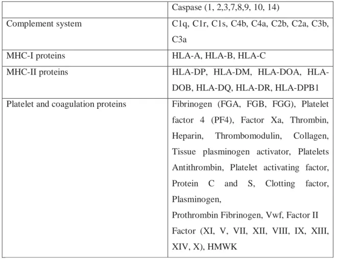
The final sepsis-related pathway that was explored was coagulopathy. A large number of proteins are involved in regulating coagulation as both pro- and anti-coagulants. These include platelet and coagulation proteins (FGA, FGB, FGG, PF4, Heparin, vWF, thrombin, thrombomodulin, collagen, tissue plasminogen activator, platelets antithrombin, protein C and S, plasminogen) as well as the coagulation factors [II (prothrombin), V, VII, VIII, IX, X, XI, XII, XIII, IV]) and proteins related to the complement system (C1q, C1r, C1s, C4b, C4aC2b, C2a, C3b, C3a). The complement system is a key player in the defense against infections. It comprises a group of plasma membrane and circulating proteins that opsonize pathogens and promote their phagocytosis. [REF]
Sepsis has profound effects on coagulation, ranging from mild alterations up to severe disseminated intravascular coagulation (DIC) (Paola Saracco et al., 2013). DIC is characterized by widespread microvascular thrombosis, which contributes to multiple organ dysfunction/failure, and subsequent consumption of platelets and coagulation factors, eventually causing simultaneous bleeding and clotting (Cheng-MingTsaoet al., 2015). “The pathogenesis of coagulopathy in sepsis is driven by an up-regulation of procoagulant mechanisms and simultaneous down-regulation of natural anticoagulants” (Simmons et al., 2017).
128 genes across the different pathways involved in sepsis
Innate immunity TLR-2, TLR-4 TLR-6, TLR-10,
CX3CR1, DEFB1, YKL-40
Adaptive immunity CHN2, CD4+, CCL-4, BTN3A2,
C9ORF95, RFX1, C21ORF56, C6ORF15, BATF, CD5, CD14, CD40, CD80, CD86, C-SMAC (CD2, CD4, CD8, CD28), P-SMAC (ICAM-1 and ICAM-2), D-P-SMAC
Proinflammatory pathway NF-KB, TNF-α, IL-1, IL-12, IL-6, IL-8,
IL-1β, IL-18, IL1R2, MyD88, RAGE,
HMGB1, NOD-1, Alpha- 1B
glycoprotein, Haptoglobin, Dipeptidyl peptidase-4
Cell replication and proliferation GSTM3C, BET, INO80, HAS-2,
WBSCR27, SRNX1, RPGRIP1, C3AR1, CEACAM1, SESN-2, HSPG2, A2AP, MTCH1
Membrane proteins Lipocalin (transporter),
Lysosome-associated membrane proteins-1, GNA12, OCLN, CLDN1
Protein folding HSP100, HSP90, HSP70, HSP60
Anti-apoptotic proteins BIRC1, BIRC2, BIRC5, BIRC6, BIRC8,
Livin, ML-IAP
Caspase (1, 2,3,7,8,9, 10, 14)
Complement system C1q, C1r, C1s, C4b, C4a, C2b, C2a, C3b,
C3a
MHC-I proteins HLA-A, HLA-B, HLA-C
MHC-II proteins DP, DM, DOA,
HLA-DOB, HLA-DQ, HLA-DR, HLA-DPB1
Platelet and coagulation proteins Fibrinogen (FGA, FGB, FGG), Platelet
factor 4 (PF4), Factor Xa, Thrombin,
Heparin, Thrombomodulin, Collagen,
Tissue plasminogen activator, Platelets Antithrombin, Platelet activating factor, Protein C and S, Clotting factor, Plasminogen,
Prothrombin Fibrinogen, Vwf, Factor II Factor (XI, V, VII, XII, VIII, IX, XIII, XIV, X), HMWK
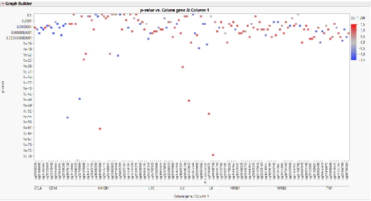
3.3- Phase 2: Methylation Arrays
The EPSIS methylation dataset is, to our knowledge, the first database of genome-wide DNA methylation data from critically-ill patients and from septic patients of any kind. The current study utilizes the EPSIS dataset to perform a targeted analysis of DNA methylation changes in sepsis-related genes.
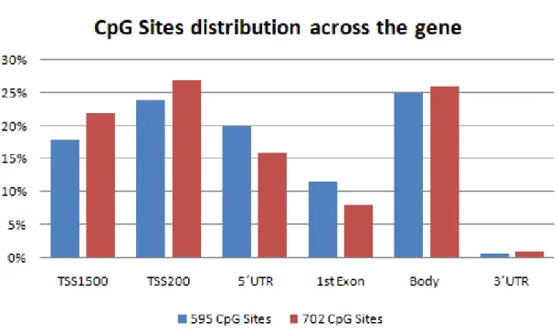
In Phase 2 of our pipeline we used the EPSIS dataset to look for sepsis-associated DNA methylation changes in the 128 genes identified in our literature search. Using the documentation provided by the manufacturer, we identified a total of 705 CpG sites located in our target genes that are also present in the llumina HumanBeadchip 450k array.
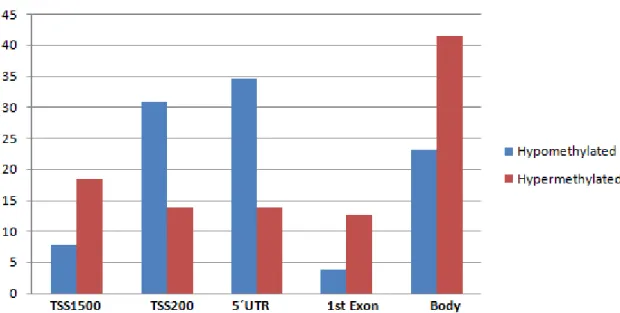
We next analyzed the EPSIS database to identify which of these 702 CpG sites showed significant methylation differences between the septic and non-septic critically-ill patients. Using a beta-value difference cutoff of > 2% and a false discovery rate of 5%, we identified a total of 595 CpG sites that were differentially methylated in the EPSIS samples. The
beta-Table 2: Reorganization of the 128 genes according to the metabolic pathway in which they are inserted and