Anatomy and morphometric aspects of the trachea of
Saimiri sciureus
Linnaeus, 1758: knowledge for emergency procedures
LUANE L. PINHEIRO1, ANA R. LIMA1, JOSÉ A.P.C. MUNIZ2, ALINE IMBELONI2, EMERSON T. FIORETO3, RODRIGO F. FONTES3, ROSA CABRAL1 and ÉRIKA BRANCO1
1Laboratório de Pesquisa Morfológica Animal – LaPMA, Universidade Federal Rural da Amazônia, Av. Perimetral, 2501, 66.077-530 Belém, PA, Brasil
2Centro Nacional de Primatas (CENP), Instituto Evandro Chagas (IEC), BR 316, Km 7, n 44, 67030-000 Ananindeua, PA, Brasil
3Departamente de Morfologia, Centro de Biologia e Ciências da Saúde, Laboratório de Biologia Celular, Universidade Federal de Sergipe (UFS), Rua Lagarto, 952, Centro, 49010390 Aracaju, SE, Brasil
Manuscript received on May 9, 2011; accepted for publication on June 22, 2011
ABSTRACT
The aim of this work is to study the morphological characteristics of the trachea of Saimiri sciureus through
quantification and measurement of the cartilaginous rings, providing information to facilitate the election of more appropriate endotracheal tube, laryngeal mask or tracheostomy tube for anesthetic and emergency procedures, as it is a species of Neotropical primates most commonly used as biological models, and little is known about their morphology. Nine animals were investigated, being 4 adults and 5 young acquired from the Centro Nacional de Primatas (National Primate Center – CENP) - Ananindeua - PA, which died from natural causes and then fixed in aqueous buffered formalin 10%. Saimiri sciureus trachea comprises
an average of 32.8 incomplete rings and an average length of 3.74 cm in young animals, while in adults
it demonstrated an average of 30.25 rings and average length of 3.67 cm. The shape of the light and its
proportion varied along the trachea. Endotracheal tube with a diameter the 2.0 – 2.5mm, laryngeal mask number 1.0 or tracheostomy tube neonatal Shiley number 3.0, can be placed in animals weighing 600g – 1.2 Kg. Given the great importance of the species studied, which is widely used as a biological model, the detailing on the morphology and morphometry of tracheal animal studies provides new approaches needed in respiratory emergency, as well as, facilitates the development of future anesthetic protocols.
Key words: emergency procedure, morphology, respiratory system, Saimiri sciureus, trachea.
Correspondence to: Érika Branco E-mail: [email protected]
INTRODUCTION
Cebidae
constitute the majority of Neotropical
primates. Among them, stands out the
Saimiriinae
subfamily, that includes the
Saimiri sciureus
Linnaeus, 1758, which is one of the Brazilian
species most used as biological model (Vaughan
1985, Aurichio 1995). Known as Common Squirrel
Monkey, it demonstrates a wide geographical
distri-bution and lives in the tops of tall trees, 30 or 40m off
the ground, often in troops of hundreds, of individuals.
Considered frugivorous and insectivorous, they are
slim and agile with a very thick tail.
S. sciureus
,
differently to other Neotropical primates, present a
well defined seasonality (Dukelow 1978).
(Annals of the Brazilian Academy of Sciences)
Trachea is a tubular fl exible organ, connective
extends from the cricoid cartilage of the larynx
to its bifurcation dorsally to the cranial region of
the heart base, demonstrating a cervical portion
and a thoracic portion. The entire trachea must
be fl exible to allow the movement of head, neck
and larynx. This fl exibility is possible due to the
cartilages which forms individual rings connected
by fi broelastic ligaments (Ettinger and Feldman
2004). In histologic terms, the trachea is composed
of three layers: mucosa, submucosa and adventitia.
The mucosa consists of respiratory epithelia,
lamina propria and an elastic lamina. Respiratory
epithelia, is a pseudo stratifi ed cylindrical ciliated,
and presents several cell types as caliciform cells
(Ross et al. 1993).
Lackness of literature on the morphology and
anatomy of
Saimiri sciureus
regarding to trachea
macroscopic and morphometric aspects, lead us to its
investigation. Moreover, these Neotropical primate
is considered an important experimental model for
biomedical research due to its phylogenetic similarities
to human. Thus, this study was designed to facilitate
the morphological knowledge of this spcecies’
trachea, in order to promote adequate anesthesia
and mechanical ventilation, both in experimental
procedures and approaches in an emergency.
MATERIALS AND METHODS
We investigated nine animals, four adults and fi ve
youngs, males and females, obtained from the
Centro Nacional de Primatas (National Primate
Center – CENP). The animals died from natural
causes, and were fi xed in aqueous solution of 10%
buffered formaldehyde and storage in the same
solution for seven days.
Following thoracotomy, trachea was removed
(including the distal end of the larynx) and right
and left main bronchi (up to the level of pulmonary
hilum). Pieces were dissected by mesoscopic,
with the aid of dissection material. Cartilage rings
were counted. A constant number of rings were
considered to divide the trachea into 3 regions
- cranial, middle and caudal, represented by a 3
parallel drawing lines perpendicular to the major
tracheal axis. The third line was limited cranially
to the carina, limiting the trachea from the main
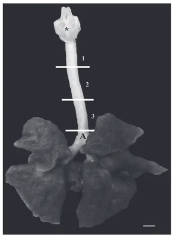
bronchi (Fig. 1).
Fig. 1 - Trachea and lung of S. sciureus showing larynx (*), carina (A) and demarcation of the cranial portion (1), middle (2) and caudal (3) portions. Scale bar: 1cm.
Tracheal length was measured from the fi rst
to the last tracheal ring in cranio-caudal direction;
trachea diameters were measured by two axis -
height (dorsal-ventral) and width (lateral-lateral)
of cranial, caudal and middle regions with the aid
of a caliper rule. Following measurements, the
tracheae were submitted to histological procedure,
embedded in paraffi n, and cuts of 5μm stained with
RESULTS
Saimiri sciureus
was observed as a tube of variable
length in young and adults, being the trachea of
young a little major due to the increase in number
of rings. In these individuals, trachea is composed
of an average of 32.8 incomplete rings and average
length of 3.74 cm. In adults, the trachea showed
an average of 30.25 also incomplete cartilaginous
rings and average length of 3.67 cm (Table I).
DISCUSSION
The literature is scarce in relation to morphological
descriptions and morphometric aspects of the
trachea in non human primates. In general, the
trachea of
Saimiri sciureus
in structure resembles
to the findings of higher primates or
Anthropoidea
.
Straus and Willian (1931) in their study of
adult primates demonstrated an absolute number
of cartilage rings what would depend, most
probably, to the length of neck. The number of
cartilage rings have been described in a range of
variation according to sex and within the species:
in
Tarsius
Storr, 1780, for example, Burmeister
(1846) described 26 rings, while Woollard (1925)
about 16; in
Daubentonia
Geoffroy, 1795, Owen
(1866) counted 26 rings, as Peters (1852) and
Zuckerkandl (1898), 22 rings; in
Lemur
Linnaeus,
1758, however, Peters (1852) described 26 rings
and Patten (1899) 32 cartilaginous rings. In our
study,
Saimiri sciureus
demonstrated an average of
30.25 rings in adult animals.
Tracheal cartilages do not form complete
rings in humans and apes, and smiliar feature was
observed in
S. sciureus
, in which they appeared to
be incomplete on its dorsal surface, demonstrating
a "C" shape. Free borders of these half-rings were
united by a membrane of connective tissue and
smooth muscle fibers (Straus and Willian 1931).
In some cases the ends of the cartilages were
superimposed. However, it was quite evident
that the rings were incomplete (Straus and
Willian 1931). The overlap is presumably a result
of changes after death, a hypothesis reinforced by
the observations of Beattie (1927) on the trachea of
Callithrix jacchus
Linnaeus, 1758, when comparing
samples fixed in formalin to the living animal that
demonstrated a distinct and clear space between the
dorsal borders of the rings. This overlap, however,
was not observed in our study.
In
Galago
Geoffroy, 1796, and
Nycticebus
Geoffroy, 1812, free borders of the rings have a
Trachea
Number of Rings Length (cm)
Young Adult Young Adult
36 28 3.50 3.30
33 30 4.00 3.60
35 31 3.60 3.90
30 32 3.80 3.90
30 - 3.80
-Average 32.8 30.25 3.74 3.67
SD 6.96 2.18 0.03 0.06
SEM 3.11 1.09 0.01 0.03
CV (%) 21.2 7.23 0.80 1.63
SD: Average Standard deviation; SEM: Standard error of mean; CV%: Coefficient of Variance.
TABLE I
Values for number of rings and length of the trachea of Saimiri sciureus.
The shape of the light was diverse along
the trachea and its proportion varied between
youth and adults (Table II and Fig. 2). In young
animals the initial segment of the trachea tended
to be rounded, becoming oval in middle and caudal
regions. In adults, the initial portion tended to be
dorsal-ventrally flattened becoming oval with a
tendency to become slightly enlarged or
spatula-shaped (Straus and Willian 1931). This condition
was not noticeable in
S. sciureus
.
Along
S. sciureus
trachea, the diameter varied
in lateral direction, tending to be more oval in the
caudal portion resembling to the shape of the light
of the trachea in humans, usually circular or oval
according to Gamsu and Webb (1982).
Histologically, the epithelium of
S. sciureus
trachea resembled to that of domestic animals, a
pseudostratifi ed cylindrical ciliated epithelia with
numerous goblet cells producing mucus (Getty
1986) and the human (Ross et al. 1993).
Maintenance of a patient airway may be
realized by of an endotracheal tube, laryngeal
mask or tracheostomy tube. Intubation of non
human primates makes this tecchnically diffi cult,
but becomes relatively easy if we use a small
curved blade design to visualized the larynx
(Tranquilli et al. 2007). Tubes with a diameter the
2.0 – 2.5mm (Portex
®) can be placed in animals
weighing 600g – 1.2 Kg.
However, Vilani et al. (2000) say that the
laryngeal mask was correctly inserted in the fi rst
trial, and its use was an excellent alternative for
the patient’s adequate ventilation. In addition,
TABLE II
Mean values and standard deviation for width (w) and height (h) (cm) in three parts of the trachea of Saimiri sciureus.
Trachea
Region Cranial Middle Caudal
Young Adult Young Adult Young Adult
w h w h w h w H w h w h
Average 0.40 0.40 0.45 0.40 0.38 0.40 0.40 0.42 0.36 0.37 0.37 0.40 Minimum 0.30 0.30 0.40 0.30 0.30 0.30 0.30 0.40 0.30 0.30 0.30 0.40 Maximum 0.50 0.50 0.50 0.50 0.40 0.50 0.50 0.50 0.40 0.40 0.40 0.40 CV % 0.07 0.07 0.04 0.08 0.04 0.07 0.08 0.05 0.05 0.04 0.05 0
SEM 0.03 0.03 0.02 0.04 0.01 0.03 0.04 0.02 0.02 0.01 0.02 0
the waken-up occurred quickly after ceasing the
administration of the inhalatory anesthetic. For the
species studied in given laryngeal mask number 1.0
(NovaMasc
®). In situations where it is not possible in
orotracheal intubation or placement of the laryngeal
mask, the alternative to keep the ventilation is the
placement of the tracheostomy tube (Fraga et al.
2009). This species should be used neonatal Shiley
tracheostomy tube number 3.0 (Mallinckrodt
®).
ACKNOWLEDGMENTS
We would like to thank the Centro Nacional de
Primatas (National Primate Center – CENP) -
Ananindeua - PA for donating the animals and the
technical support during laboratory investigation.
We also thank the Research Fellowship Program -
REUNI/UFRA for financial support.
RESUMO
Estudou-se as características morfológicas da traqueia do Saimiri sciureus através da quantificação e mensuração dos anéis cartilaginosos, a fim de fornecer informações que
facilitem a eleição mais adequada de sonda endotraqueal,
máscara laríngea ou cânula de trasqueostomia para
procedimentos anestésicos e de emergência, visto que é uma das espécies de primatas neotropicais mais
comu-mente utilizadas como modelos biológicos e pouco se sabe sobre sua morfologia. Foram utilizados nove animais, sendo quatro adultos e cinco jovens, oriundos do Centro Nacional de Primatas (CENP) - Ananindeua - PA, os quais vieram a óbito por causas naturais e, posteriormente, foram fixados em solução aquosa de formol tamponado a 10%. A
traqueia do Saimiri sciureus é composta por uma média de
32,8 anéis incompletos nos animais jovens e comprimento médio de 3,74cm, já os adultos apresentam uma média de
30,25 anéis e comprimento médio de 3,67cm. A forma da
luz e suas proporções variaram ao longo da traqueia. Sonda
endotraqueal com diâmetro de 2,0 – 2,5mm, máscara
laríngea número 1,0 ou cânula de traqueostomia neonatal Shiley número 3,0, podem ser colocados em animais pesando 600g - 1,2 K. Dada a grande importância da espécie estudada, que é amplamente utilizada como modelo
biológico, o detalhamento sobre o estudo da morfologia e morfometria traqueal animal, fornece novas abordagens necessárias em emergência respiratória, bem como facilita
o desenvolvimento de futuros protocolos anestésicos.
Palavras-chave: procedimentos emergênciais, morfologia, sistema respiratório, Saimiri sciureus, traqueia.
REFERENCES
AURICHIO P.1995. Primatas do Brasil. São Paulo: Terra Brasilis,
168 p.
BEATTIE J. 1927. The anatomy of the common marmoset (Hapale jacchus Kuhl). Proc Zool Soc Lond 3: 593-718. BURMEISTER H. 1846. Beitrage zur naheren Kenntnis der
Gattung Tarsius. Berlin: G Reimer, 140 p.
DUKELOW WR.1978. Reproduction in the squirrel monkey (Saimiri sciureus). Rec Adv Primatol 2: 195-200. ETTINGER SJ AND FELDMAN E. 2004. Tratado de medicina
interna veterinária. 5ª ed., Rio de Janeiro: Guanabara Koogan, 2156 p.
FRAGA JC, SOUZA JCK AND KRUEL J. 2009. Pediatric
tracheostomy. J Pediatr 85: 97-103.
GAMSU G AND WEBB WR.1982. Computed tomography of
the trachea: normal and abnormal. AJR 139: 321-326.
GETTY R.1986. Sisson & Grossman, Anatomia dos animais
domésticos. 5ª ed., Rio de Janeiro: Guanabara Koogan,
2000 p.
OWEN R.1866. On the aye-aye (Chiromys madagascarensis).
Trans Zool Soc Lond 5: 33-101.
PATTEN CJ.1899. The form and position of the thoracic viscera of the ruffed lemur (Lemur varius). Trans Roy Acad Med Ireland 20: 441-473.
PETERS WCH. 1852. Naturwissenschaftlicke Reise nach Mossambique. Zoologie I. Berlin: Saugethiere, 202 p.
ROSS MH, REITH EJ AND ROMRELL LJ. 1993. Histologia
Texto e Atlas. 2ª ed., São Paulo: Panamericana, 779 p.
TRANQUILLI WJ,THURMON JC AND GRIMM KA.2007. Lumb
& Jones Veterinary Anesthesia and Analgesia. 4th
ed.,
Iowa: Blackwell Publishing, 1096 p.
STRAUS JR AND WILLIAN L.1931. The Form of the Tracheal
Cartilages of Primates, with Remarks on the Supposed Taxonomic Importance. J Mammal 12: 281-285.
VAUGHAN TA.1985. Order primates. In: VAUGHAN TA (Ed.),
J Mammal. Northern Arizona University, Flagstaff, USA, p. 138-143.
VILANI RGD’OC,VILANI PD’OC,PACHALY JR,MANGINI PR, MACHADO GV AND SUSKO I.2000. Inhalatory anesthesia
with laryngeal mask in a chimpanzee (Pan troglodytes). Arch Vet Sci 5: 17-21.
WOOLLARD HH.1925. The anatomy of Tarsius spectrum. Proc
Zool Soc Lond 70: 1071-1184.
ZUCKERKANDL E. 1898. Zur Anatomie von Chiromys madagascarensis. Denkschr d k Akad Wiss Wien