This Accepted Author Manuscript is copyrighted and published by Elsevier. It is posted here by agreement between Elsevier and University of Brasilia. Changes resulting from the publishing process - such as editing, corrections, structural formatting, and other quality control
mechanisms - may not be reflected in this version of the text. The definitive version of the text was subsequently published in [Journal of Invertebrate Pathology, Volume 67, Issue 3, May 1996, Pages 309–311, doi:10.1006/jipa.1996.0047].You may download, copy and otherwise use the AAM for non-commercial purposes provided that your license is limited by the following restrictions:
(1) You may use this AAM for non-commercial purposes only under the terms of the CC-BY-NC-ND license.
(2) The integrity of the work and identification of the author, copyright owner, and publisher must be preserved in any copy.
(3) You must attribute this AAM in the following format: [agreed attribution language, including link to CC BY-NC-ND license + Digital Object Identifier link to the published journal article on Elsevier’s ScienceDirect® platform].
________________________________________________________________________
Este Manuscrito do Autor Aceito para Publicação (AAM) é protegido por direitos autorais e publicado pela Elsevier. Ele esta disponível neste Repositório, por acordo entre a Elsevier e a Universidade de Brasília. As alterações decorrentes do processo de publicação - como a edição, correção, formatação estrutural, e outros mecanismos de controle de qualidade - não estão refletidas nesta versão do texto. A versão definitiva do texto foi posteriormente publicado em [Journal of Invertebrate Pathology, Volume 67, Issue 3, May 1996, Pages 309–311,
doi:10.1006/jipa.1996.0047]. Você pode baixar, copiar e utilizar de outra forma o AAM para fins não comerciais , desde que sua licença seja limitada pelas seguintes restrições:
(1) Você pode usar este AAM para fins não comerciais apenas sob os termos da licença CC- BY- NC-ND.
(2) A integridade do trabalho e identificação do autor, detentor dos direitos autorais e editor deve ser preservado em qualquer cópia.
Virus-like Particles and Rickettsia-like Organisms in Male Germ and Cyst
Cells ofBemisia tabaci(Homoptera, Aleyrodidae)
Sônia N. Báo Elliot W. Kitajima Giuliano Callaini Romano Dallai
Insect sperms have attracted intensive attention from the cytologists, especially those
trying to correlate the large diversity of their structure and the evolutionary process. We
recently engaged in a study of male germ cell differentiation and structure in whiteflies, a less
studied though important group of insects.
During our study it became apparent that male germ and cyst cells of Bemisia tabaci
contain rickettsia-like organisms and also virus-like particles. This note describes details of
these findings. In a separate article a more comprehensive description of B. tabaci sperm is
being made.
Adult males of B. tabaci were kindly supplied by Dr. Josias C. Farias, from his colony
maintained on soybean at Centro Nacional de Pesquisa do Arroz e Feija˜ o (Goianˆ ia, GO, Brazil). Whiteflies were dissected and the testes fixed for 1 hr in 2.5% glutaraldehyde, 1.8%
sucrose in a 0.1 M phosphate buffer and then for 1 hr in 1% osmium tetroxide in the same
buffer. The samples were dehydrated with acetone and then embedded in Epon resin. After
sectioning and staining with uranyl acetate and lead citrate, the sections were examined with a
JEOL 100 C or Philips CM 10 transmission electron microscopes.
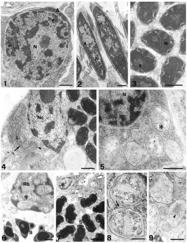
Virus-like particles (VLP). VLPs were detected in four of six of the examined male B.
tabaci. These particles were found in both germ and cyst cells (Figs. 1–4). They were present in
the nucleus of spermatids and spermatozoa, but in the cyst cells, VLPs were noted only in the
cytoplasm. In the nucleus of young spermatids, VLPs were always found interspersed in the
dense, compacted chromatin region (Fig. 1). However, as the differentiation process
continued, they were found dispersed in the less dense area (Fig. 2). Finally, in more
condensed nuclei in well-differentiated sperms, VLPs appeared aggregated, commonly in the
central region (Fig. 3). When detected, VLPs were present in practically all spermatids and
sperms, numbering up to 2 dozen/section. Similar particles were also noticed in large numbers
in the cyst cells, within huge vesicles interpreted as lysosomes (Fig. 4) mixed with myelin-like
structures. VLPs, however, were not observed free in the cytoplasm. The VLPs were rounded,
with an electron-dense core of 20– 25 nm, surrounded by an electron-lucent zone, possibly the
coat protein, 15–20 nm wide. Center-to-center distance in a regular package of the particles
Another remarkable finding was the presence of numerous rickettsia-like procarionts,
also both in the germ and cyst cells. In the germ cells these bacterioids were localized in the
cytoplasm. In early spermatids the rickettsia-like organisms appeared scattered throughout the
cytoplasm (Fig. 5). As usual, as the spermatid elongated, excess cytoplasm containing the
bacterioids was eliminated as residual body (Fig. 6); thus, in mature sperm they could not be
found. These symbionts were also present in the cytoplasm of cyst cells (Fig. 7). In sections,
they exhibited a profile that varied from round (ca. 1 mm in diameter) to elongated (2–2.5 mm
long) and surrounded by a double membrane: the outer, adjacent to the host cytoplasm, thick
and dense and the inner membrane, thinner and less dense. In about 5% of these symbionts,
besides ribosomes and DNA, fibrillar particles, 6–7 nm thick and ca. 0.5 mm long, could be
detected. They appeared in a parallel array, which in cross sections revealed an ordered,
paracrystalline arrangement (Figs. 8 and 9).
Many reports have been made of VLPs in several insect groups, without being
associated with a particular pathogenic condition, and are believed to be latent infections. The
presence of VLPs in germ cells, however, has been less frequent, and the best interpretation is
that this indicated a route of vertical transmission of the putative virus (e.g., Diptera, Schankel
and Schwalm, 1975; Afzelius et al., 1989, and Coleoptera, Kitajima et al., 1985). Rickettsia-like
organisms in insects have been commonly been found may be considered as a rule, and
though also being detected in some few cases in male germ cells (Wright et al., 1978; Wright
and Barr, 1980; Ndiaye and Mattei, 1993), their vertical transmission probably is transovarial.
In the present case, VLPs were only detected in the nucleus of the germ cells, initially
interspersed in the chromatin, but as the nuclei condensed they are expelled to the
nucleoplasm, finally being packed within the condensed chromatin and possibly being
transmitted vertically after fertilization. In the cyst cells, VLPs are only found within
lysosome-like vesicles, in huge numbers. They are possibly remnants of degraded sperm, endocyted by
cyst cells. Most of the presumed latent infection by VLPs in insects described in the literature
involves reovirus or picorna-type particles (Kim, 1980; Matthews, 1982; Kitajima, 1989). Those
found in the present study are somewhat smaller and do not fit into any of the known insect
the cytoplasm of the cyst cell. S, spermatozoa. FIGS. 8 and 9. Rickettsia-like organisms showing double membrane (arrow) and a paracrystalline array of filamentous particles (arrowhead). Bars, 0.5 mm.
The bacteria found both in germ and cyst cells have a double-layered thin envelope
and are contained in a cytoplasmic vacuole similar to rickettsia-like microorganisms described
in germ cells of mosquitoes. Rickettsia-like microorganisms are obligatory intracellular bacteria
widespread among insects. They have been implicated as causes of parthenogenesis in
Hymenoptera (Stouthamer et al., 1993) and cytoplasmic incompatibility in Homoptera,
Coleoptera, Hymenoptera, Lepidoptera, and Diptera (for review, see Rousset and Raymond,
1991). We do not know if this is also the case in Bemisia, where these intracellular bacteria are
found for the first time. They seem unnecessary to sperm function because they are
eliminated. The filamentous particles found within them morphologically resemble phages of
the Inoviridae family and may be responsible for eventual degeneration of the Rickettsiae as
reported in mosquitoes (Yen, 1976; Ndiaye and Mattei, 1993).
We conclude that the virus-like particles and rickettsia-like microorganisms represent
agents that infect the B. tabaci testes, but they apparently do not interfere in the
spermiogenesis process.
KEY WORDS: Rickettsia-like; Bemisia tabaci; viruslike particles; whiteflies; insect sperm; male
germ cell.
REFERENCES
Afzelius, B. A., Alberti, G., Dallai, R., Godula, J., and Witalinksi, W. 1989. J. Invertebr. Pathol. 53, 365–377.
Kim, K. S. 1980. J. Invertebr. Pathol. 36, 292–301. Kitajima, E. W. 1989. Mem. Int. Oswaldo Cruz 84, 9–15.
Kitajima, E. W., Kim, K. S., Scott, H. A., and Gergerich, R. C. 1985. J. Invertebr. Pathol. 46, 83– 97.
Matthews, R. E. F. 1982. Intervirology 17, 1–200. Ndiaye, M., and Mattei, X. 1993. J. Submicrosc. Cytol. Pathol. 25, 71–77.
Rousset, F., and Raymond, M. 1991. . TREE 6, 54–57. Schankel, K. R., and Schwalm, F. E. 1975. J. Invertebr. Pathol. 26, 265–268.
Stouthamer, R., Breeuwer, J. A. J., Luck, R. F., and Werren, J. H. 1993. Nature 136, 66–68.
Wright, J. D., and Barr, A. R. 1980. J. Ultrastruct. Res. 72, 52–64.