2019/2020
Bianca Sousa Barros
Avaliação Isocinética do Músculo Deltoide Após
Artroplastia Invertida do Ombro /
Isokinetic Evaluation of the Deltoid Muscle After
Reverse Shoulder Arthroplasty
Mestrado Integrado em Medicina
Área: Ciências Médicas e da Saúde
Tipologia: Dissertação
Trabalho efetuado sob a Orientação de:
Professor Doutor Manuel António Pereira Gutierres
E sob a Coorientação de:
Doutor João Bernardo Matos Nunes
Trabalho organizado de acordo com as normas da revista:
Clinical Biomechanics
Bianca Sousa Barros
Avaliação Isocinética do Músculo Deltoide Após
Artroplastia Invertida do Ombro /
Isokinetic Evaluation of the Deltoid Muscle After
Reverse Shoulder Arthroplasty
Dedico este trabalho à minha família e aos meus amigos que me apoiaram incansavelmente ao longo de todo o meu percurso.
Um agradecimento especial ao meu orientador, Professor Doutor Manuel Gutierres, e ao meu co-orientador, Doutor Bernardo Nunes, por toda a dedicação, disponibilidade e
compreensão, que me permitiram uma aprendizagem transversal única e a realização de um trabalho do qual me orgulho.
Isokinetic Evaluation of the Deltoid Muscle After Reverse Shoulder
Arthroplasty
Bianca Sousa Barros a, *, Manuel Gutierres a, b, João Bernardo Nunes b, Leandro Machado c, d,
Pedro Fonseca c, Filipa Sousa c, d
a Faculty of Medicine (FMUP), University of Porto, Portugal
b Orthopaedics and Traumatology Department, S. João University Hospital, Faculty of Medicine, University
of Porto. Porto, Portugal.
c Porto Biomechanics Laboratory (LABIOMEP), University of Porto, Portugal. d CIFI2D, Faculty of Sports (FADEUP), University of Porto, Portugal.
*Corresponding Author: Bianca Sousa Barros (email: biancabsb96@hotmail.com), Faculty of Medicine, University of Porto. Porto, Portugal.
Statement of equal authors contribution: all authors contributed equally to this manuscript. Declarations of interest: none.
Abstract
Background: Reverse Shoulder Arthroplasty depends on the Deltoid muscle to improve function and stability of the shoulder. Deltoid tension and pre- and postoperative conditions are key factors. Although good subjective results are reported, functional outcomes have shown variable improvements. The purpose of this study is to understand the biomechanical and functional influence of the Reverse Shoulder Arthroplasty in the Deltoid.
Methods: Fifteen participants after unilateral Reverse Shoulder Arthroplasty, were evaluated with isokinetic dynamometer (Abduction/Adduction and Forward Flexion/Extension), Electromyography and Constant-Murley Score. The arm without the prosthesis was considered the best performance status and used as comparison. Arm-length was measured and calculated the difference between arms. Participants were divided in two groups according to Constant-Murley Score of the arm without prosthesis: Group 0 (superior/equal to 80) and Group 1 (inferior to 80).
Findings: Significant differences in isokinetic parameters were observed, especially in Group 0, with the arm without prosthesis having better results. The Electromyography showed that Group 0 has an overall decrease of the electromyography activity in the arm with prosthesis, especially in the anterior and middle portion of the Deltoid, in Abduction and Forward Flexion. Group 1 revealed less significant differences.
Interpretation: Isokinetic evaluation combined with Electromyography is a useful tool to assess muscular and joint outcomes. This study demonstrated that Reverse Shoulder Arthroplasty has a significant effect in range of motion and strength of the shoulder joint and the Deltoid. Shoulders with the prosthesis presented worse performance, but these changes may only be significant when a higher functional level is present.
1. Introduction
Reverse Shoulder Arthroplasty (RSA) depends on the deltoid muscle to improve function and stability of the shoulder joint, using a convex glenoid and a concave humeral component. Since Paul Grammont’s concept of medializing and lowering the center of rotation of the cuff-deficient shoulder (Grammont et al., 1987), its use has been expanded to other pathologies (Jazayeri and Kwon, 2011).
The concept of RSA consists in a joint with a fixed center of rotation on the scapula and a cup on the humerus to provide a stable fulcrum. This fulcrum prevents superior humeral head migration and allows the elevation of the arm performed by the Deltoid, despite the absence of rotator cuff muscles (Hamilton et al., 2015).
With expanding indications for RSA, understanding the effect on the surrounding soft-tissues may be useful to improve the results and prevent failures. Previous studies concluded that there is a medial translation of the humerus compared to its original anatomy and shortening of the remaining rotator cuff (Ackland et al., 2015).
Deltoid tension is modified by the new center of rotation (Hamilton et al., 2013), and is paramount to the success of RSA. Its role is also critical to avoid dislocation and failure to adequately tension the Deltoid may result in prosthetic instability, which is one of the most significant complications (Lädermann et al., 2014).
Pre- and postoperative conditions of the Deltoid are key factors for the surgical outcome as arm elevation is mostly dependent on this muscle after RSA (Boudreau et al., 2007). An excessive lengthening in the Deltoid can be associated with neurologic lesions, restricted motion with fixed arm abduction, acromial or scapular spine fractures. On the other hand, an insufficient tension of this muscle may be associated with postoperative poor anterior elevation and prosthetic instability (Lädermann et al., 2014). Optimal conditions of the Deltoid may be difficult to assess pre and intra-operatively. As such, surgeons have to select the correct size and/or combination of RSA implants based mostly on experience, in order to achieve tension and stability in replaced joints (Hatta et al., 2016).
Although RSA is a good solution in cases of rotator cuff deficient shoulders, resulting in good subjective results, functional outcomes have shown variable improvements in the range of motion. Poor postoperative forward flexion can be associated with improper use, poor preoperative forward flexion, poor patient selection and postoperative complications (such as dislocation, spine scapula fracture or neurologic impairment). These conditions may be caused by inadequate Deltoid function, and the appropriate soft-tissue tension to provide the best functional outcome should restore arm length when compared with the contralateral arm (Lädermann et al., 2012, 2009).
The purpose of this study is to understand the biomechanical and functional implications in the Deltoid muscle after RSA.
2. Methods
2.1 Participants Selection
Surgical records were analyzed to retrieve patients submitted to unilateral RSA at a single institution, between September 2013 and September 2018. Inclusion criteria were defined as: 1) primary RSA through a deltopectoral approach, with a minimum 6 months of follow-up; 2) shoulder arthritis (primary or secondary), massive rotator cuff tears or proximal humerus fractures as diagnosis for surgery; 3) over 18 years-old; 4) willingness to participate and informed consent signing. Patients were excluded if they had previous surgery in the contralateral upper limb (including shoulder replacement), previous conditions that could influence humeral-length or side to side differences (such as humeral diaphyseal fracture), history of post-operative complications after RSA (neurologic injury, acromion, scapular spine or glenoid fractures, infection, instability or periprosthetic fractures), or if they missed the required evaluations. The institutional ethics committee approved the research protocol and all participants gave their written informed consent before the evaluation.
2.2. Measurements
All measurements were performed in LABIOMEP – Porto Biomechanics Laboratory, evaluating one participant at the time and in a single evaluation. The contralateral arm was defined as control-group for comparison.
The Constant-Murley Score (CMS) was assessed for each shoulder. Range of Motion (RoM) in active forward flexion and abduction was evaluated using a goniometer, with its axis placed over the joint and determining the number of degrees from neutral position with the arm at the side (Hayes et al., 2001).
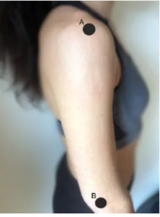
Humeral length was measured using an anthropometer, from the acromial to the radial external landmarks, as indicated in Figure 1. Each arm was measured twice, and the mean value of measurements was used for analysis (International Society for the Advancement of Kinanthropometry (ISAK), 2001).
Figure 1: The participant assumed a relaxed standing position with the arms hanging by the side and the measure is from the acromiale landmark (“A”), the point on the superior part of the acromion border in line with the most lateral aspect, to the radiale landmark, the point at the proximal and lateral border of the head of the radius (“B”).
The isokinetic evaluation combined with electromyography (EMG) was performed lastly. The isokinetic evaluation used the Biodex System 4 Pro, an isokinetic dynamometer for muscle function with fully assisted dynamometer height adjustment, front-to-back chair adjustment and side-to-side adjustment. This isokinetic dynamometer provides a constant velocity with accommodating resistant throughout a joint’s RoM. This resistance is provided at a user-defined constant speed (“Biodex System 4 Pro Specifications,” 2020). The dynamometer velocity was 60 degrees per second. In all exercises, participants were stabilized in the chair with shoulder and abdominal straps. The position of the chair, in height, front-to-back and side-to-side position, was adapted to every participant so that the anatomical center of the shoulder joint was aligned to the dynamometer axis.
There were four different protocols in the Biodex: isometric exercise at abduction, isometric exercise at forward flexion, isokinetic exercise with abduction/adduction, and isokinetic exercise for forward flexion/extension. For the abduction/adduction protocol, the dynamometer was set with 10-degree tilt. However, for the forward flexion /extension protocol the dynamometer was set in neutral tilt (0 degrees). The isometric exercises were at 45 degrees in abduction and forward flexion, executing two contractions separated by one-minute rest. The isokinetic exercise consisted in five repetitions of full ROM movements, with an interval of 0 degrees and 135 degrees, both for abduction/adduction and forward flexion /extension. The participant started with the arm not submitted to RSA and did every exercise with that arm first and then with the arm with prosthesis. Before each exercise, isometric and isokinetic, the participant was asked to try to do the exercise as a warm-up, followed by a five minutes rest before the exercise for record was performed. The order of exercises was: 1) isometric in abduction; 2) isokinetic with abduction/adduction; 3) isometric in forward flexion; 4) isokinetic in forward flexion/extension. The participants were encouraged to reach the maximal muscle and joint performance during all exercises. The following parameters were extracted for further analysis: Peak Torque, Peak Torque per Body Weight (PT/BW), Angle of Peak Torque (AngPT) in degrees, Power in Watts and Work in joules.
In addition, surface electromyography was used simultaneously with the isokinetic evaluation of the three portions of the Deltoid muscle (anterior, middle and posterior), during the abduction/adduction and forward flexion /extension exercises. Surface bipolar EMG electrodes (Dormo&Blaico SX-30, Telic S.A. Spain) were placed on the skin above the Deltoid according to the Surface Electromyography for the Non-Invasive Assessment of Muscles recommendations and with an inter-electrode distance of 20 mm (SENIAM, 2019). The skin was cleaned with a cotton swab soaked in 90% alcohol prior to electrode´s placement. The EMG signals were bandpass filtered (5-500 Hz), amplified and recorded with a BIOPAC MP100 (BIOPAC Systems, INC, USA) analog-to-digital converter operating at a 2000Hz sampling frequency. A custom written Matlab R2014a (MathWorks, MA, USA) routine was used to process the EMG data. The isokinetic dynamometer RoM was used to set four performance ranges: 0-45º, 45-90º, 90-135º 135-180º. The EMG envelopes was then normalized to its maximum value of each range, and
the average muscle activation in each range was extracted. As such, Deltoid muscle activity was recorded while the undertaking the exercises in the isokinetic evaluation.
2.3. Statistical analysis:
Statistical Analysis was performed using IBM SPSS Statistics software (IBM Corporation, NY, USA). All data are expressed as means ± standard deviation (SD). The Wilcoxon test was used to assess differences between the shoulder with RSA and the shoulder without RSA in the parameters analyzed. The probability level p<0.05 was defined as statistically significant.
3. Results
The study included 15 participants (13 women and 2 men). The mean age was 66,53 ± 6,23 years (range from 52 to 78). Eleven participants had surgery in the right shoulder and four in the left shoulder. The indications for RSA were degenerative (Primary and Secondary Arthritis) and traumatic causes. The mean time between surgery and the evaluation was 31,67 ± 16,64 months (range from 9 to 67 months).
In all statistical analysis showed below, the differences were calculated between the arm with RSA, designated by “Ipsilateral”, and the arm considered healthy, without RSA, designated by “Contralateral”.
3.1. Constant-Murley Score
Constant-Murley Score (CMS) was compared for each arm (Table 1). Side-to-side comparison of the CMS revealed that Ipsilateral shoulders had a mean CMS of 52,7 ± 10,57, compared to 77,76 ± 8,57 of the Contralateral side (p<0,001).
From the 15 participants, 13 referred no pain in Contralateral arm. In the Ipsilateral arm, 9 participants referred minor pain and 3 no pain, with only 3 referring moderate pain. In Abduction, 10 of the participants had a RoM of 121 – 150° in the Contralateral arm (4 had a RoM of 91 - 120° and 1 had >151°). However, in the Ipsilateral arm, 9 participants had a RoM of 91 - 120° for Abduction, 4 had 61 - 90° and 2 had a higher RoM of 121 - 150°. In Forward Flexion, the same tendency between arms was observed: 12 participants achieved the interval of 121 - 150° in the Contralateral arm (two 91 - 120° and one >150°). In the Ipsilateral arm, 7 achieved a RoM in the interval 91 - 120°, five at 61 - 90° and 3 at 121 - 150°. In External Rotation, all 15 participants had the maximum classification (10) for the Contralateral arm. However, for the Ipsilateral results were variable ranging from 2 to 10. In Internal Rotation, 14 participants had 10 of classification (one participant had 8) in the Contralateral arm. In the Ipsilateral arm, 2 participants had a classification of 0, 6 a classification of 2, 2 had a classification of 6 and 5 had a classification of 8. The mean difference between arms in strength is 2,04 kg.
The participants were divided in two groups based on the CMS of the arm without RSA. Therefore, the statistical analysis was performed in one group with 6 participants with CMS superior or equal to 80, designated by Group 0, and another group with 9 participants with CMS inferior to 80, designated by Group 1.
3.2. Arm length
Mean arm length was 28,56 ± 2,24 cm in the Ipsilateral arm, comparing with 27,21 ± 1,69 cm in the Contralateral one, indicating a mean difference of 1,35 ± 0,96 cm (p=0,07)
Participants in group 0 had a longer arm length in the Ipsilateral side, with a mean difference of 1,54±1,10 cm (p=0,22). Participants in Group 1 also demonstrated an Ipsilateral arm 1,22 ± 0,91 cm (mean) longer than the Contralateral (p=0,19).
3.3. Isokinetic Dynamometer Evaluation
In the Isokinetic Dynamometer Evaluation the following parameters were evaluated: Peak Torque in Newton meters (T/BW (N.m)), Peak Torque per Body Weight (PT/BW (%)), Angle of Peak Torque in degrees (AngPT (Deg)), Power in Watts (Pow(W)) and Work in Joules (Work (J)).
3.3.1. Peak Torque (T/BW):
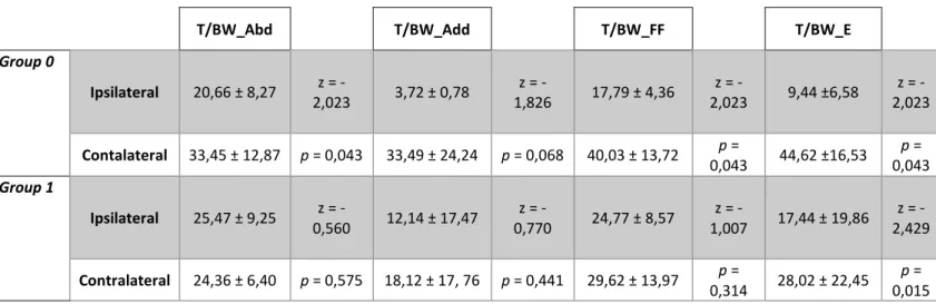
In terms of Peak Torque in Abduction/Adduction isokinetic exercise, Group 0 had significant differences between shoulders in Abduction as the Ipsilateral side was significantly lower
(p=0,043). In Group 1, no significant differences between arms were found. Peak Torque in Adduction did not demonstrate significant differences between Ipsilateral and Contralateral sides, both in Group 0 and 1.
In Forward Flexion/Extension isokinetic exercise, Group 0 also demonstrated that Peak Torque was significantly lower in the Ipsilateral side both in Forward Flexion (p=0,043) and Extension (p=0,043). In Group 1, Peak Torque in the Ipsilateral side was significantly lower in Extension (p=0,015). (Table 1 Suppl)
3.3.2. Peak Torque per Body weight (PT/BW), Time of Peak Torque (TimePT), Angle of Peak Torque (AngPT), Power and Work:
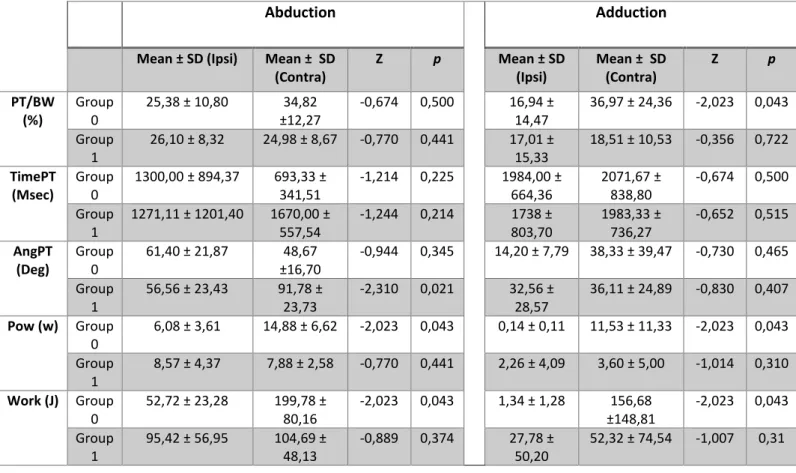
In the Abduction/Adduction isokinetic exercise, in Group 0, the Ipsilateral shoulder had significantly lower results regarding PT/BW in Adduction (p=0,043), Power in Abduction and Adduction (p=0,043 for both) and Work in Abduction and Adduction (p=0,043). All the other evaluated parameters were not significantly different between sides.
In group 1, there were no significant differences between sides except in the AngPT in Abduction, which was lower in the Ipsilateral shoulder (p=0,021). (Table 2 Suppl)
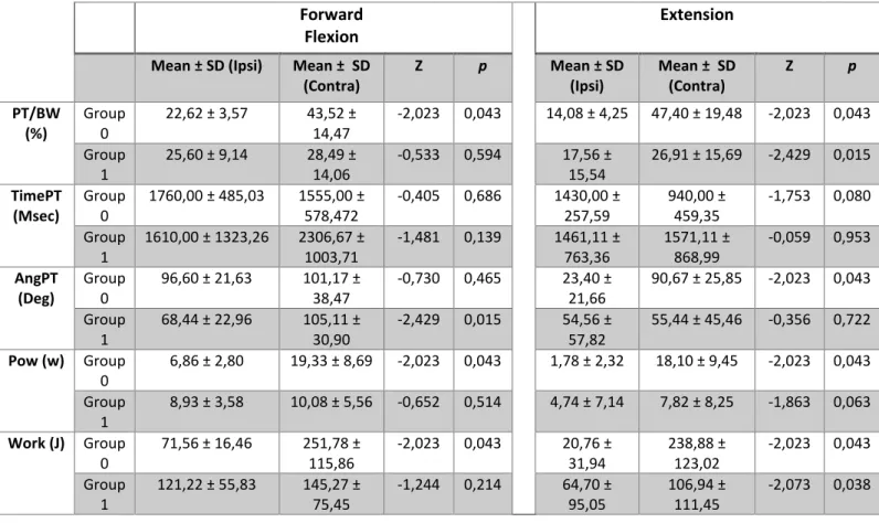
Forward Flexion/Extension isokinetic evaluation demonstrated that in Group 0, the Ipsilateral shoulder was significantly lower in PT/BW in Forward Flexion and Extension (p=0,043 for both), AngPT in Extension (p=0,043), Power in Forward Flexion and Extension (p=0,043 for both), Work in Forward Flexion and Extension (p=0,043 for both).
In Group 1, the Ipsilateral shoulder had significantly inferior values of PT/BW in Extension (p=0,015), AngPT in Forward Flexion (p=0,015), and Work in Extension (p=0,038). No other side-to-side differences were found in the remaining parameters. (Table 3 Suppl)
3.4. Electromyography
3.4.1. Anterior, Medial and Posterior Deltoid, in Abduction/Adduction:
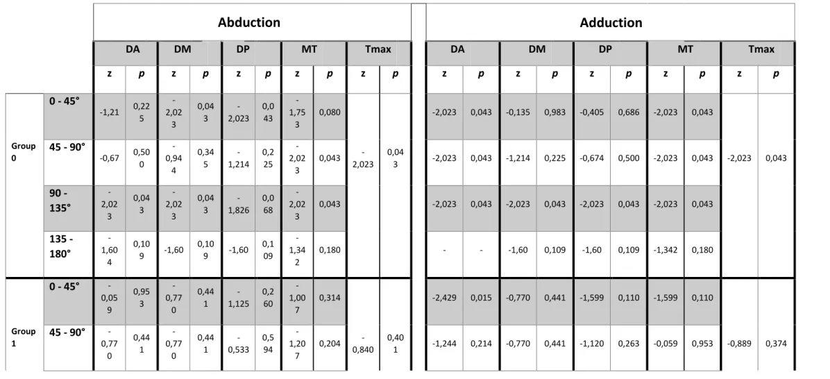
In Group 0, electromyographic assessment of the Anterior Deltoid highlighted significant differences between shoulders in the following movements and range of performances: Abduction in 90 - 135° (p=0,043), Adduction in 90 - 135° (p=0,043) and in 0 - 45° (p=0,043).
In the Middle Deltoid portion, significant side to side differences were found in Abduction in 0 - 45° (p=0,043) and in 90 - 135° (p=0,043), and Adduction in 90 - 135° (p=0,043).
In terms of the Posterior portion of the Deltoid muscle, the parameters with significant differences are Abduction in 0 - 45° (p=0,043) and Adduction in 90 - 135° (p=0,043).
The Mean Torque (MT) also had significant differences between the shoulders in Abduction in 45 - 90° (p=0,043) and in 90 - 135° (p=0,043), Adduction in 90 - 135° (p of 0,043), in 45 - 90° (p=0,043) and in 0 - 45° (p=0,043). The Maximum Torque (Tmax) had significant differences in Abduction and Adduction (p=0,043 for both).
Group 1 demonstrated statistically significant differences between shoulders in Adduction in 0 - 45° (p=0,015) for the Middle portion of the Deltoid muscle.
All statistically significant differences mentioned above are related to a higher electric activity in the Contralateral shoulder, except for two parameters in Group 0: Anterior Deltoid in Adduction in 90 - 135° and for the Middle portion of the Deltoid in Adduction in 0 - 45°, where the arm submitted to RSA had higher activity. (Tables 4, 6 and 8 Suppl)
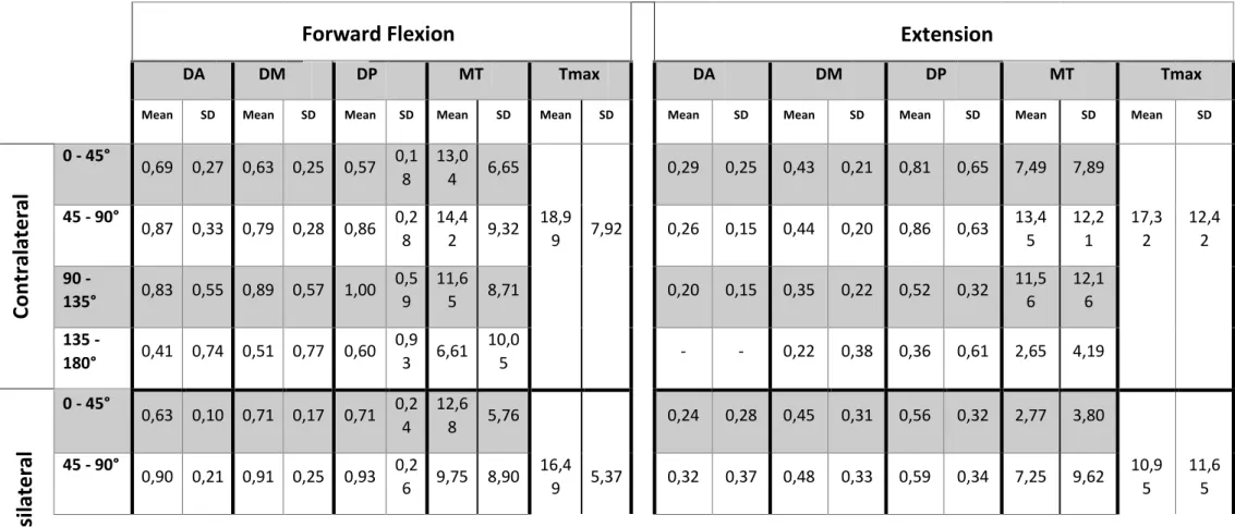
3.4.2. Anterior, Middle and Posterior Deltoid, in Forward Flexion/Extension:
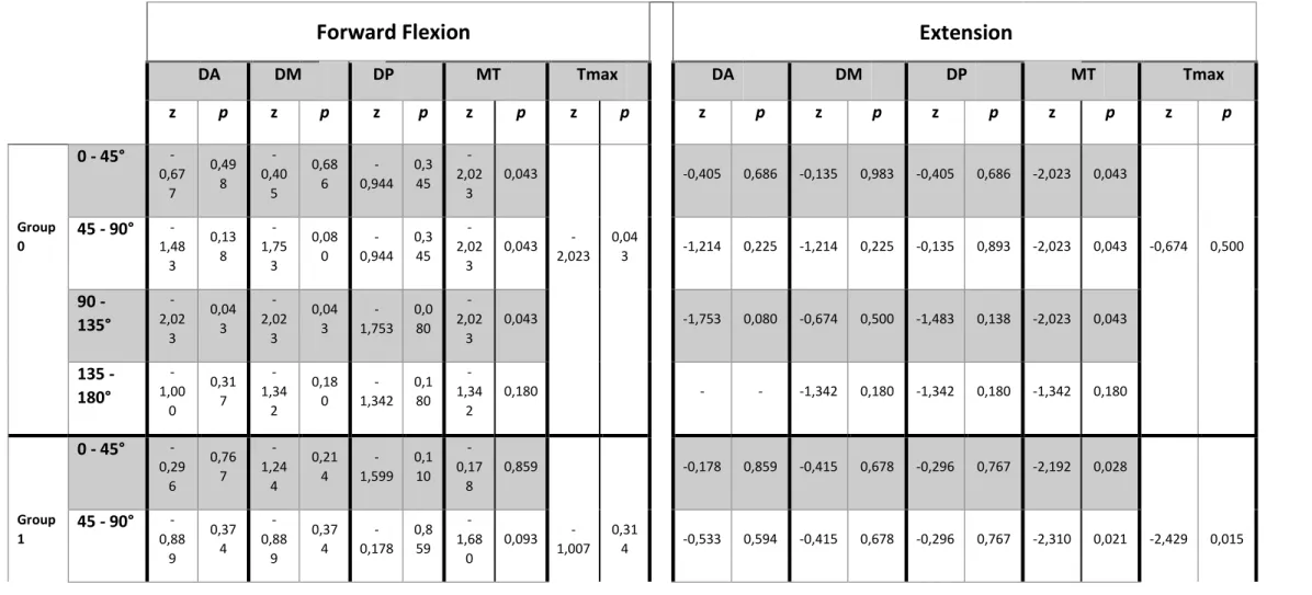
In the Anterior Deltoid, Group 0 had significant differences between shoulders in Forward Flexion in 0 - 135° (p=0,043).
In the Middle portion of the Deltoid muscle, there were significant differences in Forward Flexion in 90 - 135° (p=0,043).
The MT also had significant differences in Forward Flexion in 0 - 45° (p=0,043), in 45 - 90° (p=0,043), in 90 - 135° (p=0,043), and in Extension in 90 - 135° (p=0,043), in 45 - 90° (p=0,043) and in 0 - 45° (p=0,043).
Group 1 had significant differences between shoulders in terms of MT in Forward Flexion in 90 - 135° (p=0,012), in Extension in 45 - 90° (p=0,012) and in 0 - 45° (p=0,012). The Tmax was also significant better in the Contralateral arm in Extension (p=0,015).
Once again, all statistically significant differences mentioned above are related to a higher electric activity in the arm not submitted to RSA in both groups. (Tables 5, 7 and 9 Suppl)
4. Discussion
The purpose of this study was to evaluate the implications of RSA in the Deltoid muscle function, using its isokinetic profile and electromyography data. Isokinetic muscle tests are not frequently used in clinical practice, especially when associated with EMG, which results in limited literature available. The understanding of the biomechanics of the reverse shoulder anatomy is crucial, although there are few studies regarding the individual properties of the Deltoid adaptation after RSA (Fischer et al., 2019; Rugg et al., 2019; Walker et al., 2016). Most studies mainly evaluate isometric muscle strength. However, most functional activities are dynamic which makes imperative to have an isokinetic evaluation that can be related to functional and clinical outcome (Alta et al., 2014, 2012).
From a clinical standpoint, this study demonstrated that CMS in RSA shoulders were lower than the contralateral side, but as no baseline data were available, this may also result from a worse pre-operative overall function. In fact, pre-operative RoM appears to have a great influence in post-operative RoM (Friedman et al., 2019). Although pain level was very similar for both sides, RoM in Abduction and Forward Flexion were decreased in the RSA shoulder, as well as the strength assessed in the CMS.It is important to notice that the expectation for functional and RoM results have to be set case-by-case, depending on the pathology indicative for RSA, the performance status and the possibility of rehabilitation for the Deltoid and periscapular musculature (Boudreau et al., 2007). There is an overall RoM decreased in RSA shoulders comparing to healthy controls but these differences may not be as important clinically, as an acceptable RoM is achieved and pain relief is significant. In fact, RSA seems to be effective in pain relief and allows a satisfactory functional outcome (Jeon and Rhee, 2018), even though pre-lesion function seems not achievable, as we found in our study. In addition, a previous study suggests that the limited RoM after RSA can be a result of lack of joint torque generation (Bergmann et al., 2008), showing the importance of an isokinetic evaluation.
In this study, the contralateral arm was defined as the control for comparison. Because of the exclusion criteria, the contralateral arm should be the best performance status for each participant, as all individuals who had had any history of previous or current shoulder pathologies
and surgeries were excluded, as it could interfere with its strength and RoM. According to Constant et al, the cut-off value of 80 points in the CMS is considered as a normal result in the age range of this sample (Constant et al., 2008). Based on this, we attempted to identify objective isokinetic side-to-side differences in participants that had higher contralateral shoulder function (Group 0, CMS superior or equal to 80) and those with lower contralateral function (Group 1, CMS inferior to 80). Overall, patients in Group 0 demonstrated more statistically significant differences between shoulders, with the contralateral arm having a better isokinetic and EMG performance than the RSA one.
Isokinetic evaluation enables a safe, objective and reliable assessment of muscular performance. It provides a dynamic measurement of isolated joint motions and muscular contributions, and helps to assess underlying muscular strength and strength balance (Ellenbecker and Davies, 2000). As previous studies using isokinetic evaluation in samples with similar mean age and evaluating abduction/adduction and/or forward flexion/extension, we used dynamometer velocity set of 60 degrees per second (Alta et al., 2012; Wang et al., 2016). All participants were able to execute the exercises, which reveals as an appropriate velocity.
In this study, patients in Group 0 demonstrated higher Peak Torque in Abduction, Forward Flexion and Extension in the contralateral shoulder, but in patients with lower contralateral function, only Peak Torque in Extension was significantly affected. Deltoid muscle is mostly responsible for abduction, with a role in forward flexion by assisting Pectoralis Major in drawing the arm forward and with a minor role in extension by assisting Latissimus dorsi and Teres Minor (Susan Standring DSc, 2016). After RSA, the lever arm of the Deltoid is improved so that Forward Flexion and Abduction can be achieved without the stabilizing role of the rotator cuff (Boudreau et al., 2007). Our findings suggest that although RSA appears to decrease the maximum strength of the Deltoid, this may only be significant in patients with a higher than normal overall shoulder function. A previous study showed that pre-operative shoulder strength and RoM have a major role in the post-operative outcome, as individuals with poor function previous to the prosthesis will have worse functional outcomes with RSA (Li et al., 2020).
Other parameters of the isokinetic evaluation also revealed this tendency, that more significant differences were only reached when a higher contralateral function was present. In the scapula plane, several parameters (PT/BW, Work, Power) were significantly lower in the RSA, both in Abduction and Adduction, but only in Group 0. Group 1 demonstrated lower Angle of PT in Abduction in the RSA shoulder, pointing out that the maximum strength generation is reached in a lower angle, and this holds even comparing with shoulders with poorer overall function. Nevertheless, this finding must be interpreted with caution, as no significant difference was found for the same parameter in Group 0, where side-to-side differences seem to be more aggravated. In the sagittal plane, many parameters were significantly decreased in the RSA shoulder of patients in Group 0 (PT/BW, AngPT, Power and Work), both in Forward Flexion and Extension. Group 1 also presented more significant differences in this plane, which may reveal that strength in the sagittal plane may be more significantly affected.
EMG analysis completed Deltoid evaluation and also demonstrated more significant side-to-side differences in Group 0. Deltoid electric activity in the Anterior and Middle portion was lower in the RSA shoulder, regarding Abduction and Forward Flexion in the 0-135º range, which is also consistent with the movements where Deltoid has a more important role (Fischer et al., 2019; Schwartz et al., 2013). This pattern was confirmed in the Mean Torque and in the Maximum Torque. Group 1 failed to demonstrate significant differences in the EMG, and this may represent that RSA can restore Deltoid electrical activity to levels similar to the contralateral shoulder in lower demand patients (with a lower overall shoulder function).
Alta et al. observed that participants with RSA have significant lower Torques in Abduction when compared to healthy subjects. Even though there are differences between this study and ours (such as the comparison with healthy participants versus using the contralateral arm and not specifically evaluation the Deltoid activity), these results corroborate our hypothesis that RSA does not totally restore shoulder’s RoM and muscle strength (Alta et al., 2012). As mentioned before, there is very limited literature with studies that used similar protocols including isokinetic assessment complemented by EMG. This reveals as a limitation to have data to compare our findings and conclusions, as more studies using this kind of protocol are necessary.
RSA shoulders generally demonstrated a poorer isokinetic performance. Some studies highlighted that humeral length after RSA may influence Deltoid function due to slackening or fatigue induced by overtensioning (Lädermann et al., 2012; Li et al., 2020). In our study, humeral length was preserved after RSA, but there were significant differences in the isokinetic and EMG evaluation that may be conditioned by RSA design and surgical intervention.
This study has limitations that must be carefully interpreted. The limited number of participants may induce bias, and no baseline evaluation was available for comparison. However, isokinetic objective evaluation may be difficult before surgery, as many patients suffer from painful conditions that preclude an exhaustive functional assessment. Time elapsed from surgery was not homogenous between patients, and this may have an effect as some studies report a decrease in Deltoid function with time (Pegreffi et al., 2017). In addition, serial evaluation of this sample may contribute to the understanding of temporal effects and improve the reliability of assessment. In further studies, larger samples and standardized timings of evaluation would be more appropriate for improvement. The cut-off value of 80 for the CMS was used for separating two groups. The difference of mean CMS of each group was approximately 10 points, which may be argued that Group 0 did not have a clinically significant poor overall shoulder function of the Contralateral arm. Notwithstanding, this may be interpreted that RSA shoulders can achieve a nearly equivalent performance to the contralateral side if shoulder function is not severely decreased, but may fail to do so when function is exceedingly good.
5. Conclusion
Isokinetic evaluation combined with EMG is a useful and applicable tool to assess muscular and joint outcomes after RSA. This study demonstrated that this procedure has a significant effect in pain, RoM and strength of the shoulder joint and the Deltoid muscle. This effect was more relevant in patients with a better overall function of the contralateral shoulder, and may be only marginal when contralateral shoulders have a poorer function. In conclusion, isokinetic evaluation complemented with EMG and clinical scores (CMS) allowed us to have a more consistent and reliable understanding of the function of the Deltoid muscle after RSA.
Acknowledgments:
The authors of this article would like to express their gratitude to the Porto Biomechanics Laboratory (LABIOMEP) administration for allowing the evaluation of the participants in their facilities.
References
Ackland, D.C., Patel, M., Knox, D., 2015. Prosthesis design and placement in reverse total shoulder arthroplasty. J. Orthop. Surg. Res. 10, Published 2015 Jul 2. doi:10.1186/s13018-015-0244-2
Alta, T.D.W., Veeger, D., De Toledo, J.M., Janssen, T.W.J., Willems, W.J., 2014. Isokinetic strength differences between patients with primary reverse and total shoulder prostheses: Muscle strength quantified with a dynamometer. Clin. Biomech. 29, 965–970. doi:10.1016/j.clinbiomech.2014.08.018
Alta, T.D.W., Veeger, H.E.J., Janssen, T.W.J., Willems, W.J., 2012. Are shoulders with a reverse shoulder prosthesis strong enough? A pilot study shoulder. Clin. Orthop. Relat. Res. 470, 2185– 2192. doi:10.1007/s11999-012-2277-8
Bergmann, J.H.M., De Leeuw, M., Janssen, T.W.J., Veeger, D.J.H.E.J., Willems, W.J., 2008. Contribution of the reverse endoprosthesis to glenohumeral kinematics. Clin. Orthop. Relat. Res. 466, 594–598. doi:10.1007/s11999-007-0091-5
Biodex System 4 Pro Specifications [WWW Document], 2020. URL www.biodex.com
Boudreau, S., Boudreau, E.D., Higgins, L.D., Wilcox, R.B., 2007. Rehabilitation following reverse total shoulder arthroplasty. J. Orthop. Sports Phys. Ther. 37, 734–743. doi:10.2519/jospt.2007.2562
Constant, C.R., Gerber, C., Emery, R.J.H., Søjbjerg, J.O., Gohlke, F., Boileau, P., 2008. A review of the Constant score: Modifications and guidelines for its use. J. Shoulder Elb. Surg. 17, 355– 361. doi:10.1016/j.jse.2007.06.022
Ellenbecker, T.S., Davies, G.J., 2000. The Application of Isokinetics in Testing and Rehabilitation of the Shoulder Complex. J. Athl. Train. 35, 338–350.
Fischer, C., Flammer, S., Kauczor, H.U., Zeifang, F., Schmidmaier, G., Kunz, P., 2019. Preoperative deltoid assessment by contrast-enhanced ultrasound (CEUS) as predictor for shoulder function after reverse shoulder arthroplasty: a prospective pilot study. Arch. Orthop. Trauma Surg. doi:10.1007/s00402-019-03281-w
Friedman, R.J., Eichinger, J., Schoch, B., Wright, T., Zuckerman, J., Flurin, P.-H., Bolch, C., Roche, C., 2019. Preoperative parameters that predict postoperative patient-reported outcome measures and range of motion with anatomic and reverse total shoulder arthroplasty. JSES Open Access 3, 266–272. doi:10.1016/j.jses.2019.09.010
Grammont, P., Laffay, J., Deries, X., 1987. Study and development of a new shoulder prosthesis [in French]. Rheumatologie 39, 407–418.
Hamilton, M.A., Diep, P., Roche, C., Flurin, P.H., Wright, T.W., Zuckerman, J.D., Routman, H., 2015. Effect of reverse shoulder design philosophy on muscle moment arms. J. Orthop. Res. 33, 605–613. doi:10.1002/jor.22803
Hamilton, M.A., Roche, C.P., Diep, P., Flurin, P.H., Routman, H.D., 2013. Effect of prosthesis design on muscle length and moment arms in reverse total shoulder arthroplasty. Bull. NYU Hosp. Jt. Dis. 71, S31-35.
Hatta, T., Giambini, H., Sukegawa, K., Yamanaka, Y., Sperling, J.W., Steinmann, S.P., Itoi, E., An, K.N., 2016. Quantified mechanical properties of the deltoid muscle using the shear wave elastography: Potential implications for reverse shoulder arthroplasty. PLoS One 11, e0155102. doi:10.1371/journal.pone.0155102
Hayes, K., Walton, J.R., Szomor, Z.L., Murrell, G.A.C., 2001. Reliability of five methods for assessing shoulder range of motion. Aust. J. Physiother. 47, 289–294. doi:S0004-9514(14)60274-9
International Society for the Advancement of Kinanthropometry (ISAK), 2001. International Standards for Anthropometric Assessment. International Society for the Advancement of Kinanthropometry, Underdale, SA, Australia.
Jazayeri, R., Kwon, Y.W., 2011. Evolution of the reverse total shoulder prosthesis. Bull. NYU Hosp. Jt. Dis. 69, 50–55.
Jeon, Y.S., Rhee, Y.G., 2018. Factors associated with poor active anterior elevation after reverse total shoulder arthroplasty. J. Shoulder Elb. Surg. 27, 786–793. doi:10.1016/j.jse.2017.10.027 Lädermann, A., Edwards, T.B., Walch, G., 2014. Arm lengthening after reverse shoulder arthroplasty: A review. Int. Orthop. 38, 991–1000. doi:10.1007/s00264-013-2175-z
Lädermann, A., Walch, G., Lubbeke, A., Drake, G.N., Melis, B., Bacle, G., Collin, P., Edwards, T.B., Sirveaux, F., 2012. Influence of arm lengthening in reverse shoulder arthroplasty. J. Shoulder Elb. Surg. 21, 336–341. doi:10.1016/j.jse.2011.04.020
Lädermann, A., Williams, M.D., Melis, B., Hoffmeyer, P., Walch, G., 2009. Objective evaluation of lengthening in reverse shoulder arthroplasty. J. Shoulder Elb. Surg. 18, 588–595. doi:10.1016/j.jse.2009.03.012
Li, H.R., Yoon, S. hyun, Lee, D., Chung, H., 2020. Relation between preoperative electromyographic activity of the deltoid and upper trapezius muscle and clinical results in patients treated with reverse shoulder arthroplasty. J. Shoulder Elb. Surg. 29, 195–201. doi:10.1016/j.jse.2019.05.032
Pegreffi, F., Pellegrini, A., Paladini, P., Merolla, G., Belli, G., Velarde, P.U., Porcellini, G., 2017. Deltoid muscle activity in patients with reverse shoulder prosthesis at 2-year follow-up. Musculoskelet. Surg. 101, 129–135. doi:10.1007/s12306-017-0516-6
Rugg, C.M., Coughlan, M.J., Lansdown, D.A., 2019. Reverse Total Shoulder Arthroplasty: Biomechanics and Indications. Curr. Rev. Musculoskelet. Med. 12, 542–553. doi:10.1007/s12178-019-09586-y
Schwartz, D.G., Kang, S.H., Lynch, T.S., Edwards, S., Nuber, G., Zhang, L.Q., Saltzman, M., 2013. The anterior deltoid’s importance in reverse shoulder arthroplasty: A cadaveric biomechanical study. J. Shoulder Elb. Surg. 22, 357–364. doi:10.1016/j.jse.2012.02.002
SENIAM, 2019. Surface ElectroMyoGraphy for the Non-Invasive Assessment of Muscles [WWW Document]. Proj. SENIAM. URL www.seniam.org
Susan Standring DSc, P., 2016. Gray’s anatomy 41st edition: The anatomical basis of clinical practice, Gray’s Anatomy.
Walker, D.R., Struk, A.M., Matsuki, K., Wright, T.W., Banks, S.A., 2016. How do deltoid muscle moment arms change after reverse total shoulder arthroplasty? J. Shoulder Elb. Surg. 25, 581– 588. doi:10.1016/j.jse.2015.09.015
Wang, A., Doyle, T., Cunningham, G., Brutty, M., Campbell, P., Bharat, C., Ackland, T., 2016. Isokinetic shoulder strength correlates with level of sports participation and functional activity after reverse total shoulder arthroplasty. J. Shoulder Elb. Surg. 25, 1464–1469. doi:10.1016/j.jse.2016.01.025
Tables
Table 1: Costant-Murley Score (CMS) for the arm with RSA designated by Constant_ipsi and the
arm without RSA designated by Constant_contra. The bottom line shows the difference between CMS between arms.
Minimum Maximum Mean Standard
Desviation
Constant_ipsi 37,54 70,67 52,729 10,56938
Constant_contra 61,50 91,83 77,7617 8,57314
Supplementary Material
Table 1 Suppl: “T/BW_Abd” – Peak Torque in Abduction, “T/BW_Add” – Peak Torque in Adduction, “T/BW_FF” – Peak Torque in Forward Flexion and “T/BW_E” – Peak Torque in Extension. There are presented values of mean ± standard deviation, and values of z and p related to the Wilcoxon test performed to assess differences between the arm with RSA (“Ipsilateral”) and the arm without RSA (“Contralateral”), for Group 0 and Group 1.
T/BW_Abd T/BW_Add T/BW_FF T/BW_E
Group 0 Ipsilateral 20,66 ± 8,27 z = -2,023 3,72 ± 0,78 z = -1,826 17,79 ± 4,36 z = -2,023 9,44 ±6,58 z = -2,023 Contalateral 33,45 ± 12,87 p = 0,043 33,49 ± 24,24 p = 0,068 40,03 ± 13,72 p = 0,043 44,62 ±16,53 p = 0,043 Group 1 Ipsilateral 25,47 ± 9,25 z = -0,560 12,14 ± 17,47 z = -0,770 24,77 ± 8,57 z = -1,007 17,44 ± 19,86 z = -2,429 Contralateral 24,36 ± 6,40 p = 0,575 18,12 ± 17, 76 p = 0,441 29,62 ± 13,97 p = 0,314 28,02 ± 22,45 p = 0,015
Table 2 Suppl: Peak Torque per Body Weight (“PT/BW (%)”), Angle of Peak Torque in degrees (“AngPT (Deg)”), Power in Watts (“Pow(W)”) and Work in Joules (“Work (J)”) in Abduction and Adduction isokinetic exercise in group 0 and group 1. There are presented values of mean ±
standard deviation for the arm with RSA (“Ipsi”) and the arm without RSA (“Contra”), and values of z and p related to the Wilcoxon test performed to assess differences between arms.
Abduction Adduction
Mean ± SD (Ipsi) Mean ± SD (Contra) Z p Mean ± SD (Ipsi) Mean ± SD (Contra) Z p PT/BW (%) Group 0 25,38 ± 10,80 34,82 ±12,27 -0,674 0,500 16,94 ± 14,47 36,97 ± 24,36 -2,023 0,043 Group 1 26,10 ± 8,32 24,98 ± 8,67 -0,770 0,441 17,01 ± 15,33 18,51 ± 10,53 -0,356 0,722 TimePT (Msec) Group 0 1300,00 ± 894,37 693,33 ± 341,51 -1,214 0,225 1984,00 ± 664,36 2071,67 ± 838,80 -0,674 0,500 Group 1 1271,11 ± 1201,40 1670,00 ± 557,54 -1,244 0,214 1738 ± 803,70 1983,33 ± 736,27 -0,652 0,515 AngPT (Deg) Group 0 61,40 ± 21,87 48,67 ±16,70 -0,944 0,345 14,20 ± 7,79 38,33 ± 39,47 -0,730 0,465 Group 1 56,56 ± 23,43 91,78 ± 23,73 -2,310 0,021 32,56 ± 28,57 36,11 ± 24,89 -0,830 0,407 Pow (w) Group 0 6,08 ± 3,61 14,88 ± 6,62 -2,023 0,043 0,14 ± 0,11 11,53 ± 11,33 -2,023 0,043 Group 1 8,57 ± 4,37 7,88 ± 2,58 -0,770 0,441 2,26 ± 4,09 3,60 ± 5,00 -1,014 0,310 Work (J) Group 0 52,72 ± 23,28 199,78 ± 80,16 -2,023 0,043 1,34 ± 1,28 156,68 ±148,81 -2,023 0,043 Group 1 95,42 ± 56,95 104,69 ± 48,13 -0,889 0,374 27,78 ± 50,20 52,32 ± 74,54 -1,007 0,31
Table 3 Suppl: Peak Torque per Body Weight (“PT/BW (%)”), Angle of Peak Torque in degrees (“AngPT (Deg)”), Power in Watts (“Pow(W)”) and Work in Joules (“Work (J)”) in Forward Flexion and Extension isokinetic exercise in Group 0 and Group 1. There are presented values of mean
± standard deviationfor the arm with RSA (“Ipsi”) and the arm without RSA (“Contra”), and values of z and p related to the Wilcoxon test performed to assess differences between arms.
Forward Flexion
Extension
Mean ± SD (Ipsi) Mean ± SD (Contra) Z p Mean ± SD (Ipsi) Mean ± SD (Contra) Z p PT/BW (%) Group 0 22,62 ± 3,57 43,52 ± 14,47 -2,023 0,043 14,08 ± 4,25 47,40 ± 19,48 -2,023 0,043 Group 1 25,60 ± 9,14 28,49 ± 14,06 -0,533 0,594 17,56 ± 15,54 26,91 ± 15,69 -2,429 0,015 TimePT (Msec) Group 0 1760,00 ± 485,03 1555,00 ± 578,472 -0,405 0,686 1430,00 ± 257,59 940,00 ± 459,35 -1,753 0,080 Group 1 1610,00 ± 1323,26 2306,67 ± 1003,71 -1,481 0,139 1461,11 ± 763,36 1571,11 ± 868,99 -0,059 0,953 AngPT (Deg) Group 0 96,60 ± 21,63 101,17 ± 38,47 -0,730 0,465 23,40 ± 21,66 90,67 ± 25,85 -2,023 0,043 Group 1 68,44 ± 22,96 105,11 ± 30,90 -2,429 0,015 54,56 ± 57,82 55,44 ± 45,46 -0,356 0,722 Pow (w) Group 0 6,86 ± 2,80 19,33 ± 8,69 -2,023 0,043 1,78 ± 2,32 18,10 ± 9,45 -2,023 0,043 Group 1 8,93 ± 3,58 10,08 ± 5,56 -0,652 0,514 4,74 ± 7,14 7,82 ± 8,25 -1,863 0,063 Work (J) Group 0 71,56 ± 16,46 251,78 ± 115,86 -2,023 0,043 20,76 ± 31,94 238,88 ± 123,02 -2,023 0,043 Group 1 121,22 ± 55,83 145,27 ± 75,45 -1,244 0,214 64,70 ± 95,05 106,94 ± 111,45 -2,073 0,038
Table 4 Suppl: Differences between arms in the electromyographic assessment of the Anterior (DA), Middle (DM) and Posterior (DP) portions of the Deltoid
Muscle during the Abduction/Adduction isokinetic exercise. Differences are represented by values of z and p related to the Wilcoxon test. It is also represented the values for the Mean Torque (“MT”) and the Maximum Torque (“Tmax”). Each parameter is evaluated for a specific interval of amplitude (o - 45°, 45 - 90°, 90 - 135° and 135 – 180° in Abduction, and the reverse in Adduction).
DA DM DP MT Tmax DA DM DP MT Tmax z p z p z p z p z p z p z p z p z p z p 0 - 45° -1,21 0,22 5 -2,02 3 0,04 3 -2,023 0,0 43 -1,75 3 0,080 -2,023 0,043 -0,135 0,983 -0,405 0,686 -2,023 0,043 Group 0 45 - 90° -0,67 0,50 0 -0,94 4 0,34 5 -1,214 0,2 25 -2,02 3 0,043 -2,023 0,04 3 -2,023 0,043 -1,214 0,225 -0,674 0,500 -2,023 0,043 -2,023 0,043 90 - 135° -2,02 3 0,04 3 -2,02 3 0,04 3 -1,826 0,0 68 -2,02 3 0,043 -2,023 0,043 -2,023 0,043 -2,023 0,043 -2,023 0,043 135 - 180° -1,60 4 0,10 9 -1,60 0,10 9 -1,60 0,1 09 -1,34 2 0,180 - - -1,60 0,109 -1,60 0,109 -1,342 0,180 0 - 45° -0,05 9 0,95 3 -0,77 0 0,44 1 -1,125 0,2 60 -1,00 7 0,314 -2,429 0,015 -0,770 0,441 -1,599 0,110 -1,599 0,110 Group 1 45 - 90° -0,77 0 0,44 1 -0,77 0 0,44 1 -0,533 0,5 94 -1,20 7 0,204 -0,840 0,40 1 -1,244 0,214 -0,770 0,441 -1,120 0,263 -0,059 0,953 -0,889 0,374
Abduction
Adduction
90 - 135° -0,84 5 0,39 8 -1,18 3 0,23 7 -1,183 0,2 37 -1,52 1 0,128 -1,352 0,176 -1,014 0,310 -1,014 0,310 -0,845 0,398 135 - 180° -1,21 4 0,22 5 -0,94 4 0,34 5 -0,944 0,3 45 -0,73 0 0,465 - - -0,674 0,500 -1,214 0,225 -1,214 0,225
Table 5 Suppl: Differences between arms in the electromyographic assessment of the Anterior (DA), Middle (DM) and Posterior (DP) portions of the Deltoid
Muscle during the Forward Flexion/Extension isokinetic exercise. Differences are represented by values of z and p related to the Wilcoxon test. It is also represented the values for the Mean Torque (“MT”) and the Maximum Torque (“Tmax”). Each parameter is evaluated for a specific interval of amplitude (o - 45°, 45 - 90°, 90 - 135° and 135 – 180° in Abduction, and the reverse in Adduction).
DA DM DP MT Tmax DA DM DP MT Tmax z p z p z p z p z p z p z p z p z p z p 0 - 45° -0,67 7 0,49 8 -0,40 5 0,68 6 -0,944 0,3 45 -2,02 3 0,043 -0,405 0,686 -0,135 0,983 -0,405 0,686 -2,023 0,043 Group 0 45 - 90° -1,48 3 0,13 8 -1,75 3 0,08 0 -0,944 0,3 45 -2,02 3 0,043 -2,023 0,04 3 -1,214 0,225 -1,214 0,225 -0,135 0,893 -2,023 0,043 -0,674 0,500 90 - 135° -2,02 3 0,04 3 -2,02 3 0,04 3 -1,753 0,0 80 -2,02 3 0,043 -1,753 0,080 -0,674 0,500 -1,483 0,138 -2,023 0,043 135 - 180° -1,00 0 0,31 7 -1,34 2 0,18 0 -1,342 0,1 80 -1,34 2 0,180 - - -1,342 0,180 -1,342 0,180 -1,342 0,180 0 - 45° -0,29 6 0,76 7 -1,24 4 0,21 4 -1,599 0,1 10 -0,17 8 0,859 -0,178 0,859 -0,415 0,678 -0,296 0,767 -2,192 0,028 Group 1 45 - 90° -0,88 9 0,37 4 -0,88 9 0,37 4 -0,178 0,8 59 -1,68 0 0,093 -1,007 0,31 4 -0,533 0,594 -0,415 0,678 -0,296 0,767 -2,310 0,021 -2,429 0,015
90 - 135° -0,42 0 0,67 4 -1,26 0 0,20 8 -1,120 0,2 63 -2,52 1 0,012 -0,420 0,674 -0,280 0,779 -0,420 0,674 -0,631 0,528 135 - 180° -0,33 8 0,73 5 -0,16 9 0,86 6 0,000 1 -0,10 5 0,917 - - -0,169 0,866 -0,169 0,866 -0,931 0,352
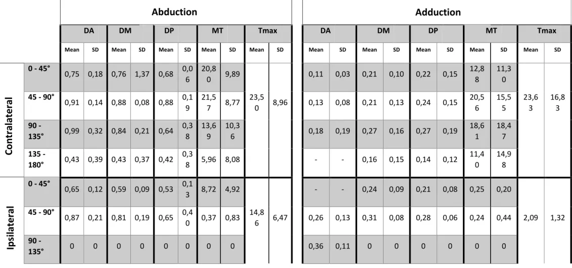
Table 6 Suppl: Electromyographic results of the Anterior (DA), Middle (DM) and Posterior (DP) portions of the Deltoid Muscle in Group 0, in the Abduction/Adduction isokinetic exercise represented by values of mean and Standard Desviation (“SD”). It is also represented the Mean Torque (“MT”) and the Maximum Torque (“Tmax”). Each parameter is evaluated for a specific interval of amplitude (o - 45°, 45 - 90°, 90 - 135° and 135 – 180° in Abduction, and the reverse in Adduction).
DA DM DP MT Tmax DA DM DP MT Tmax
Mean SD Mean SD Mean SD Mean SD Mean SD Mean SD Mean SD Mean SD Mean SD Mean SD 0 - 45° 0,75 0,18 0,76 1,37 0,68 0,0 6 20,8 0 9,89 0,11 0,03 0,21 0,10 0,22 0,15 12,8 8 11,3 0 45 - 90° 0,91 0,14 0,88 0,08 0,88 0,1 9 21,5 7 8,77 23,5 0 8,96 0,13 0,08 0,21 0,13 0,24 0,15 20,5 6 15,5 5 23,6 3 16,8 3 90 - 135° 0,99 0,32 0,84 0,21 0,64 0,3 8 13,6 9 10,3 6 0,18 0,19 0,27 0,16 0,27 0,19 18,6 1 18,4 7 135 - 180° 0,43 0,39 0,43 0,37 0,42 0,3 8 5,96 8,08 - - 0,16 0,15 0,14 0,12 11,4 0 14,9 8 0 - 45° 0,65 0,12 0,59 0,09 0,53 0,1 3 8,72 4,92 - - 0,24 0,09 0,21 0,08 0,25 0,20 45 - 90° 0,87 0,21 0,81 0,19 0,65 0,4 0 0,37 0,83 14,8 6 6,47 0,26 0,13 0,31 0,08 0,28 0,06 0,24 0,44 2,09 1,32 90 - 135° 0 0 0 0 0 0 0 0 0,36 0,11 0 0 0 0 0 0
Abduction
Adduction
Ipsi
lat
er
al
Contra
la
ter
al
135 -
Table 7 Suppl: Electromyographic results of the Anterior (DA), Middle (DM) and Posterior (DP) portions of the Deltoid Muscle in Group 1, in the
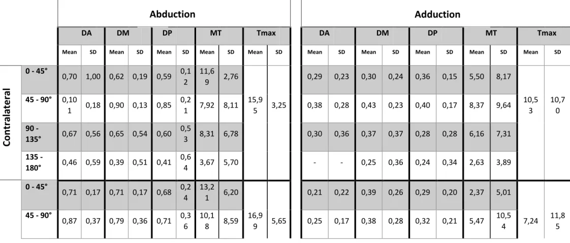
Abduction/Adduction isokinetic exercise represented by values of mean and Standard Desviation (“SD”). It is also represented the Mean Torque (“MT”) and the Maximum Torque (“Tmax”). Each parameter is evaluated for a specific interval of amplitude (o - 45°, 45 - 90°, 90 - 135° and 135 – 180° in Abduction, and the reverse in Adduction).
DA DM DP MT Tmax DA DM DP MT Tmax
Mean SD Mean SD Mean SD Mean SD Mean SD Mean SD Mean SD Mean SD Mean SD Mean SD 0 - 45° 0,70 1,00 0,62 0,19 0,59 0,1 2 11,6 9 2,76 0,29 0,23 0,30 0,24 0,36 0,15 5,50 8,17 45 - 90° 0,10 1 0,18 0,90 0,13 0,85 0,2 1 7,92 8,11 15,9 5 3,25 0,38 0,28 0,43 0,23 0,40 0,17 8,37 9,64 10,5 3 10,7 0 90 - 135° 0,67 0,56 0,65 0,54 0,60 0,5 3 8,31 6,78 0,30 0,36 0,37 0,37 0,28 0,28 6,16 7,31 135 - 180° 0,46 0,59 0,39 0,51 0,41 0,6 4 3,67 5,70 - - 0,25 0,36 0,24 0,34 2,63 3,89 0 - 45° 0,71 0,17 0,71 0,17 0,68 0,2 4 13,2 1 6,20 0,21 0,22 0,39 0,26 0,29 0,20 2,37 5,01 45 - 90° 0,87 0,37 0,79 0,36 0,71 0,3 6 10,1 8 8,59 16,9 9 5,65 0,25 0,17 0,38 0,28 0,32 0,21 5,47 10,5 4 7,24 11,8 5
Abduction
Adduction
Contra
la
ter
al
90 - 135° 0,66 0,93 0,40 0,40 0,34 0,3 6 3,43 6,05 0,12 0,13 0,22 0,27 0,20 0,24 3,82 7,49 135 - 180° 0,12 0,25 0,14 0,31 0,13 0,3 3 1,27 3,17 0 0 0,12 0,29 0,32 0,21 0,99 2,98
Ips
ilat
er
al
Table 8 Suppl: Electromyographic results of the Anterior (DA), Middle (DM) and Posterior (DP) portions of the Deltoid Muscle in Group 0, in the Forward
Flexion/Extension isokinetic exercise represented by values of mean and Standard Desviation (“SD”). It is also represented the Mean Torque (“MT”) and the Maximum Torque (“Tmax”). Each parameter is evaluated for a specific interval of amplitude (o - 45°, 45 - 90°, 90 - 135° and 135 – 180° in Forward Flexion, and the reverse in Extension)
.
DA DM DP MT Tmax DA DM DP MT Tmax
Mean SD Mean SD Mean SD Mean SD Mean SD Mean SD Mean SD Mean SD Mean SD Mean SD 0 - 45° 0,64 0,15 0,65 0,12 0,55 0,2 4 23,2 5 10,0 1 0,08 0,03 0,24 0,16 0,41 0,12 16,0 3 7,71 45 - 90° 0,91 0,18 0,93 0,18 0,87 0,3 1 26,1 0 10,8 8 28,4 5 10,4 8 0,08 0,03 0,23 0,17 0,35 0,14 26,9 1 10,5 6 17,2 9 32,1 4 90 - 135° 0,90 0,15 0,94 0,19 1,03 0,3 9 22,3 8 9,11 0,10 0,04 0,22 0,15 0,28 0,09 26,1 2 14,0 8 135 - 180° 0,32 0,45 0,45 0,54 0,61 0,8 2 12,0 8 14,3 7 - - 0,07 0,12 0,12 0,15 11,3 3 13,0 7 0 - 45° 0,59 0,19 0,64 0,20 0,63 0,1 8 10,6 9 4,60 0,09 0,06 0,30 0,30 0,31 0,18 3,04 3,38 45 - 90° 0,76 0,23 0,75 0,27 0,78 0,1 6 2,05 3,53 12,7 6 3,80 0,16 0,09 0,36 0,33 0,32 0,17 4,36 5,87 6,55 6,56
Forward Flexion
Extension
Ipsi
lat
er
al
Contra
la
ter
al
90 - 135° 0,15 0,33 0,30 0,41 0,32 0,4 5 0,96 2,16 0,05 0,06 0,11 0,15 0,11 0,15 1,13 2,52 135 - 180° 0 0 0 0 0 0 0 0 0 0 0 0 0 0 0 0
Table 9 Suppl: Electromyographic results of the Anterior (DA), Middle (DM) and Posterior (DP) portions of the Deltoid Muscle in Group 1, in the Forward
Flexion/Extension isokinetic exercise represented by values of mean and Standard Desviation (“SD”). It is also represented the Mean Torque (“MT”) and the Maximum Torque (“Tmax”). Each parameter is evaluated for a specific interval of amplitude (o - 45°, 45 - 90°, 90 - 135° and 135 – 180° in Forward Flexion, and the reverse in Extension).
DA DM DP MT Tmax DA DM DP MT Tmax
Mean SD Mean SD Mean SD Mean SD Mean SD Mean SD Mean SD Mean SD Mean SD Mean SD 0 - 45° 0,69 0,27 0,63 0,25 0,57 0,1 8 13,0 4 6,65 0,29 0,25 0,43 0,21 0,81 0,65 7,49 7,89 45 - 90° 0,87 0,33 0,79 0,28 0,86 0,2 8 14,4 2 9,32 18,9 9 7,92 0,26 0,15 0,44 0,20 0,86 0,63 13,4 5 12,2 1 17,3 2 12,4 2 90 - 135° 0,83 0,55 0,89 0,57 1,00 0,5 9 11,6 5 8,71 0,20 0,15 0,35 0,22 0,52 0,32 11,5 6 12,1 6 135 - 180° 0,41 0,74 0,51 0,77 0,60 0,9 3 6,61 10,0 5 - - 0,22 0,38 0,36 0,61 2,65 4,19 0 - 45° 0,63 0,10 0,71 0,17 0,71 0,2 4 12,6 8 5,76 0,24 0,28 0,45 0,31 0,56 0,32 2,77 3,80 45 - 90° 0,90 0,21 0,91 0,25 0,93 0,2 6 9,75 8,90 16,4 9 5,37 0,32 0,37 0,48 0,33 0,59 0,34 7,25 9,62 10,9 5 11,6 5
Forward Flexion
Extension
Ipsi
lat
er
al
Contra
la
ter
al
90 - 135° 0,68 0,43 0,65 0,43 0,71 0,4 7 4,96 6,29 0,18 0,22 0,33 0,33 0,44 0,41 8,57 12,0 8 135 - 180° 0,45 0,47 0,47 0,50 0,47 0,4 9 5,67 5,50 0 0 0,26 0,32 0,33 0,41 5,40 8,26
CLINICAL BIOMECHANICS
A journal affiliated to the International Society of Biomechanics, the American Society of Biomechanics, the European Society of Biomechanics, the Taiwanese Society for Biomechanics and the Socit de Biomcanique.
AUTHOR INFORMATION PACK
TABLE OF CONTENTS
.XXX
.• Description
• Audience
• Impact Factor
• Abstracting and Indexing
• Editorial Board
• Guide for Authors
p.1
p.1
p.2
p.2
p.2
p.3
ISSN: 0268-0033DESCRIPTION
.Clinical Biomechanics is an international multidisciplinary journal of biomechanics with a focus on
medical and clinical applications of new knowledge in the field.
The science of biomechanics helps explain the causes of cell, tissue, organ and body system disorders, and supports clinicians in the diagnosis, prognosis and evaluation of treatment methods and technologies. Clinical Biomechanics aims to strengthen the links between laboratory and clinic by publishing cutting-edge biomechanics research which helps to explain the causes of injury and disease, and which provides evidence contributing to improved clinical management.
A rigorous peer review system is employed and every attempt is made to process and publish top-quality papers promptly.
Clinical Biomechanics explores all facets of body system, organ, tissue and cell biomechanics, with
an emphasis on medical and clinical applications of the basic science aspects. The role of basic science is therefore recognized in a medical or clinical context. The readership of the journal closely reflects its multi-disciplinary contents, being a balance of scientists, engineers and clinicians.
The contents are in the form of research papers, brief reports, review papers and correspondence, whilst special interest issues and supplements are published from time to time.
Disciplines covered include biomechanics and mechanobiology at all scales, bioengineering and use of tissue engineering and biomaterials for clinical applications, biophysics, as well as biomechanical aspects of medical robotics, ergonomics, physical and occupational therapeutics and rehabilitation. The journal is affiliated to the European Society of Biomechanics, the American Society of Biomechanics, the International Society of Biomechanics, the Taiwanese Society of Biomechanics, and the Socit de Biomcanique.
AUDIENCE
.Biomechanists, bioengineers, orthopaedic physicians, physiotherapists, ergonomists and rheumatologists.
IMPACT FACTOR
.2018: 1.977 © Clarivate Analytics Journal Citation Reports 2019
ABSTRACTING AND INDEXING
.
Sports Documentation Monthly Bulletin and British Medicine PubMed/Medline Current Contents Embase Ergonomics Abstracts Bioengineering Now Reference Update
Sociedad Iberoamericana de Informacion Cientifica (SIIC) Data Bases Scopus
EDITORIAL BOARD
.Editor
Amit Gefen, Tel Aviv University Iby and Aladar Fleischman Faculty of Engineering, 69978, Tel Aviv, Israel Managing Editor
D. McStrafick, 30 Queen Street, PR8 2AG, Huddersfield, HD1 2SP Associate Editors
Dan Bader, Associate Editor for Soft Tissue Biomechanics, University of Southampton, Southampton, UK,
England
Lynne Bilston, University of New South Wales, Sydney, New South Wales, Australia Josué Sznitman, Technion Israel Institute of Technology, Haifa, Israel
Alon Wolf, Associate Editor for Posture and Motion Analysis, Technion Israel Institute of Technology Faculty
of Mechanical Engineering, Haifa, Israel
Editorial Board
L. Blankevoort, AMC Orthopaedics, Amsterdam, Netherlands M. Bobbert, VU Amsterdam, Amsterdam, Netherlands
A. van den Bogert, Cleveland State University, Cleveland, Ohio, United States P. Büchler, University of Bern, Bern, Switzerland
J. Cabri, Norwegian University of Sport and Physical Education, Oslo, Norway
C.K. Cheng, BeiHang University School of Biological Science and Medical Engineering, Beijing, China L. Cheze, Lyon Catholic University, Lyon, France
J. van Dieen, VU Amsterdam, Amsterdam, Netherlands V. Feipel, ULB Faculty of Motor Sciences, Brussels, Belgium S. Ferguson, University of Bern, Bern, Switzerland
T. Finni, University of Jyvaskyla, Jyväskylä, Finland
H. Graichen, Asklepios Orthopaedic Hospital Lindenlohe, Department of Arthroplasty and General Orthopaedic
Surgery, Schwandorf, Germany
A. Heiner, University of Iowa, Iowa City, Iowa, United States A. Karduna, University of Oregon, Eugene, Oregon, United States D. Lacroix, The University of Sheffield, Sheffield, United Kingdom K. Mabuchi, Kitasato University School of Medicine, Sagamihara, Japan W. S. Marras, OHIO STATE UNIVERSITY, Columbus, Ohio, United States
P. McNair, Auckland University of Technology School of Business, Auckland, New Zealand V. Medved, University Hospital Centre Zagreb, Zagreb, Croatia
O.G. Meijer, VU Amsterdam, Amsterdam, Netherlands F.C. Su, National Cheng Kung University, Tainan, Taiwan P. Swider, Toulouse 1 University Capitole, Toulouse, France P. Thoreux, Paris 13 University, Villetaneuse, France R. Vauhnik, University of Ljubljana, Ljubljana, Slovenia D. Veeger, VU Amsterdam, Amsterdam, Netherlands
M. Viceconti, The University of Sheffield, Sheffield, United Kingdom K. Wenger, Augusta University, Augusta, Georgia, United States
GUIDE FOR AUTHORS
.Your Paper Your Way
We now differentiate between the requirements for new and revised submissions. You may choose to submit your manuscript as a single Word or PDF file to be used in the refereeing process. Only when your paper is at the revision stage, will you be requested to put your paper in to a 'correct format' for acceptance and provide the items required for the publication of your article.
To find out more, please visit the Preparation section below.
Page charges
This journal has no page charges.
Submission checklist
You can use this list to carry out a final check of your submission before you send it to the journal for review. Please check the relevant section in this Guide for Authors for more details.
Ensure that the following items are present:
One author has been designated as the corresponding author with contact details: • E-mail address
• Full postal address
All necessary files have been uploaded:
Manuscript:
• Include keywords
• All figures (include relevant captions)
• All tables (including titles, description, footnotes)
• Ensure all figure and table citations in the text match the files provided • Indicate clearly if color should be used for any figures in print
Graphical Abstracts / Highlights files (where applicable) Supplemental files (where applicable)
Further considerations
• Manuscript has been 'spell checked' and 'grammar checked'
• All references mentioned in the Reference List are cited in the text, and vice versa
• Permission has been obtained for use of copyrighted material from other sources (including the Internet)
• A competing interests statement is provided, even if the authors have no competing interests to declare
• Journal policies detailed in this guide have been reviewed
• Referee suggestions and contact details provided, based on journal requirements For further information, visit our Support Center.
Author Checklist
Author Checklist is mandatory. Please click here to download the form and attach during submission process
BEFORE YOU BEGIN Ethics in publishing
Please see our information pages on Ethics in publishing and Ethical guidelines for journal publication.
Declaration of interest
All authors must disclose any financial and personal relationships with other people or organizations that could inappropriately influence (bias) their work. Examples of potential competing interests include employment, consultancies, stock ownership, honoraria, paid expert testimony, patent applications/registrations, and grants or other funding. Authors must disclose any interests in two places: 1. A summary declaration of interest statement in the title page file (if double-blind) or the manuscript file (if single-blind). If there are no interests to declare then please state this: 'Declarations of interest: none'. This summary statement will be ultimately published if the article is accepted.
2. Detailed disclosures as part of a separate Declaration of Interest form, which forms part of the journal's official records. It is important for potential interests to be declared in both places and that the information matches. More information.
Submission declaration and verification
Submission of an article implies that the work described has not been published previously (except in the form of an abstract, a published lecture or academic thesis, see 'Multiple, redundant or concurrent publication' for more information), that it is not under consideration for publication elsewhere, that its publication is approved by all authors and tacitly or explicitly by the responsible authorities where the work was carried out, and that, if accepted, it will not be published elsewhere in the same form, in English or in any other language, including electronically without the written consent of the copyright-holder. To verify originality, your article may be checked by the originality detection service Crossref Similarity Check.
Preprints
Please note that preprints can be shared anywhere at any time, in line with Elsevier's sharing policy. Sharing your preprints e.g. on a preprint server will not count as prior publication (see 'Multiple, redundant or concurrent publication' for more information).
Use of inclusive language
Inclusive language acknowledges diversity, conveys respect to all people, is sensitive to differences, and promotes equal opportunities. Articles should make no assumptions about the beliefs or commitments of any reader, should contain nothing which might imply that one individual is superior to another on the grounds of race, sex, culture or any other characteristic, and should use inclusive language throughout. Authors should ensure that writing is free from bias, for instance by using 'he or she', 'his/her' instead of 'he' or 'his', and by making use of job titles that are free of stereotyping (e.g. 'chairperson' instead of 'chairman' and 'flight attendant' instead of 'stewardess').
Author contributions
For transparency, we encourage authors to submit an author statement file outlining their individual contributions to the paper using the relevant CRediT roles: Conceptualization; Data curation; Formal analysis; Funding acquisition; Investigation; Methodology; Project administration; Resources; Software; Supervision; Validation; Visualization; Roles/Writing - original draft; Writing - review & editing. Authorship statements should be formatted with the names of authors first and CRediT role(s) following. More details and an example
Authorship
All authors should have made substantial contributions to all of the following: (1) the conception and design of the study, or acquisition of data, or analysis and interpretation of data, (2) drafting the article or revising it critically for important intellectual content, (3) final approval of the version to be submitted. Collaborators who do not satisfy the criteria for authorship can be listed as 'contributors' under the Acknowledgments section.
Changes to authorship
Authors are expected to consider carefully the list and order of authors before submitting their manuscript and provide the definitive list of authors at the time of the original submission. Any addition, deletion or rearrangement of author names in the authorship list should be made only before the manuscript has been accepted and only if approved by the journal Editor. To request such a change, the Editor must receive the following from the corresponding author: (a) the reason for the change in author list and (b) written confirmation (e-mail, letter) from all authors that they agree with the addition, removal or rearrangement. In the case of addition or removal of authors, this includes confirmation from the author being added or removed.
Only in exceptional circumstances will the Editor consider the addition, deletion or rearrangement of authors after the manuscript has been accepted. While the Editor considers the request, publication of the manuscript will be suspended. If the manuscript has already been published in an online issue, any requests approved by the Editor will result in a corrigendum.
Clinical trial results
In line with the position of the International Committee of Medical Journal Editors, the journal will not consider results posted in the same clinical trials registry in which primary registration resides to be prior publication if the results posted are presented in the form of a brief structured (less than 500