Recebido 26 de junho de 2018. Aceito 18 de julho de 2019.
Vaginal microbiota of nulliparous and multiparous cows and their resistance to
antimicrobials
[Microbiota vaginal de vacas nulíparas e multíparas e sua resistência a antimicrobianos]
“Artigo Científico/Scientific Article”
Lucas Pereira Silva
1, Sebastião Ávila Ramos
2, Cícero Cerqueira Cavalcanti Neto
1,
Tania Marta Carvalho dos Santos
1, Jorge Alberto Cavalcante de Oliveira
1,
Micheline Thais dos Santos
1, João Manoel da Silva
1*, José Soares Brito Neto
1,
Yamina Coentro Montaldo
11Microbiology Laboratory, Center of Agricultural Sciences, Federal University of Alagoas, Alagoas-AL, Brazil. 2Regional Council of Veterinary Medicine, Alagoas-AL, Brazil.
*Corresponding author/Autor para correspondência: E-mail: [email protected] Abstract
Infertility and prenatal mortality are the most important causes of production losses in domestic animals. The presence of pathogenic agents in the female reproductive system of cattle can negatively influence the reproductive performance of the herd. The objective of this study was to isolate, quantify, and characterize the components of the vaginal microbiota of multiparous and nulliparous mongrel cows, focusing on proper health and reproductive management. Samples were collected from 20 healthy animals: 10 nulliparous and 10 multiparous cows. The inoculation was performed simultaneously on four different culture media: BBL CHROM agar Candida, BBL Bile Esculin Agar Slants, Baird-Parker Agar, and MacConkey Agar bile salts. The isolated microorganisms were identified according to morphological and biochemical features. Microorganisms were detected in 80% of the nulliparous and 10% of the multiparous cows, with the following frequency: 88.78% of bacteria had morphophysiological characteristics of Staphylococcus spp., 11.22% of Escherichia coli, and 120 isolated yeast colonies which were identified as Candida tropicalis (69%), C. albicans (24%), and C. krusei. Seventeen isolates of Staphylococcus spp. (53.12%) presented sensitivity to all antimicrobials tested: 65.6% to amoxillin, 46.9% to erythromycin, 71.8% to rifampicin, and 46.9% to tetracycline. The rate of resistance to antimicrobials (MDR) was 0.125. The presence of typical microorganisms was detected. The MDR indicated that the isolates did not show multiresistance, with only two (6.25%) resistant to more than one antimicrobial simultaneously. The bacterial isolates studied were sensitive to the antimicrobial drugs tested, demonstrating the potential for use of these active ingredients.
Keywords: Enterococcus; Staphylococcus aureus; Escherichia coli; Candida sp.; antimicrobials. Resumo
Infertilidade e mortalidade pré-natal são as causas mais importantes de perdas na produção em animais domésticos. A presença de agentes patogênicos no sistema reprodutor feminino de bovinos pode influenciar negativamente o desempenho reprodutivo do rebanho. O objetivo desse estudo foi isolar, quantificar e caracterizar os componentes da microbiota vaginal de vacas nulíparas e multíparas e testar in vitro a suscetibilidade dos isolados patogênicos a antimicrobianos. Foram coletadas amostras vaginais de 20 animais considerados clinicamente saudáveis, sendo 10 nulíparas e 10 multíparas. A inoculação foi realizada simultaneamente em quatro diferentes meios de cultura: BBL CHROM ágar Candida, BBL Bile Esculin Agar Slants, Baird-Parker Agar e MacConkey Agar bile salts. Os micro-organismos isolados foram identificados de acordo com características morfológicas e bioquímicas. Foram detectados micro-organismos em 80% das nulíparas e 10% multíparas, sendo observadas as seguintes freqüências: 88,78% das bactérias com características morfofisiológicas de Staphylococcus spp. e 11,22% de Escherichia coli, 120 colônias isoladas de leveduras que foram identificadas: Candida tropicalis (69%), C. albicans (24%) e C. krusei. Dezessete isolados de Staphylococcus spp. (53,12%) apresentaram sensibilidade para todos os antimicrobianos testados, 65,6% para amoxilina, 46,9% para eritromicina, 71,8% para rifampicina e 46,9% para tetraciclina, o MDA
verificado foi de 0,125. Foi detectada a presença de micro-organismos típicos. O MDA indica que os isolados não possuem multi-resistência, porém, apenas dois (6,25%) sçao resistentes a mais de um antimicrobiano simultaneamente. Os isolados bacterianos estudados são sensíveis aos antimicrobianos testados, demonstrando o potencial de uso desses princípios ativos.
Palavras-chave: Enterococcus; Staphylococcus aureus; Escherichia coli; Candida sp.; antimicrobianos.
Introdução
Although there are some physical barriers that prevent opportunistic pathogens from colonizing the genital tract, females can become susceptible to microorganisms that pass through the cervix and reach the uterus (Rocha et al., 2004). The normal microorganisms of the vagina become pathogenic when animals exhibit a compromised immune system due to stress caused by various factors, such as temperature changes, poor nutrition, and end of pregnancy, demonstrating an opportunistic character at the source of infections (Kuntze and Aurich, 1995; Lianjuan et al., 1995; Aires de Sousa et al., 2007; Borges and Paschoal, 2012).
Under normal conditions, the composition of the vaginal microbiota has a variable number of microorganisms, which are also found in feces and skin, and due to their invasive properties, may be in the uterus in a small number of healthy animals (Ramaswamy et al., 1991). Cows do not suffer continuous contamination from such agents; however, they colonize the genital tract if there is an opportunity.
The normal microbiota includes bacteria, fungi, and protozoa that live inside or on healthy animals without causing disease. Included in this saprophytic biota are many pathogens and microorganisms with opportunistic potential. The microbiota of each tissue is a miniature ecosystem that is affected by a variety of endogenous and exogenous factors.
The resident microbiota has a definitive role in maintaining health and normal functions in animals. Its importance is such that it is considered one of the factors affecting natural resistance to infectious diseases. Organisms in the resident microbiota are harmless and can be beneficial in its normal location in the host and in the absence of abnormalities (Rosemberg, 2015).
The transient microbiota consists only of non-pathogenic or only potentially pathogenic members, are hosted on the skin or mucous membrane for hours, days, or weeks, are from the environment, do not cause disease, and do not establish themselves on the surface. Members of
transient microbiota are usually of low importance, while the resident microbiota remain unchanged in the event of proliferation and can cause diseases (Bouters and Vandeplasscge, 1977; Otero et al., 2000).
Although females have physical barriers that prevent colonization of the genital tract by opportunistic pathogens, they can become susceptible to microorganisms passing through the cervix and reaching the uterus (Rocha et al., 2004). During the postpartum period, there is migration of microorganisms in the vulva and vagina to the cervix and uterus.
The normal vaginal microbiota consists mostly of bacteria and, to a lesser extent, fungi, with the most common bacteria being aerobic, facultative pathogens, including Staphylococcus, hemolytic Streptococcus, and anaerobic gram-negative coliforms (Alba and Silveira, 2006; Martinez-Fernandez et al., 2006; Ocando et al., 2010). Facultative and obligate anaerobic bacteria can act synergistically, allowing growth and increasing pathogenicity. Among the most cited are Lactobacillus,
Fusobacterium, and Peptostreptococcus.
Based on this information, the objective of this study was to evaluate the microbiota present in cervicovaginal content of nulliparous and multiparous cows, aiming towards a proper orientation of the health and reproductive management, and thus making it possible to increase the number of healthy cows.
Material and Methods
The experiments were carried out with approval of the Animal Ethics Committee (AEC) of Federal University of Alagoas, with registration number 80/2017. The study was conducted in two farms located in the municipality of Chã Preta, in the state of Alagoas, latitude 15° 09' 19" S, longitude 36° 17' 46" W, altitude 463m. The climate is tropical and there is a dry season, according to the Koppen classification. Both properties have apparently similar characteristics
with respect to reproductive and nutritional management and sanitary control. The animals used were destined for meat production, and the sample consisted of crossbred females of different age groups, but within reproductive age, raised in an extensive system. The herd was constituted of 20 animals divided into two groups (Farm A with
ten nulliparous cows with an average of 24±4 months of age, and Farm B with ten multiparous cows with ages between 48 and 72 months), ten on each property (Table 1). The selective sampling was made based on the animals’ history and examination of the genital tract.
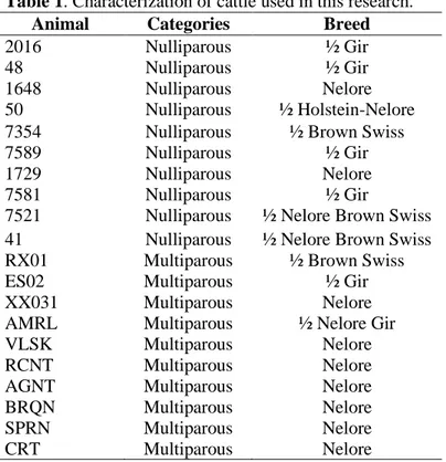
Table 1. Characterization of cattle used in this research.
Animal Categories Breed
2016 Nulliparous ½ Gir
48 Nulliparous ½ Gir
1648 Nulliparous Nelore
50 Nulliparous ½ Holstein-Nelore
7354 Nulliparous ½ Brown Swiss
7589 Nulliparous ½ Gir
1729 Nulliparous Nelore
7581 Nulliparous ½ Gir
7521 Nulliparous ½ Nelore Brown Swiss
41 Nulliparous ½ Nelore Brown Swiss
RX01 Multiparous ½ Brown Swiss
ES02 Multiparous ½ Gir
XX031 Multiparous Nelore
AMRL Multiparous ½ Nelore Gir
VLSK Multiparous Nelore
RCNT Multiparous Nelore
AGNT Multiparous Nelore
BRQN Multiparous Nelore
SPRN Multiparous Nelore
CRT Multiparous Nelore
Specula made from PVC pipes and measuring 37 cm length and 0.32 cm diameter were used. The specula had the edged rounded off using sandpaper to not injure the animals, were autoclaved at 1 atm pressure for 20 minutes, and then kept protected until ready to use.
Swabs were made with PVC, measuring 42 cm each. At one end the swabs owned the OB®
absorbent. All were moistened with sterile saline solution.
Samples were collected in the month of May, coinciding with the first official round of vaccinations against foot-and-mouth disease (FMD) in Brazil, to facilitate access to the animals. Samples of vaginal contents were collected with sterile Swabs moistened with saline solution.
During the collection, the perineal region was wiped down with a paper towel, then the labia were held open and the tubular sterile speculum was introduced until the bottom of the vaginal sack, performing rotational movements to obtain material. After collection, the tampons were placed in sterile tubes containing modified Stuart medium,
packed in ice, and sent to the laboratory for analysis.
Inoculation was carried out simultaneously in four different culture media: BBL CHROMagar Candida medium for isolation and identification of Candida albicans, C. tropicalis, and C. krusei (C. albicans colonies appear with a green-light to medium green color, C. tropicalis appear greenish blue to metallic blue, and C. krusei appear a clear pink color with a whitish border); BBL Bile Esculin Agar Slants for presumptive identification of Enterococcus species (diffuse blackening on the agar surface or, in some cases, a brown or black halo around the colonies); BD Baird-Parker Agar medium, which is moderately selective for differentiation, used for the isolation and enumeration of S. aureus (glossy black colonies, convex, with an opaque ring, surrounded by a clear halo, standing out on the opacity of the medium); MacConkey Agar with Crystal violet, NaCl, and 0.15% bile salts is recommended for selective isolation and differentiation of coliforms and other enteric pathogens.
After presumptive identification, bacterial colonies were submitted to the Gram method for confirmation using their morphology and staining characteristics, and evidence of catalase. According to the presented features, the isolates were submitted to the relevant identification procedures for each group.
Three out of five typical colonies of S. aureus (ATCC 25923 and ATCC 29213 were used to compare the strains of S. aureus) isolated from each plate with the Baird-Parker medium were placed into tubes containing Brain Heart Infusion broth (BHI Broth). The mass of cells with the broth were transferred to a tube with Trypticase Soy Agar (TSA) slant. Broth media were incubated at 35 ºC for 24 hours.
From the subculture grown in BHI broth, a coagulase test was carried out, and from the subculture grown on agar TSA, a catalase proof. For calculating the results, cultures with coagulase reaction levels 3 and 4, or cultures with coagulase reaction 1 and 2, but with positive reactions to the catalase test, that were Gram-positive, and formed clusters of cocci were considered coagulase-positive staphylococci.
To test DNase from this culture in BHI was made stretch marks, with horizontal lines, on medium agar DNAse (HIMEDIA, M482), which was subjected to incubation for 24 hours at a temperature of 36 °C ± 1 °C. After the incubation period, toluidine blue solution 0.1% was added to bacterial growth. The solution causes depolymerization of the middle DNA, showing a pink halo around the growth line.
Cultures considered coagulase-positive staphylococci were tested for sensitivity to single antimicrobial agents via the modified Kirby-Bauer disc diffusion test method. The cultures were transferred to test tubes containing Muller-Hinton broth and incubated at 37 °C to provide a turbidity equivalent to 0.5 MacFarland standard, seeded in Petri dishes containing Muller Hinton agar, to which discs containing antibiotic were applied, a light pressure contact allowed between the disc and the surface of the inoculated medium.
The evaluated antimicrobials was were amoxillin (10µg), bacitracin (10 U.I.), cephalothin (30μg), ciprofloxacin (30µg), doxycycline (30µg), enrofloxacin (5µg), erythromycin (15µg), gentamycin (10µg), lincomycin (2µg), kanamycin (30 mcg), norfloxacin (10µg), oxacillin (1µg), penicillin (10μg), rifampicin (30µg), tetracycline (30µg), and vancomycin (30µg). The rate of
resistance to antimicrobials (MDR) was determined (12) by dividing the number of groups of antimicrobials to which the isolates were resistant by the total number of groups of antimicrobials tested, with multiresistance considered at values above 0.4.
Results
Based on the data obtained, it was observed that the reproduction state of the animals influenced the growth of microorganisms. From the 20 animals, 205 bacterial isolates and 120 yeast isolates were obtained.
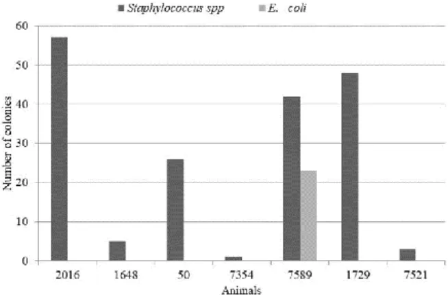
Although the farms appear to have a similar hygienic and sanitary management, it was observed that farm A has deficiencies which allowed a higher index of infected nulliparous than multiparous cows. Microorganisms were detected in 80% of nulliparous and 10% of multiparous cows (Figure 1); the following frequency was observed: 88.78% of the isolated bacteria presented morphophysiological characteristics of Staphylococcus spp., and 11.22%, of E. coli. These results were observed with the differential selective culture medium used (Figure 1). Enterococcus was not present. The majority seen in nulliparous cows is in function of the hygienic and sanitary management practices of the farms, since the animals were in different farms that had different management practices.
Figure 1. Number of colonies with typical characteristics of
Staphylococcus and Escherichia. coli isolated from nulliparous cows undergoing vaginal microbiological evaluation.
Of the 182 isolated cultures of Baird-Parker, 121 had arrangements shaped as grapes (staphylococci) and Gram +, 39 presented positive catalase reaction, of which 32 were positive for coagulase (Figure 2), 12.50% of these microorganisms were level 2, 65.62% were level 3, and 21.80% were level 4 (Figure 3).
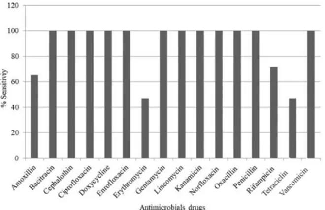
The antimicrobial resistance profiles of the isolates can be observed in Figure 4. Seventeen isolates (53.12%) presented sensitivity to all antimicrobial drugs tested: 65.6% to amoxillin, 46.9% to erythromycin, 71.8% to rifampicin, and 46.9% to tetracycline. Rate of resistance to antimicrobials was observed (125) where isolates did not show multidrug resistance, however, only two (6.25%) were resistant to more than one antimicrobial simultaneously.
One hundred and twenty yeast colonies were isolated and identified (using the Chromagar Candida®) as C. tropicalis, (69%) C. albicans
(24%), and C. krusei (Figure 5).
Figure 2. Characterization of typical colonies of
Staphylococcus aureus isolated in Baird Parker culture medium obtained from nulliparous cows.
Figure 3. Frequency of coagulase-positive cultures in
accordance with the colonies isolated from the Baird Parker medium reaction levels, obtained from bovine nulliparous females undergoing vaginal microbiological evaluation.
Figure 4. Percentage of sensitivity for Staphylococcus aureus
isolates via disk diffusion test in Petri dishes containing the agar Mueller Hinton.
Figure 5. Frequency of Candida yeasts isolated from
nulliparous and multiparous cows undergoing vaginal microbiological evaluation (identified through Chromagar Candida®).
Discussion
No differences were detected regarding microorganisms or their frequency among farms. The predominant occurrence of Staphylococcus has been commonly reported in similar survey studies on the vaginal microbiota of cows. Prevalence of members of these genera has been described in a survey of the microbiota present in cervical-vaginal mucosa of girolando cows (Rocha et al., 2004). In a study of the vaginal microbiota of cows associated with uterine infections, researchers also detected the presence of
Staphylococcus spp. and E. coli, with
predominance of the first (Gani et al., 2008). These studies suggested that, unlike the gastrointestinal microbiota, where the basic potential beneficial functions are related to the rich biodiversity, the vaginal ecosystem is characterized
by low microbial diversity, which consists almost exclusively of Lactobacillus (Branco et al., 2010).
The predominant enterobacteria in the mucosa of the vulva and vagina are derived from the gastrointestinal tract and have invasive properties in tissues that can cause infectious processes and movement of inflammatory cells into tissues. The presence of S. aureus is possibly due to the fact that this microorganism is commonly found in the skin and mucous membranes (Silva et al., 2011). When combined with other microorganisms, they may promote greater proliferation of pathogenic bacteria, because they produce the enzyme penicillinase that inactivates most penicillin-based treatments, resulting in inefficiency of these antibiotics as well as of other protectant bacteria. Other factor is the management of the farms, which can contribute to proliferation of pathogenic bacteria.
Yeasts are commensal organisms, living in the genital and gastrointestinal tracts, and in other mucous membranes of mammals. The genus Candida is frequently isolated from domestic and wild animals. In general, they do not cause any harm to their hosts, however, because of disturbances in physical, chemical, and immunological protections, these microorganisms can become pathogenic (Garcia et al., 2007).
A previous research analyzing 306 samples (Florião and Fraga, 2014) from the cervical-vaginal fluid of 17 heifer calves and 17 cows obtained 16 (7.77%) isolates of Candida sp. This microorganism has been associated with bovine mastitis and in some cases, predominates in relation to other yeasts.
C. albicans is the species most often involved in cases of candidiasis in animals (Jin and Lin, 2004; Jadhav and Pal, 2006; Kivaria and Noordhuizen, 2007). However, other Candida species such as C. tropicalis, C. parapsilosis, among others, are also referred to as agents in such infections (Pressler et al., 2003; Ozawa et al., 2005; Kivaria and Noordhuizen, 2007).
Knowledge of the normal microbiota is a necessary prerequisite to knowing the ecology of infectious diseases. The host and its characteristic microbiota are usually in a state of dynamic equilibrium, providing favorable conditions for the maintenance of the microbial population and not reacting against it. Several populations interact without harming the host and can benefit the host. The cervix and the vagina region have a small number of bacteria that can be isolated from normal
microbiota: Streptococcus, coliform bacteria, yeasts and fungi (Florião and Fraga, 2014).
Under normal conditions, the vaginal microbiota shows varied composition and number, and the microorganisms are also present on the skin and feces. Cows do not suffer continuous contaminations with these agents; however, these agents colonize the genital tract when there is an opportunity. After birthing, the vaginal microbiota can invade the uterus via the cervix. Most of the bacteria isolated from the vaginal content is constituted by Gram-negative gastrointestinal tract bacteria.
Abortion, infertility and other impairments in the reproductive tract of animals may be caused by various microorganisms belonging or not to the natural microbiota. Many of these microorganisms affect animals when the environment is not ideal for optimal health conditions and can manifest at any stage of the estrous cycle (Kaltungo and Musa, 2013; Szacawa et al., 2018).
Changes in the vaginal environment may cause changes in the microbiota, such as vaginal pH change during pregnancy or oestrus. Microbiota components can be opportunistic and play an important role in the development of infection in the upper reproductive tract, decreasing reproductive performance and therefore being responsible for significant economic losses (Suárez et al., 2006).
Based on the data obtained, it was observed that the experimental groups showed different effects of the microbiota. In multiparous cows, the presence of Candida was observed in only one animal out of ten. In nulliparous females, the presence of Candida in five animals and S. aureus in seven animals was seen among the ten submitted for evaluation, where only the animal number 7589 had Candida and S. aureus.
It is believed that the highest rates of microorganisms found in nulliparous females are due to the sanitary and nutritional management on the property because the experimental groups originated from different properties, despite being neighbors. However, the fact that nulliparous cows have presented higher microbial infection rate also drew attention because, in this situation, the authors expected them to be less exposed to microorganisms.
In contrast, animals reared under pasture conditions are constantly being challenged by disease-causing agents such as bacteria, viruses, fungi, and parasites. In this context, it is necessary
that the immune system of these animals is healthy and prepared to combat such attacks, thereby raising the productive potential of the animal. As the immune system needs adequate nutritional levels of protein, energy, minerals and vitamins for best performance, an appropriate nutritional management is indispensable. Some trace minerals such as zinc, copper, selenium, and manganese stand out for being part of enzymes that participate in the formation and action of the defense cells of the body. These trace minerals, because they are found in low concentrations in Brazilian fodder, need to be supplemented in the diet (Borges and Paschoal, 2012).
Based on the reproductive data of heifers after the evaluation period, 70% of nulliparous cows showed effective pregnancy and 30% did not become pregnant. This suggests the interference of microorganisms not in the reproductive process.
The results obtained for antimicrobial sensitivity were consistent with those reported by other authors. Susceptibility to various classes of antimicrobials between isolates of bovines was observed by several authors (Lange et al., 1999; Tollersrud et al., 2000). In the present study, 53.12% of isolates were susceptible to all antimicrobials tested, a susceptibility rate similar to that found by other Brazilian researchers.
The MDR obtained (0.125) indicated that the isolates did not show multiresistance, however, two (6.25%) were resistant to more than one antimicrobial simultaneously. The multidrug resistance of microorganisms is deriving from the improper use of antimicrobial drugs, presenting a potential risk to public health which may hinder the treatment of diseases in animals and humans (Silva et al., 2011).
Conclusion
It is verified that inefficient management favors a greater rate of infected animals, as demonstrated in farm A. Typical microorganisms were detected in all studied groups, with bacteria mostly prevalent, and highest frequency in nulliparous cows with 80% of the total microorganism count. The bacterial isolates studied are sensitive to the groups of antimicrobial drugs tested, demonstrating the potential for use of these active ingredients, as well as the availability of various choices, given the absence of multidrug resistance.
Conflict of Interest
The authors declare that they have no financial or personal relationship that may have inappropriately influenced them in writing this article.
Ethics Committee
The research was approved by the Ethical Committee of Animal Use (ECAU) of the Federal University of Alagoas under number 80/2017.
References
Aires-de-Sousa, M.; Parente, C.E.S.R.; Vieira-da-Mota, O.; Bonna, I.C.F.; Silva. D.A.; Lencatre, H. Characterization of Staphylococcus aureus isolates from buffalo, bovine, ovine and caprine milk samples collected in Rio de Janeiro, Brazil. Applied
and Environmental Microbiology, 73(12):
3845-9, 2007.
Alba, L.O.; Silveira, E.A. Bovine vaginal leucorrhea of non-inflammatory character and its clinical significance. REDEVET, Revista
Electronica de Veterinaria, 10: 1-29, 2006.
Borges, L.E.M.; Paschoal, J.J. Influence of beneficial micro-minerals Cu, Mn, Se and Zn in the bovine immune system. Caderno da
Pos-Graduação da FAZU, 3: 1-11, 2012.
Bouters, R.; Vandeplassche, M. Postpartum infection in cattle: Diagnosis and preventive and curative treatment. Journal of the South
African Veterinary Association, 48(4): 237–
239, 1977.
Florião, M.M.; Fraga, M.E. Mycoflora of cervical-vaginal fluid of an organic cattle breeding in tropical region. Revista Brasileira de
Medicina Veterinária, 36(1): 85-89, 2014.
Gani, M.O.; Amin, M.M.; Alam, M.G.S.; Kayesh, M.E.H.; Karim, M.R.; Samad, M.A.; Islam, M.R. Bacterial flora associated with repeat breeding and uterine infections in dairy cows.
Bangladesh Journal of Veterinary Medicine, 6(1): 79-86, 2008.
Garcia, M.E.; Lanzarot, P.; Lopez Rodas, V.; Costas, E.; Blanco, J.L. Fungal flora in the trachea of birds from a wildlife rehabilitation centre in Spain. Veterinarni Medicina, 52(10): 464-470, 2007.
Jadhav, V.J.; Pal, M. Canine mycotic stomatitis due
to Candida albicans. Revista
Iberoamericana de Micología, 23(4):
Jin, Y.; Lin, D. Fungal urinary tract infections in the dog and cat: a retrospective study 2001-2004. Journal of the American Animal
Hospital Association, 41(6): 373-381, 2005.
Kaltungo, B.Y.; Musa, I.W. A Review of Some Protozoan Parasites Causing Infertility in Farm Animals. ISRN Tropical Medicine, 2013.
Kivaria, F.M.; Noordhuizen, J.P.T.M. A retrospective study of the aetiology and temporal distribution of bovine clinical mastitis in smallholder dairy herds in the Dar es Salaam region of Tanzania. Veterinary
Journal, 173(3): 617-622, 2007.
Kuntze, A.; Aurich, J. Der
Endometritis-Pyometra-Komplex bei tieren. Berlin:
Gustav Fischer, 1995. 125p.
Lange, C.; Cardoso, M.; Senczek, D.; Schwarz, S. Molecular subtyping of Staphylococcus aureus isolates from cases of bovine mastitis in Brazil. Veterinary Microbiology, 67(2): 127–141, 1999.
Lianjuan, M.; Yuemin, L.; Xun. M. Microbial flora of the vagina of cows after parturition.
Chinese Journal of Veterinary Science and Technology, 25(5): 26-27, 1995.
Martínez-Fernández, A.; Silveira, E.A.; López, O.F. Las infecciones uterinas en la he mbra bovina. REDEVET, Revista Electronica de
Veterinaria, 7(10): 1-40, 2006.
Ocando, J.B.; Nava, S.Z.; Nava, J.; Martinez, G.P. Perfil de la flora bacteriana vaginal: Un riesgo potencial para la reproducción de vacas criollo limonero. Revista Cientifica de Maracaibo, 20(3): 12-23, 2010.
Otero, C.; Saavedra, L.; Silva de Ruiz, C.; Wilde, O.; Holgado, A.R.; Nader-Macías, M.E. Vaginal bacterial microflora modifications during the growth of healthy cows. Letters of
Applied Microbiology, 31(3): 251-254,
2000.
Ozawa, H.; Okabayashi, K.; Kano, R.; Watari, T.; Watanabe, S.; Hasegawa, A. Rapid identification of Candida tropicalisfrom canine cystitis. Mycopathology, 160(2): 159-162, 2005.
Pressler, B.M.; Vaden, S.L.; Lane, I.F.; Cowgill, L.D.; Dye, J.A. Candida spp. urinary tract infections in 13 dogs and seven cats: predisposing factors, treatment, and outcome.
Journal of American Animal Hospital Association, 39(3): 263-270, 2003.
Ramaswamy, V.; Andrew, M.; Roy, P. Aerobic microbes of cervico-vaginal mucus from repeat breeders bovines and their antibiogram.
Singapore Veterinary Journal, 14(15):
60-65, 1991.
Rocha, A.A.; Gambarini, M.L.; Andrade, M.A.; Oliveira Filho, B.D.; Gomes, F.A. Cervico-vaginal microbiota around the parturition time. Ciência Animal Brasileira, 5(4): 215-220, 2004.
Rosemberg, R. Detecting the emergence of novel, zoonotic viruses pathogenic to humans.
Cellular and Molecular Life Sciences,
72(6): 1115-1125, 2015.
Silva, V.F.; Damasceno, T.E.F.; Souza, N.J.D.; Franco, I.; Costa, M.M. Cervical-vaginal microbiota of crossbred sheep in Petrolina/PE, Brazil, and its susceptibility to antibiotics. Pesquisa Veterinária Brasileira, 31(7): 586-590, 2011.
Suárez, G.; Zunino, P.; Carol, H.; Ungerfeld, R. Changes in the aerobic vaginal bacterial mucous load and assessment of the susceptibility to antibiotics after treatment with intravaginal sponges in anestrous ewes.
Small Ruminant Research, 63(1-2): 39-43,
2006.
Szacawa, E.; Jawor, P.; Dudek, K.; Bednarek, D.; Stefaniak, T. Screening for Mollicutes microorganisms in perinatal calf mortality cases in Polish dairy herds. Polish Journal of
Veterinary Sciences, 21(3): 441-444, 2018.
Tollersrud, T.; Kenny, K.; Reitz, A.J.; Lee, J. Genetic and serologic evaluation of capsule production by bovine mammary isolates of
Staphylococcus aureus and other
Staphylococcus spp. from Europe and the United States. Journal of Clinical Microbiology, 38(8): 2998-3003, 2000.