Universidade do Minho Escola de Engenharia Departamento de Inform´atica
Catarina Freitas de Sousa Santos
Development of a Database and web tool
for the in silico characterization of plasmid data
Universidade do Minho Escola de Engenharia Departamento de Inform´atica
Catarina Freitas de Sousa Santos
Development of a Database and web tool
for the in silico characterization of plasmid data
Master dissertation
Master Degree in Bioinformatics
Dissertation supervised by
Professor Miguel Francisco Almeida Pereira Rocha Professora Cl ´audia Sofia Soares de Oliveira
A C K N O W L E D G E M E N T S
Firstly, I would like to thank my supervisors, Professor Miguel Rocha and Professor Cl´audia Oliveira, without whose guidance this process could not be completed. I would especially like to thank Professor Cl´audia Oliveira, for being such a positive influence and a constant presence throughout this journey. You have always had the right thing to say, the power to calm me down and been an inspiration and an enourmous role model to me.
I have to thank my parents the most, for being the best parents I could ask for and for the inconditional love and support and enduring my (not so) occasional stress-induced insanity.
To my godmother, Carmen, and my cousin, Susana: thank you for being a part of my life and always supporting me.
To D´ebora, Inˆes, Joana, Filipa and Rita: thank you for being great friends and always being a call away.
And last, but definitely not least, to Pedro, for being my happy place. Thank you, from the bottom of my heart, for always being there for me.
A B S T R A C T
Bacterial plasmids are mobile genetic structures capable of conferring selective advantages to their hosts, such as resistance to antibiotics, virulence genes and tolerance to pollutants.
By associating with other genetic elements, like integrons and transposons, plasmids pro-vide a platform for genetic recombination and for gene transfer between different bacterial species, allowing them to colonize multiple environments and guaranteeing their persis-tence.
Although there are over 4000 complete plasmid sequences available in GenBank, most have absent or non-standardized (disorganized) information regarding their isolation source, environment and year and country of isolation. Furthermore, a prediction about their mo-bility and incompamo-bility group is also lacking.
The goals of this thesis are, besides completing the missing information about plasmid data, the development of a repository of fully sequenced plasmids and the development of easy-to-use web tools for the characterization of plasmid data regardless of their source environment and bacterial host. For the development of these tools, Shiny was used, which is a package from the R scientific computing environment.
The present work is organized as follows: the core concepts related to plasmids are described, their background is characterized and a critical analysis of the available web tools for plasmid classification is carried out. Then, the adopted approach and the development (implementation, outcomes) of the database and web tool are explained. Lastly, the main conclusions are highlighted.
R E S U M O
Os plasm´ıdeos bacterianos s˜ao estruturas gen´eticas m ´oveis capazes de conferir vantagens seletivas ao seu hospedeiro, tais como resistˆencia a antibi ´oticos, genes de virulˆencia e tolerˆancia a poluentes.
Ao associar-se a outros elementos gen´eticos, como integr ˜oes e transpos ˜oes, os plasm´ıdeos constituem uma plataforma para a recombinac¸˜ao gen´etica e transferˆencia de genes entre diferentes esp´ecies bacterianas, permitindo que colonizem m ´ultiplos ambientes e garantindo a sua persistˆencia.
Embora existam acima de 4000 sequˆencias completas de plasm´ıdeos dispon´ıveis no Gen-Bank, a maioria apresenta informac¸˜ao ausente ou n˜ao sistematizada (desorganizada) em relac¸˜ao `a sua fonte de isolamento, ambiente e pa´ıs e ano de isolamento.
Os objetivos desta tese s˜ao, para al´em de completar a informac¸˜ao em falta sobre plasm´ıdeos, o desenvolvimento de um reposit ´orio de plasm´ıdeos completamente sequenciados e a disponibilizac¸˜ao de ferramentas online facilmente utiliz´aveis para a caracterizac¸˜ao de plasm´ı-deos independentemente da sua fonte de isolamento e hospedeiro bacteriano. Para o de-senvolvimento destas ferramentas, foi utilizado o Shiny, que ´e um package do sistema de computac¸˜ao cient´ıfica R.
Este trabalho est´a organizado da seguinte forma: os principais conceitos relacionados com os plasm´ıdeos s˜ao apresentados, a sua hist ´oria ´e caracterizada e ´e efetuada uma an´alise das ferramentas online existentes para a classificac¸˜ao de plasm´ıdeos. Depois, a abordagem utilizada e a ferramenta desenvolvida s˜ao explicadas. Finalmente, as principais conclus ˜oes s˜ao destacadas.
C O N T E N T S
1 i n t r o d u c t i o n 1
1.1 Motivation 1
1.2 Objectives 1
1.3 Organization of the Thesis 2
2 s tat e o f t h e a r t 3
2.1 Plasmid Definition and Main Characteristics 3
2.1.1 Plasmid Replication 5
2.1.2 Replication Control 8
2.1.3 Plasmid Stability and Partition 9
2.1.4 Antibiotic Resistance and Virulence 10
2.2 Mobile Genetic Elements 11
2.2.1 Horizontal Gene Transfer 12
2.2.2 Plasmid Mobility Groups and Mating Pair Formation 14
2.3 Plasmid Incompatibility 15
2.4 Plasmids at the NCBI Database 19
2.5 Available Databases and Programs for Mobile Genetic Elements 20
3 d e v e l o p m e n t 22
3.1 Construction and Structure of the Plasmid Database 22
3.2 Populating the Database 24
3.2.1 Obtaining the Plasmid Files 24
3.2.2 Getting the Taxonomy Information 27
3.2.3 Classifying According to Incompatibility and Mobility 27
3.3 Shiny 29
3.4 The PlasmidClassifier Application 32
3.4.1 Lists/Search Tool and Interactive Plots Tab 38
3.4.2 Plasmid Classification Panel 40
4 r e s u lt s a n d d i s c u s s i o n 48
4.1 Plasmid Classification Results 48
4.2 The PlasmidClassifier Application 50
4.2.1 Generated Plots and Tables 50
4.2.2 Classification Results 51
5 c o n c l u s i o n s a n d f u t u r e w o r k 58
5.1 Conclusions 58
Contents v
5.2 Prospect for future work 60
References 62
a at ta c h m e n t s 67
a.1 User Report Without Matches 68
a.2 User Report With a Single Match 72
L I S T O F A C R O N Y M S
BLASTBasic Local Alignment Search Tool. CRANComprehensive R Archive Network. ctRNAcountertranscribed RNA.
EEREnhanced Entity-Relationship. HGTHorizontal Gene Transfer.
IDEIntegrated Development Environment. IncIncompatibility.
IS Insertion sequences.
MGEMobile Genetic Elements. MpfMating Pair Formation. mrsMultimer Resolution System. ORFsOpen Reading Frames. oriOrigin of replication.
oriVOrigin of vegetative replication. pRNAprimer RNA.
PSI-BLAST Position-Specific Iterative Basic Local Alignment Search Tool. PSKPost-Segregational Killing.
RCR Rolling Circle Replication. RepReplication Iniciation. RMRestriction Modification. SQLStructured Query Language. TnTransposons.
T4CPType 4 Coupling Protein. T4SSType IV Secretion System. TAToxin-Antitoxin.
TETransposable elements. ui User-Interface.
WGSWhole Genome Shotgun.
L I S T O F F I G U R E S
Figure 1 Interaction between some Mobile Genetic Elements 11 Figure 2 Lineage distribution of the complete plasmid sequences in the NCBI
Plasmid Genome Database. 20
Figure 3 Enhanced Entity-Relationship (EER) diagram depicting the structure
of the database. 23
Figure 4 Tooltip used in the Start Classification Process button 31
Figure 5 Disabled textInput 31
Figure 6 General aesthetics of the PlamidClassifier Application 35
Figure 7 Homepage of the PlamidClassifier application 36
Figure 8 Definitions Tab of the PlamidClassifier application 37 Figure 9 About/Contacts Tab of the PlamidClassifier application 38 Figure 10 Side panel of the Lists/Search Tool and Interactive Plots Tab highlighting the available filtes and transferability options 39 Figure 11 Plot options available in the application with an empty plot in the
background 40
Figure 12 Appearance of the Plamid Classification panel upon initialization 41 Figure 13 Output in the Sequence tab for existing sequence (A) without or (B)
by uploading it 42
Figure 14 Application response when an unexisting sequence file is inputted 43 Figure 15 Sequence details (A) without and (B) after Classification process 44
Figure 16 Adding a new sequence to the Database 45
Figure 17 Sequences in the seqRepository table 47
Figure 18 Distribution Plots depicted in the PlasmidClassifier app when no
fil-ters are selected 53
Figure 19 Proportion Plots drawn in the PlasmidClassifier app when no filters
are selected, using the ggplot2 package 54
Figure 20 First 10 entries of the Plasmid Table available in the Lists/Search and Interactive Plots panel when no filters are selected 55 Figure 21 First 10 entries of the Taxonomy Table available in the Lists/Search and Interactive Plots panel when no filters are selected 56 Figure 22 First 10 entries of the MOB Table available in the Interactive Plots panel
when no filters are selected 57
L I S T O F TA B L E S
Table 1 Common plasmid initiator proteins 5
Table 2 Mobilization, occurrence, host range and general characteristics of
known plasmid Incompatibility groups 17
Table 3 Archetype plasmid, GenBank number, replicon typing (amplicon size and target gene region) and bibliography of known plasmid
In-compatibility groups 18
Table 4 Prefixes found in the Plasmid Genome Database 19
Table 5 Websites and references for the web tools described in section 2.5 21 Table 6 File types found at the NCBI FTP repository concerning plasmid
data 24
Table 7 Functions used in the Home, Definitions and About/Contacts tabs 33 Table 8 Functions used in the Lists/Search Tool & Interactive Plots tabsetPanel 33
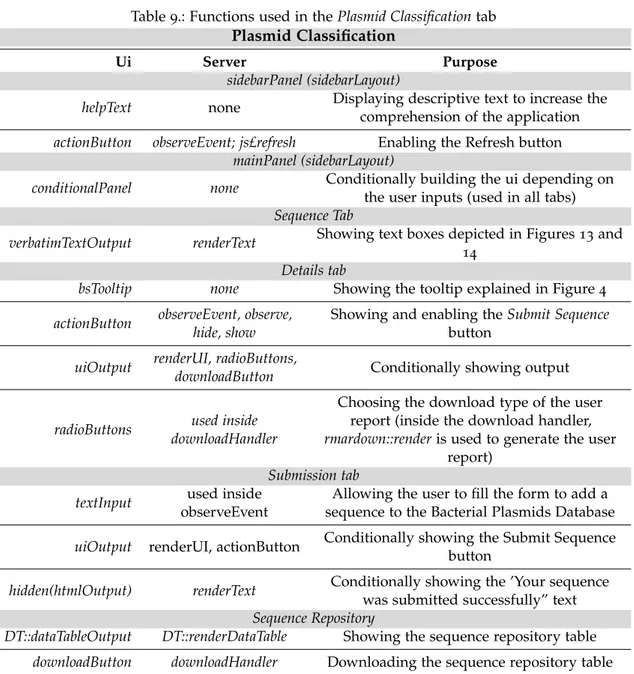
Table 9 Functions used in the Plasmid Classification tab 34
Table 10 Comparison between the results obtained in this study and Shintani
et al. (2015) and Orlek et al. (2017) 49
Table 11 Transferability outcomes for the results in common between this
study and Shintani et al. (2015) 49
1
I N T R O D U C T I O N
1.1 m o t i vat i o n
Bacterial plasmids are genetic structures that have essential roles in bacterial adaptation (Heuer and Smalla, 2012). By associating with other genetic structures, like integrons and transposons, plasmids provide a platform for genetic recombination and for gene transfer between different bacterial species (Ochman et al., 2000; Heuer and Smalla, 2012). So far, over 10000 plasmid sequences – of which over 4000 are complete – are available in GenBank. They have diverse genetic structures, and can be classified according to their encoded ac-cessory traits (like, for example, resistance and catabolic plasmids), mobility (conjugative, mobilizable and non-mobilizable), or incompatibility groups (from IncA to Z).
In recent years, there have been important efforts to analyze plasmid sequence data, es-sentially from clinical origin, due to their important involvement with epidemiology of multidrug resistant bacteria (Carattoli et al., 2014). However, plasmid importance should not be limited to bacterial hosts associated with hospital environments. Similarly with what has been proved for resistance genes (Gibson et al., 2014), plasmid encoded functions and genetic diversity are constrained by ecology (Binh et al., 2008). The number of plasmid se-quences in the database has been growing (Shintani et al., 2015; Smillie et al., 2010), but only approximately 47% (our study, unpublished results) can be associated with clinical related environments1
. The remaining plasmids, coming from other environmental compartments such as soil, water, food, extreme environments, and wastewater have been poorly com-pared and explored. A comprehensible and unified approach to catalogue non-clinical related plasmids is crucial to properly explore plasmid diversity and functions.
1.2 o b j e c t i v e s
The main goal of this thesis is the development of a repository of fully sequenced plasmids, gathering information about their ecology and the development of easy-to-use web tools for
1 Clinical, clinical potential and veterinary
1.3. Organization of the Thesis 2
the characterization of plasmid data regardless of their source environment and bacterial host.
In details, the scientific/technological objectives are to: (i) identify and review existing tools within the field of plasmid sequence analysis, (ii) review gene targets for plasmid classification/characterization by similarity searches, (iii) create a plasmid database with ecological information and replicon classifications and (iv) create a framework for the plas-mid web tool resource. The latter will include a list tab with additional data regarding plasmid ecology and a search tool to explore it and allow the in silico detection for plasmid characterization (mobility, incompatibility and putative transferability).
1.3 o r g a n i z at i o n o f t h e t h e s i s
This study is divided into five main chapters, all containing one or more sections:
1) Introduction, including the motivation behind the chosen theme and the objectives to achieve;
2) State of the Art, comprising the main concepts involving plasmids, that is:
• The background and definition of plasmid and its key features – replication, stability and partition and selective advantages for its host;
• The concept of mobile genetic elements, including horizontal gene transfer, mo-bility and mating pair formation;
• The types of plasmid data available at the NCBI database;
• The existing tools for the in silico characterization of plasmid data (and other mobile genetic elements).
3) Development, which includes the stages of designing the created tools:
• Creating the database with ecological information retrieved from NCBI and su-plementing it with information from related publications and with the results from similarity searches in terms of incompatibility, mobility and putative trans-ferability;
• Description of the software used to carry out the similarity searches and develop the web tool and the illustration of features of the application.
4) Results, regarding the obtained classification by similarity searches and developed application;
5) Conclusions and Future Work, indicating the main conclusions drawn from this work and the next steps for the improvement of the developed tools.
2
S TAT E O F T H E A R T
2.1 p l a s m i d d e f i n i t i o n a n d m a i n c h a r a c t e r i s t i c s
The term plasmid was first used to describe extrachromosomal genetic elements by Leder-berg in 1952 (Kado, 2015). Theretofore, designations such as R, F and T factors and episome were used.
Extrachromosomal genetic elements capable of conferring antibiotic resistance to their host were called R factors, those which were conjugative (see section 2.2) were identified as T factors and the ones carrying the genetic element needed for conjugation were known as F factors (Kado, 2015; Novick et al., 1976; Summers, 1996). Episome described a genetic element that can replicate integrated into or independent of the host chromosome (that is, autonomously in the cytoplasm). This term was used to label all the known bacterial genetic elements (including the three factors just described), which was very confusing, since it was too restrictive and, instead of defining a taxonomic entity, it merged the definition of plasmids and phages, among other elements (Kado, 2015; Novick et al., 1976).
To avoid this dubiety, plasmid became the conventionally accepted designation, episome was discarded as its synonym and the R and F factors are now known as Resistance (R) and F plasmids (Kado, 2015; Novick et al., 1976). In addition to these definitions, two other denominations are also noteworthy: cryptic and Col plasmids, which denote a plasmid without any specific traits and a colicin-producing plasmid, respectively (Novick et al., 1976).
Presently, a plasmid is defined as a double-stranded deoxyribonucleic acid (DNA) ex-trachromosomal genetic entity, transferable between species and capable of self-replicating (Carattoli, 2009; Carattoli et al., 2014; Novick et al., 1976).
Plasmids, mostly found in the Bacteria domain, are typically circular but occasionally linear, their size can vary from one to thousands of base pairs and their GC content also suffers major fluctuations (Novick, 1987; del Solar et al., 1998).
One of their most important specific features, which differs according to each host, is the copy number, defined as the number of molecules/concentration of a plasmid per cell, which must be maintained at a constant level so that the plasmid can persevere. For this
2.1. Plasmid Definition and Main Characteristics 4
to happen, depending on the number of copies the plasmid requires, different strategies are adopted (see subsection 2.1.1) (Novick et al., 1976; Novick, 1987; del Solar et al., 1998; Summers, 1996). Note that a plasmid can be identified as low-copy, medium-copy or high-copy, with numbers ranging from 1 to 10, 10 to 20 and 20 to over 700, respectively (Fricke et al., 2009).
Plasmids rely on two main functions to survive: persistence and proliferation. The former requires that the replication rate of the plasmid is the same as its host’s division rate, de-pending on the control of replication and, for low copy numbers, on the partition system, to guarantee that at least one copy is received by the daughter cell (see subsection 2.1.1). The latter, explained in section 2.2, is the capacity of a plasmid to transfer from one cell to another (Summers, 1996).
Although usually not being essential, these entities are systematically inherited due to being able to confer a selective advantage to the host, such as antibiotic resistance and virulence genes (see subsection 2.1.4) and tolerance to pollution, production of iron (to overcome its scarcity) and other environmental constraints, allowing them to spread to different surroundings (Carattoli et al., 2014; Kado, 2015).
Plasmids have previously been classified as parasitic, implying that they harm their host, or as symbionts, sharing a reciprocal exchange of advantages with the host (Norman et al., 2009; Summers, 1996). However, they are currently classified as selfish, due to the ability to autonomously replicate and maintain themselves in a cell, spreading within the genome or between genomes (see section 2.2) and being often acquired by sexual transmission. The degree of selfishness can vary, as they can benefit themselves while increasing or not the host’s fitness, since their persistence solely depends on their replication rate. These are also capable of influencing other plasmids, as they will try to prevent the entry of other plasmids in the cell (Kado, 1998; Liu et al., 2015; Norman et al., 2009).
Because of their unique capabilities, plasmids are acknowledged as of great interest from a biotechnological standpoint, as they have caused major breakthroughs in molecular bi-ology. The goals of researchers include studying their structure and genes, identifying their replication and partition systems and understanding the phenotypes they confer (e.g. tumor-inducing, virulence and antibiotic, heavy metal and radiation resistance), in order to use them in multiple procedures, such as reporter systems, genetic engineering and the development of gene vectors (Kado, 2015).
2.1. Plasmid Definition and Main Characteristics 5
2.1.1 Plasmid Replication
Plasmids are characterized by the presence of one or more specific regions encoding func-tions capable of activating and controlling replication, called replicons (Carattoli et al., 2014). Two types of replicons should be highlighted: (i) basic replicon, the smallest DNA sequence (usually 1-3kbp) that allows replication while preserving the regulatory scheme; and (ii) minimal replicon, the smallest DNA portion enabling replication, even if regulation is flawed (Lilly and Camps, 2015; Summers, 1996). Various plasmids have only one basic replicon, while others – multireplicon plasmids – display several replicons (for example, IncF plas-mids), although often only one is active at a time. The reason for this difference is unknown, but it is inferred that having multiple replicons may enable plasmids to colonize numerous hosts by allowing them to avoid incompatibility with the resident plasmid (see section 2.3) (Summers, 1996).
Plasmid replication can be divided into three stages: initiation, elongation, and termi-nation. The first stage, which is different for each replicon, is reliant on plasmid-encoded properties, namely the origin of replication (ori) and, generally, a replication initiation (Rep) protein, some of which represented in Table 1 (del Solar et al., 1998; Kues and Stahl, 1989). In the cases where the Rep proteins exist, the origin of replication includes directly re-peated DNA sequences with specific spacing, named iterons, which are the binding sites for the Rep proteins and are essential for replication control (Lilly and Camps, 2015; del Solar et al., 1998; Summers, 1996). Iteron spacing (iterons can be separated by other sequences or be contiguous) is of special importance, since it allows the recognition of specific DNA sequences by being at a distance that matches the helical periodicity of the DNA double helix (Lilly and Camps, 2015; del Solar et al., 1998). Because initiation is characteristic of each replicon, replication control takes place at that stage (del Solar et al., 1998).
Table 1.: Common plasmid initiator proteins (Adapted from (Kado, 2015))
Initiator Replication mode RepA Rolling circle; theta type
RepA1 Theta type
RepB Rolling circle
RepC Rolling circle; theta type
RepD Rolling circle
RepE Theta type
π(pir) Theta type
2.1. Plasmid Definition and Main Characteristics 6
As mentioned above, replication initiates at a location known as the origin of vegetative replication (oriV), which is commonly classified in one of four different ways: (i) the mini-mal cis-acting region that can support autonomous replication of the plasmid, (ii) the region where DNA strands are melted to initiate the replication process, (iii) the position/base at which the first deoxyribose base is added to the leading-strand RNA primer and/or, more strictly, (iv) the por-tion of sequence that is targeted by replicapor-tion initiapor-tion factors in trans (Lilly and Camps, 2015; Shintani et al., 2015; del Solar et al., 1998; Summers, 1996).
Albeit competent in self-replication, plasmids still have to synchronize their replication with the growth and division of their host cell, having to replicate a certain number of times per generation (usually one) to persist, since an uncontrolled plasmid concentration can have severe consequences for both the plasmid and its host (del Solar et al., 1998; Summers, 1996).
The number of plasmids per cell is controlled during the initiation stage of replication with the help of trans-activators/inhibitors, generally Rep replicases1
, encoded by plasmids (Norman et al., 2009). For an excessive copy number, to avoid an increased metabolic burden to the host, jeopardizing its viability, the average replication for generation is kept at less than one by employing trans-activators. Inversely, for insufficient copy numbers, which can lead to consecutive loss until the cell is plasmid-free (see section 2.1.3), thus requiring closer regulation, the average replication for generation is maintained at more than one with the aid of trans-inhibitors (Kues and Stahl, 1989; Norman et al., 2009; Summers, 1996).
There are three main types of plasmid replication: rolling circle, strand displacement and theta type, all of which observed in both Gram Positive and Gram Negative bacteria and explained in the next subsections.
Rolling Circle Replication
Rolling circle replication (RCR) has only been found on plasmids with less than 10kbp and it is prototyped by the pT181, pC194, pMV158 and pUB110 plasmids, isolated from organisms of the Staphylococcus genus (Shintani et al., 2015; del Solar et al., 1998). This method has a unique initiation mechanism: it is initiated from a 3’-OH primer produced by nicking one strand of the plasmid, which allows the host DNA polymerases to initiate the leading strand replication (Norman et al., 2009; Ruiz-Maso et al., 2015). Here, in order to cleave (split) and join plasmid DNA, Rep proteins have transferase2
enzymatic activity (del Solar et al., 1998).
1 Class of enzymes that catalyze the synthesis of an RNA molecule.
2.1. Plasmid Definition and Main Characteristics 7
Strand Displacement Replication
The name strand displacement is due to, in this process, the synthesis of each one of the strands taking place continuously and causing the displacement of the complementary strand. This type of replication is characteristic of the broad host IncQ family, namely of their archetype plasmid, RSF1010 (Shintani et al., 2015; del Solar et al., 1998). In this case, replication has two origins, ssiA and ssiB, one per DNA strand, and initiation is promoted by three Rep encoded proteins: (i) RepA, which has 5’ → 3’ helicase3 activity, (ii) RepB, which catalyzes the starting point for DNA synthesis, and (iii) RepC, which recognizes iterons, together allowing bidirectional replication to occur (del Solar et al., 1998).
Theta Type Replication
During this type of replication, which can happen uni- or bidirectionally, the images ob-tained by electronic microscopy (EM) resemble the greek letter θ (Lilly and Camps, 2015; Norman et al., 2009; del Solar et al., 1998). Theta type replication has been mostly studied in gram negative bacteria, although cases of theta plasmids being isolated from gram positive bacteria have also been reported (Norman et al., 2009; del Solar et al., 1998). In this method, similarly to the process in chromosomal replication, the leading strand, synthesized contin-uously, and lagging strand, synthesized discontincontin-uously, are replicated coordinately (Lilly and Camps, 2015; del Solar et al., 1998). This method can start from one to multiple origins and it includes synthesis of a primer RNA (pRNA), initiation of DNA synthesis by cova-lent extension to the pRNA and, generally, a plasmid-encoded Rep protein (del Solar et al., 1998).
There are four classes of theta type replication, from A to D. Plasmids from class A in-clude iterons (differing for each plasmid), that is, they rely on Rep proteins for replication initiation (see subsection 2.1.1), namely RepA for R1, pSC101, pPS10 and P1 (even though the Rep proteins of these three plasmids are homonymous, their characteristics are differ-ent), Trf1 for RK1 and π for R6K (plasmids of reference) (Lilly and Camps, 2015). Class B includes the archetype plasmid ColE1 and ColE1-like plasmids, which do not require Rep proteins, but rely on their host factors, such as DNA Polymerase I, for replication initia-tion and primer synthesis and only rely on antisense RNA for replicainitia-tion control (Lilly and Camps, 2015; Shintani et al., 2015; del Solar et al., 1998). Finally, classes C, containing ColE2 and ColE3 plasmids, and D – with large, low-copy streptococcal plasmids that replicate in a broad range of Gram-positive bacteria, such as pAMβ1, pIP501 and pSM19035 – have similarities with both A and B classes (del Solar et al., 1998).
2.1. Plasmid Definition and Main Characteristics 8
2.1.2 Replication Control
The goal of replication control is to maintain the plasmid copy number at a steady state, which must be in accordance with their host (Kues and Stahl, 1989; del Solar et al., 1998; Summers, 1996). In order to define and keep a specific copy number, plasmids employ neg-ative regulatory circuits, which should have a virtually imperceptible concentration upon host colonization; after that point, avoiding fluctuations in plasmid concentration, as men-tioned in subsection 2.1.1, involves adjusting the replication rate (del Solar et al., 1998; Summers, 1996). This regulation is the reason for plasmid incompatibility (see section 2.3), causing plasmids with similar regulation mechanisms to segregate within the host popula-tion (del Solar et al., 1998).
Classification of control systems is usually made according to the nature of their main replication inhibitor, which can be a protein (λ−dv, not described in natural plasmids), a small antisense RNA (ColE1, pT181 and R1), both a protein and an antisense RNA (pMV158 and pIP501), or a set of short DNA repeats, that is, DNA sites for binding initiator proteins (P1, F, RK2 and R6K) (del Solar et al., 1998; Summers, 1996).
The control by antisense RNA, found in both low and high copy plasmids, consists of a short antisense inhibitor binding close to the 5’ end of a region (longer transcript) that plays a major role in the replication process and precludes its activity, either by binding to the mRNA of a Rep protein in R1 or pT181 plasmids (prohibiting translation) or by blocking the formation of an RNA II-DNA hybrid (pre-primer RNA for replication) at the origin of replication in the ColE1 plasmid; in both cases, these inhibitors are called countertranscribed RNAs (ctRNAs) (del Solar et al., 1998; Summers, 1996).
Control by both a protein (transcriptional repressor) and antisense RNA is typical for the R1, pIP501 and pMV158 plasmids. For the latter, a repressor protein, named CopG is binded and inhibits transcription for a single promoter for the copG and repB genes and a small ctRNA is also involved and, for both these elements, mutations and deletions can increase the plasmid copy number (del Solar et al., 1998).
Lastly, besides posing as binding sites for the Rep proteins, iterons from the theta type replication also play a role in replication control when they are located at a site that is not the origin of replication. In this type of control, it is postulated that, because plasmids are able to detect the concentration of Rep proteins (titration), the frequency of replication initiation is limited. An alternative hypothesis, which considers the iteron concentration in control of replication rate, is that, when Rep proteins bind to the iteron located at the origin of replication, initiation only takes place if the copy number is low. As it increases, Rep proteins start interacting with each other and, as a consequence of cell growth, the intended plasmid concentration per cell is achieved (del Solar et al., 1998).
2.1. Plasmid Definition and Main Characteristics 9
2.1.3 Plasmid Stability and Partition
Generally, the term plasmid stability is applied from a segregational perspective (Friehs, 2004). As highlighted throughout subsection 2.1.1, plasmids must be kept at a specific number per host cell. For an effective control mechanism, that is, a mechanism that inhibits replication for plasmids with an excessive copy number and stimulates it for plasmids with less than the average copy number, the only factor in maintaining stability is guaranteeing that there is, at least, one copy, i.e., an average of one, of the plasmid per cell (Novick et al., 1976; Summers and Sherratt, 1985; Summers, 1996). However, instability is not only caused by faulty replication, but also, for example, by damaging insertions of foreign DNA, enzymatic degradation and homologous recombination that leads to the formation of plasmid multi-mers (Norman et al., 2009), forcing the plasmid to resort to different strategies to persevere. Plasmids usually impose a metabolic burden on their host. Hence, if the plasmid does not encode a specific characteristic that the host is lacking, a cured, that is, a plasmid-free lineage will be privileged by the host, which means that, if no selective pressure is applied, all the plasmid copies in that cell will be gradually eliminated (Friehs, 2004; Norman et al., 2009).
These entities can attain stability by various strategies: multimer resolution, active partition-ing, plasmid addiction and, less frequently, restriction-modification (RM) systems (Baxter and Funnell, 201; Norman et al., 2009; Summers and Sherratt, 1985; Summers, 1996).
Plasmids can be distributed by random or active partitioning. Random partition can only be used by high copy plasmids, because it relies on random diffusion and will eventually cause the generation of a plasmid-free cell with a speed inversely proportional to the number of copies per cell, thus being highly dangerous for low-copy plasmids. Plasmids with a low average concentration per cell must be distributed actively, normally by a nucleoprotein complex, named segrosome, moving plasmids to a specific position before cell division, resembling the eukaryotic mitotic division (Baxter and Funnell, 201; Norman et al., 2009; Summers and Sherratt, 1985; Summers, 1996).
Multimer resolution systems (mrs), well studied in the ColE1 plasmid (Xer-cer system) consist of site-specific recombinase systems formed to avoid the damage caused by plasmid multimer formation, capable of negatively affect segregation (Norman et al., 2009).
Stability can also be accomplished by plasmid addiction, which, for competition purposes, eliminates plasmid-free lineages by post-segregational killing (PSK) or addiction systems. Customarily, this mechanism, denominated toxin-antitoxin (TA), encompasses two genes with products with different effects: the first can be responsible for growth limitation or death of the host cell, whereas the second softens these effects. Hence, stability can only be attained if the two genes are simultaneously present. One example of this method can be
2.1. Plasmid Definition and Main Characteristics 10
observed in the RK2 plasmid, where host replication is inhibited by a toxin that suppresses gyrase unless it is coupled with its complementary antitoxin protein (Norman et al., 2009). Finally, restriction modification (RM) systems, although typically composed of selfish ele-ments, can confer stability to the DNA molecule in which they are inserted (Norman et al., 2009).
2.1.4 Antibiotic Resistance and Virulence
As the number of multiresistant organisms arises at a concerning pace, the comprehension of plasmid structure and functions becomes of even greater importance, not only in the medical and veterinary fields, but also from a technological point of view.
Reports of drug resistance trace back to 1907 when Trypanosoma brucei was first registered as resistant to the effect of para-rosaniline and 1912, when Streptococcus pneumoniae started resisting to ethyldihydrocupreine hydrochloride (Summers, 1996). However, multiple an-tibiotic resistance was first linked to plasmids in Japan in the early 1950s after an outbreak of dysentery, caused by Shigella dysenteriae (Ramirez et al., 2015; Summers, 1996).
Although multiple species of bacteria may endanger public health, the currently most threatening are those represented in the ESKAPE acronym, the culprits of most hospital in-fections by their persistence and resistance: Enterococcus faecium, Staphylococcus aureus, Kleb-siella pneumoniae, Acinetobacter baumannii, Pseudomonas aeruginosa and Enterobacter (Ramirez et al., 2015).
Antibiotic resistance is, normally, more related with the plasmid than with the host chro-mosome (Summers, 1996). Plasmids may confer resistance to a wide range of antibiotics, including, but not limited to: tetracycline, phenicol, oxazolidinone, macrolide-lincosamide-streptogramin, aminoglycoside-aminocyclitol-streptothricin, pleuromutilin, mupirocin, fu-sidic acid, trimethoprim, vancomycin and peniciline (Ramirez et al., 2015).
Virulence can be defined as the ability to cause disease. This, however, is evidence of poor adaptation to the host, since a plasmid that establishes a long, stable relationship of parasitism limits its violent reactions, thus reducing the metabolic burden of the host. It is believed that plasmid-encoded virulence is involved in processes such as adhesion, toxinogenesis and serum resistance (Summers, 1996).
Virulence can be observed in both spore-forming and non spore-forming bacteria, be-ing implicated in several ilnesses, such as enteric and gastroenteritis syndromes, tetanus, botulism and gas gangrene (Adams et al., 2015).
2.2. Mobile Genetic Elements 11
2.2 m o b i l e g e n e t i c e l e m e n t s
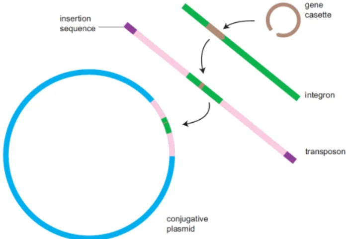
Mobile Genetic Elements (MGE) are identified as DNA sequences, with size ranging from one to thousands of base pairs, capable of recombining and transferring within the host cell or between genomes (Leplae et al., 2004). There are several types of MGE, some of which illustrated in Figure 1, such as plasmids, bacteriophages, viruses, genomic/pathogenicity islands and integron-associated gene cassettes and transposable elements (TE), including insertion sequences (IS) and transposons (Tn) (Leplae et al., 2004; Piotrowska and Popowska, 2015; Norman et al., 2009; Smillie et al., 2010). When collectively considered in the genome, these elements are called the mobilome (Piotrowska and Popowska, 2015).
An insertion sequence (IS) is the most basic transposable element, consisting only of short sequences of inverted repeats with a transposase gene in between them. Transposons are more complex, containing genes for the expression of different genotypes in addition to the transposase (Piotrowska and Popowska, 2015).
Genomic islands are chimeric genes or entire groups of genes (Leplae et al., 2004). Inte-grons are genomic elements incapable of replication, often associated with plasmids and transposons (becoming mobile integrons, since these entities stimulate their propagation), capable of capturing and expressing gene cassettes. These elements, thought to be the pri-mary way of obtaining antibiotic resistance, are composed of a gene coding for an integrase (intI), a specific recombination site (attI) and one or two promoters (Pc) controlling the expression of the captured gene (Moura et al., 2009; Piotrowska and Popowska, 2015).
The physical deslocation of DNA requires the intervention of four main enzymes, namely recombinases, which allow homologous recombination, transposases, enabling the movement and insertion of transposons, integrases, facilitating the insertion of elements into integrons and resolvases, resolving Holiday junctions resulting from recombination (Norman et al., 2009).
Figure 1.: Depiction of the interaction between some Mobile Genetic Elements, showing the elements necessary for a gene cassette to be acquired by a plasmid (Retrieved from Norman et al. (2009))
2.2. Mobile Genetic Elements 12
In order to identify their role and interactions in specific environmental niches, various prokaryotic-specific designations should be taken into consideration. On one hand, regard-ing their frequency, core genome describes the set of genes omnipresent in all strains of a given species, dispensable/ flexible genome respects genes present in some strains of a species, but not in all of them, and pan genome is the sum of the previous two. However, since all of these terms disregarded interaction of genetic elements between species, a new de-nomination, supergenome, was created by Norman et al. (2009), defined as the total pool of genes readily available to a prokaryotic organism within a specific environment. On the other hand, concerning their availability, private pool identifies genes that are encoded by the host chromosome (available only for the resident plasmid) and communal pool character-izes genes produced by mobile genetic elements (available to most prokaryotes) (Norman et al., 2009).
2.2.1 Horizontal Gene Transfer
Bacterial genomes can obtain new genes by horizontal gene transfer (HGT), also known as bacterial sex, allowing genetic variation. Horizontal gene transfer occurs either by introduc-tion via MGE or by direct uptake and incorporaintroduc-tion of DNA by recombinaintroduc-tion (Norman et al., 2009). This mechanism can lead to evolution and it is important from microbiology, ecology and pathogenically transfer perspectives, since it ensures plasmid persistence and it is responsible for the acquisition of genes encoding antibiotic resistance and virulence (Norman et al., 2009; Smillie et al., 2010).
The effectiveness and selective advantages of horizontal gene transfer depend on a mul-tiplicity of factors, such as the nature of the gene, their host restrictions and their co-inhabitant genetic elements (Norman et al., 2009; Smillie et al., 2010). Informational genes, such as those involved in transcription, translation and related processes usually belong to large molecular complexes, have a smaller probability of having been successfully trans-ferred horizontally than operational genes, that is, genes involved in amino acid biosynthe-sis and other housekeeping functions, which are generally compact operons (Norman et al., 2009).
There are two sets of genes involved in plasmid propagation: (i) the set of mobility (MOB) genes, allowing conjugative DNA processing, which is indispensable; and (ii) the mating pair formation (Mpf) complex (membrane-associated), which is a form of a type 4 secretion system (T4SS), providing the mating channel (Smillie et al., 2010). Plasmids can be classified, according to the incidence of these sets, as conjugative or self-transmissible, if the MOB set is present and they encode their own Mpf genes, as mobilizable, if they have the MOB set but rely on other genetic elements to obtain the Mpf genes, and as nonmobilizable if they fit in neither of those categories (Garcillan-Barcia et al., 2009; Norman et al., 2009;
2.2. Mobile Genetic Elements 13
Smillie et al., 2010). Conjugative plasmids, generally with a size over 30kb, are maintained at a low copy number per cell (typically less than ten copies per cell), while mobilizable plasmids, which are of a considerably smaller size (<15kb), can reach hundreds of copies per cell (Garcillan-Barcia et al., 2009; Norman et al., 2009).
Horizontal transfer can proceed by one of three manners: transformation, transduction and conjugation.
Conjugation can be defined as the combination of two functions, mating pair formation (Mpf) and RCR (rolling-circle replication), and it is composed of several steps. The first step is the formation (Mpf) of a channel connecting the donor to the recipient, generally through the synthesis of a type IV secretion system (T4SS), where the coupling is made by filaments named conjugation pili (the morphology of these filaments determines the conjugation host range and the medium of transfer). The second step is the formation of the relaxosome, which contains the single strand plasmid DNA, the relaxase (see subsection 2.2.2) and some other proteins. The final step is rolling-circle replication to synthesize a second strand in the donor and in the recipient (Norman et al., 2009). Conjugative plasmids are also capable of replicating by the theta-type mechanism during vegetative growth (Carattoli, 2009).
Transformation and transduction are likely to favor small plasmids and have a lower im-pact on the plasmid transfer rate, since they promote mainly intraspecies transfers, whereas conjugation can link remotely distant organisms (Norman et al., 2009; Smillie et al., 2010).
Transformation is the uptake of DNA into cells from the surrounding environment, depend-ing on the existence of plasmid and/or chromosomal DNA fragments in the environment, requiring only a recipient and a mechanism for the insertion of DNA (Norman et al., 2009; Smillie et al., 2010).
Transduction is the transportation of DNA through bacteriophages, dependent on a phage replicating within the donor and, during DNA packaging, DNA fragments can be incor-porated into the phage capsid (Norman et al., 2009; Smillie et al., 2010). This method is limited by the size of the phage (plasmids must have a genome with a size under or equal to it) (Smillie et al., 2010).
2.2. Mobile Genetic Elements 14
2.2.2 Plasmid Mobility Groups and Mating Pair Formation
The encoded transfer genes vary among different types of plasmids: while conjugative plasmids display the complete set of transfer genes, that is, MOB and a T4SS, mobilizable plasmids only include a simple MOB region, allowing them to be transported by other conjugative plasmids and merely consist of a vegetative origin of replication oriV, a relaxase protein and one or more nicking-accessory proteins. The MOB machinery only requires the presence of a relaxase, the protein responsible for the initiation and termination of conjugative DNA processing. These proteins are of small size and contain two or more protein domains, always including a domain at the N-terminus of the protein and at the C-terminus, with the latter generally including a DNA helicase and ligase or other domains. With some exceptions, replicases also contain a 3H (histidine triad) motif, which they use to bind divalent cations (Garcillan-Barcia et al., 2009). Mobilizable plasmids carry only the relaxosomal components (see subsection 2.2.1). Inversely, conjugative plasmids additionally include the type IV coupling protein (T4CP) – used to bind the relaxosome and the transport channel – and the components of the mating channel needed for T4SSs (Smillie et al., 2010).
Plasmids can be assigned into one of six MOB groups, namely MOBC, MOBF, MOBH, MOBP, MOBQ, and MOBV, depending on their amino acid sequence (Garcillan-Barcia et al., 2009; Shintani et al., 2015). This classification was able to organize each relaxase into a specific protein family, that is, a set of proteins sharing a biological function and similar in their sequence, for the exception of the MOBP group, still considered unfinished (Garcillan-Barcia et al., 2009; Smillie et al., 2010). Members of MOBF and MOBH are mostly found in large conjugative plasmids, while the other families are predominantly encountered in small mobilizable plasmids (Garcillan-Barcia et al., 2009).
Recently, Smillie et al. (2010) classified T4SS bacteria into four mating pair formation (Mpf) groups depending on their protein homology, and each one was named according to the archetype T4SS of the group: vir system for MPFT, F for MPFF, R64 for MPFI and ICEHIN1056 for MPFG.
As a concluding remark, it should be noted that these classification systems can be used to identify the mobilization potential of a plasmid, since a specific behavior accompanied by the existence or absence of certain elements (like the presence of a Mpf group in con-jugative plasmids), the similarity with known relaxases and the size of the plasmid (large for nonmobilizable, intermediate for conjugative and small for mobilizable) should lead to the recognition of its transfer system (Garcillan-Barcia et al., 2009; Smillie et al., 2010).
2.3. Plasmid Incompatibility 15
2.3 p l a s m i d i n c o m pat i b i l i t y
The term incompatibility was first used in the early 1960s for the F plasmid and, one decade later (1976), a formal classification scheme based on incompatibility was proposed by Novick (Carattoli, 2009; Couturier et al., 1988; Novick et al., 1976). Despite being a well accepted plasmid classification scheme, there are still some limitations, namely if the plas-mid is not transmissible or it does not contain a suitable marker gene. These difficulties are surpassed by using a set of reference miniplasmids belonging different Inc groups and having a gene for galactose utilization (Couturier et al., 1988).
Plasmid incompatibility is defined as the impossibility of two plasmids with the same replication origin (ori) and partition system to coexist stably in the same cell line or host without external selection (Carattoli, 2009; Novick et al., 1976; Novick, 1987; Shintani et al., 2015), that is, two plasmids are considered incompatible if the introduction of a second plas-mid leads to the elimination of the first (resident). Hence, incompatibility can be perceived as an evidence of relatedness between the plasmids that share the same replication controls (Couturier et al., 1988).
There are two types of incompatibility between two plasmids: symmetric and vectorial. The former implies that the possibility of losing any of the plasmids is equiprobable, whereas the latter postulates that one of the plasmids has a higher probability of being excluded than the other. These are, usually, used in different contexts: symmetric incom-patibility is commonly applicable to co-existing single replicons with similar replication and maintenance functions, while vectorial incompatibility is often associated with problems in replication (Novick, 1987).
In order to test and classify incompatibilities, the conventional method is to introduce, either by conjugation, transduction or transformation (see section 2.2), a plasmid into a strain which already carries another plasmid. If the resident plasmid is excluded, then the two plasmids belong to the same Inc group (Couturier et al., 1988). However, due to the fact that classification is based on the amino acid sequence of the replicon initiation (rep) protein (replicon typing), testing is not always necessary, since the same incompatibility group will share the same rep protein and should be assigned the same name (Shintani et al., 2015). Note that the assignment of incompatibility should take into account that the plasmids within the same host are able to mutually interact in vivo (Couturier et al., 1988).
Several factors influence plasmid incompatibility. The first is the origin of replication (ori), since two plasmids that rely on the same replication strategy cannot reside on the same cell. Another constraint is competition for replication factors, with the plasmids with selective advantages such as antibiotic resistance, less toxicity and faster replication (which should always be inversely proportional to the copy number) having a higher probability
2.3. Plasmid Incompatibility 16
of succeeding. The last factor is the copy number. On one hand, when two plasmids are compatible, each one produces a replication inhibitor that does not affect the other’s replication, maintaining their normal copy numbers and both are able to persist. On the other hand, if incompatible plasmids produce the inhibitor, the host cell cannot differentiate between the origins of replication and the replication of both will be affected: the total number of plasmid copies in the cell will be less than the sum of the total number of plasmid copy numbers (this happens because each plasmid adjusts its replication rate and copy number to the total inhibitor concentration). As such, replication will only be resumed when the pre-replication copy number is restored, that is, when one of the plasmids is no longer in the cell (Summers, 1996; Velappan et al., 2007).
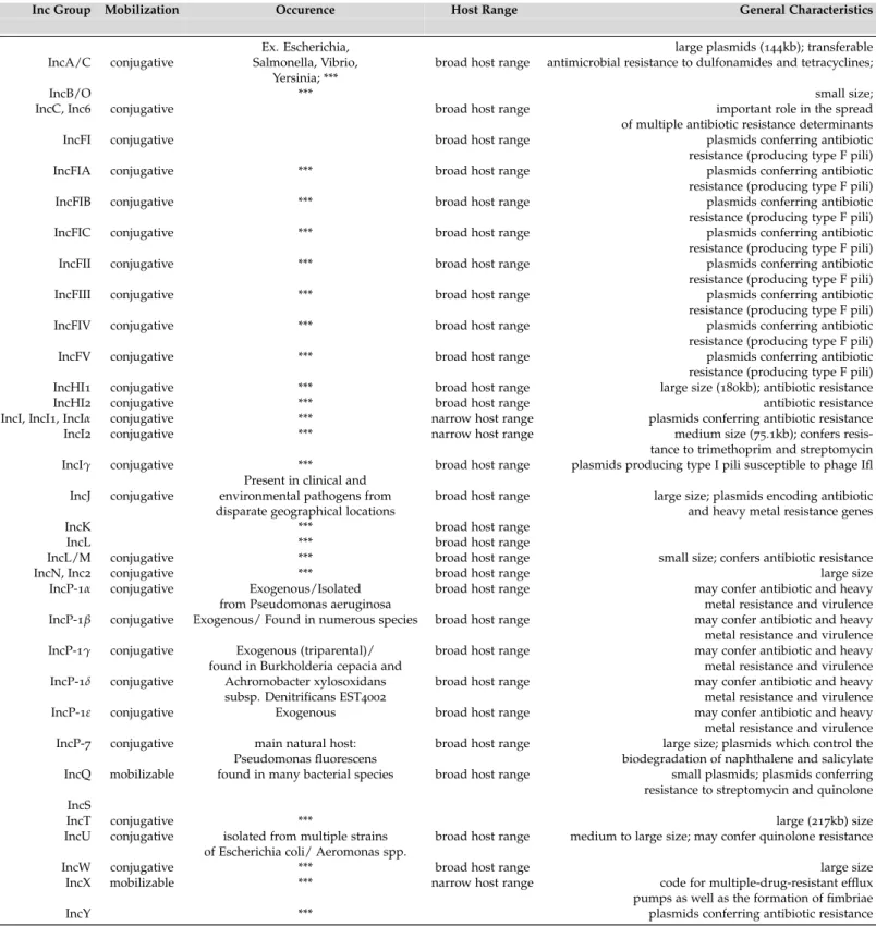
The assignment of a plasmid to a certain incompatibility group determines its hosts and interactions and possible opportunities (e.g. acquisition of new traits) (Izmalkova et al., 1987). Most of the presently known incompatibility groups are represented in Tables 2 and 3. This information was adapted, not only from the references listed in Table 3, but also from Carattoli (2009); Clark et al. (2016); Garcillan-Barcia et al. (2009); Norman et al. (2009) and Shintani et al. (2015).
2.3. Plasmid Incompatibility 17
Table 2.: Mobilization, occurrence, host range and general characteristics of known plasmid Incom-patibility groups. Note that the members of the Resistance Plasmid Families in Enterobacte-riaceae are marked as ***. The references on which this Table is based are listed in Table 3.
Inc Group Mobilization Occurence Host Range General Characteristics
Ex. Escherichia, large plasmids (144kb); transferable
IncA/C conjugative Salmonella, Vibrio, broad host range antimicrobial resistance to dulfonamides and tetracyclines;
Yersinia; ***
IncB/O *** small size;
IncC, Inc6 conjugative broad host range important role in the spread
of multiple antibiotic resistance determinants
IncFI conjugative broad host range plasmids conferring antibiotic
resistance (producing type F pili)
IncFIA conjugative *** broad host range plasmids conferring antibiotic
resistance (producing type F pili)
IncFIB conjugative *** broad host range plasmids conferring antibiotic
resistance (producing type F pili)
IncFIC conjugative *** broad host range plasmids conferring antibiotic
resistance (producing type F pili)
IncFII conjugative *** broad host range plasmids conferring antibiotic
resistance (producing type F pili)
IncFIII conjugative *** broad host range plasmids conferring antibiotic
resistance (producing type F pili)
IncFIV conjugative *** broad host range plasmids conferring antibiotic
resistance (producing type F pili)
IncFV conjugative *** broad host range plasmids conferring antibiotic
resistance (producing type F pili)
IncHI1 conjugative *** broad host range large size (180kb); antibiotic resistance
IncHI2 conjugative *** broad host range antibiotic resistance
IncI, IncI1, IncIα conjugative *** narrow host range plasmids conferring antibiotic resistance
IncI2 conjugative *** narrow host range medium size (75.1kb); confers
resis-tance to trimethoprim and streptomycin
IncIγ conjugative *** broad host range plasmids producing type I pili susceptible to phage Ifl
Present in clinical and
IncJ conjugative environmental pathogens from broad host range large size; plasmids encoding antibiotic
disparate geographical locations and heavy metal resistance genes
IncK *** broad host range
IncL *** broad host range
IncL/M conjugative *** broad host range small size; confers antibiotic resistance
IncN, Inc2 conjugative *** broad host range large size
IncP-1α conjugative Exogenous/Isolated broad host range may confer antibiotic and heavy
from Pseudomonas aeruginosa metal resistance and virulence
IncP-1β conjugative Exogenous/ Found in numerous species broad host range may confer antibiotic and heavy
metal resistance and virulence
IncP-1γ conjugative Exogenous (triparental)/ broad host range may confer antibiotic and heavy
found in Burkholderia cepacia and metal resistance and virulence
IncP-1δ conjugative Achromobacter xylosoxidans broad host range may confer antibiotic and heavy
subsp. Denitrificans EST4002 metal resistance and virulence
IncP-1ε conjugative Exogenous broad host range may confer antibiotic and heavy
metal resistance and virulence
IncP-7 conjugative main natural host: broad host range large size; plasmids which control the
Pseudomonas fluorescens biodegradation of naphthalene and salicylate
IncQ mobilizable found in many bacterial species broad host range small plasmids; plasmids conferring
resistance to streptomycin and quinolone IncS
IncT conjugative *** large (217kb) size
IncU conjugative isolated from multiple strains broad host range medium to large size; may confer quinolone resistance
of Escherichia coli/ Aeromonas spp.
IncW conjugative *** broad host range large size
IncX mobilizable *** narrow host range code for multiple-drug-resistant efflux
pumps as well as the formation of fimbriae
2.3. Plasmid Incompatibility 18
Table 3.: Archetype plasmid, GenBank number, replicon typing (amplicon size and target gene re-gion) and bibliography of known plasmid Incompatibility groups.
Inc Group Archetype plasmid GenBank Number Replicon typing Bibliography Amplicon size Target gene region
IncA/C pRA1 FJ705807 465bp repA region (plasmid replication protein
repA)
(Harmer and Hall, 2016; Novick et al., 1976)
IncB/O pMU720 M28718 159bp RNAI (miscRNA) (Novick et al., 1976)
IncC, Inc6 R4Oa, R55 (Harmer and Hall, 2016; Novick
et al., 1976)
IncFI F, R386 M11322 traA (conjugative transfer pilin protein
TraA) (Novick et al., 1976)
IncFIA pmk115 J01724 462bp iterons (Carattoli, 2005)
IncFIB unnamed M26308 702bp repA (plasmid replication protein RepA) (Carattoli, 2005)
IncFIC 262bp repA2 (plasmid replication protein A2) (Carattoli, 2005)
IncFII R100, R1 (Novick et al., 1976)
IncFIII ColB-K98 traT region (conjugative transfer surface
exclusion protein TraT) (Novick et al., 1976)
IncFIV R124
IncFV Folac (Novick et al., 1976)
IncHI1 R27 (=TP117) AF250878 471bp parA-parB (plasmid partition proteins) (Carattoli, 2005; Novick et al.,
1976)
IncHI2 R478 BX664015 644bp iterons (Carattoli, 2005; Gilmour et al.,
2004)
IncI, IncI1, IncIα ColIb-P9,∆, R144 M20413 139bp RNAI (miscRNA) (Carattoli, 2005; Novick et al.,
1976)
IncI2 R721 NC 002525 repA (plasmid replication protein RepA) (Komano, 1990; Novick et al., 1976)
IncIγ R621 NC 015965 (Novick et al., 1976)
ColIb-IM1420
IncJ R391 U13633 (McGrath et al., 2006; Novick et al.,
1976)
IncK M93063 160bp RNAI (miscRNA) (Carattoli, 2005; Novick et al.,
1976)
IncL R471a AF027768 (Novick et al., 1976)
IncL/M pMU407.1 U27345 785bp repA,B,C (plasmid replication proteins) (Carattoli, 2005)
IncN, Inc2 N3, R15 NC 003292 559bp repA (plasmid replication protein RepA) (Carattoli, 2005; Novick et al.,
1976)
IncP-1α RP4 AAA26427 281bp trfA region
(Bahl et al., 2009; Dealtry et al.,
2014; Popowska and
Krawczyk-Balska, 2013; Novick et al., 1976)
IncP-1β R751 U67194 282bp trfA region
(Bahl et al., 2009; Dealtry et al.,
2014; Popowska and
Krawczyk-Balska, 2013)
IncP-1γ pQKH54 AM157767 283bp trfA region
(Bahl et al., 2009; Dealtry et al.,
2014; Popowska and
Krawczyk-Balska, 2013)
IncP-1δ pEST4011 AY540995 284bp trfA region
(Bahl et al., 2009; Dealtry et al.,
2014; Popowska and
Krawczyk-Balska, 2013)
IncP-1ε p3-408 285bp trfA region
(Bahl et al., 2009; Dealtry et al.,
2014; Popowska and
Krawczyk-Balska, 2013)
IncP-7 pCAR1 AB088420 524bp rep region (Dealtry et al., 2014; Izmalkova
et al., 1987)
IncQ RSF1010 NC 001740 703bp strA and strB (steptomycin resistance
-streptomycin phosphotransferase A and B)
(Gotz et al., 1996; Pezzella et al.,
2004; Novick et al., 1976)
IncS R478 BX664015 (Novick et al., 1976)
IncT Rts1 K00053 750bp repA (plasmid replication protein RepA) (Novick et al., 1976)
IncU RA3 DQ401103 250bp repAp, repB, repB1p/repB2p, repB2p, repA
(plasmid replication proteins)
(Garcillan-Barcia et al., 2009; Carattoli, 2005)
IncW R388 U12441 242bp repA (plasmid replication protein RepA) (Carattoli, 2005)
IncX R6K Y00768 376bp ori y (protein coding) (Garcillan-Barcia et al., 2009;
Carattoli, 2005)
2.4. Plasmids at the NCBI Database 19
2.4 p l a s m i d s at t h e n c b i d ata b a s e
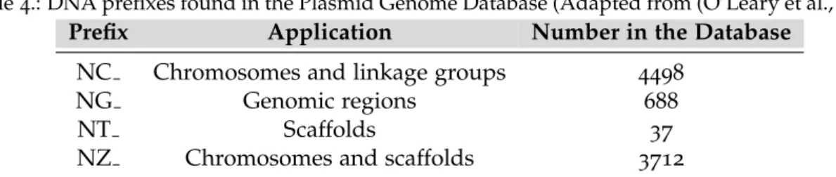
In January of 2017, there were 8935 plasmid reference sequences (RefSeq) available in the NCBI Plasmid Genome Database (ftp://ftp.ncbi.nlm.nih.gov/genomes/refseq/plasmid/), from which 4498 are of high importance, since they are complete (closed) sequences (see Ta-ble 4) (O’Leary et al., 2015).
The number of complete sequences, however, has suffered considerable fluctuations throughout the years, with many entries added and some removed. Between January of 2009 and August of 2014, this number has exponentially grown from 1600-1700 (Smillie et al., 2010; Norman et al., 2009) to 4602 (Shintani et al., 2015). Finally, since March of 2016 (4514 sequences, our study), 16 sequences have been uploaded to the database and 32 have been excluded due to missing RNA genes, deriving from environmental sources, length of the genome being too large or too small, having many frameshifted proteins, being from a mixed culture or reasons not stated (Clark et al., 2016).
Table 4.: DNA prefixes found in the Plasmid Genome Database (Adapted from (O’Leary et al., 2015))
Prefix Application Number in the Database
NC Chromosomes and linkage groups 4498
NG Genomic regions 688
NT Scaffolds 37
NZ Chromosomes and scaffolds 3712
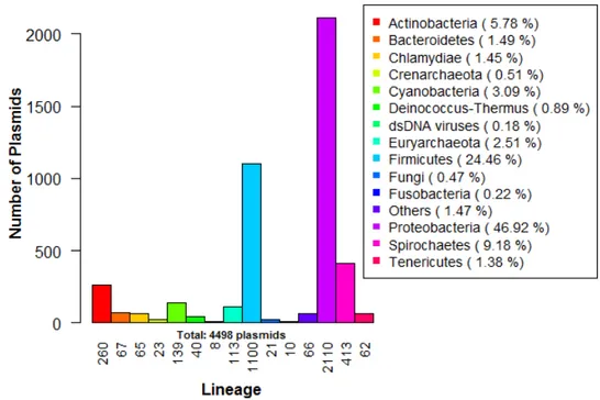
The fully sequenced plasmids present in the database are quite discrepant in terms of type, lineage, size and GC content. From these, 139 are from Archaea, 4312 from Bacteria, 38 from Eukaryota, 8 from viruses and, finally, one last sequence from ”other sequences plas-mids”. From the identified phyla, the most abundant are Proteobacteria (2110 hits, 46.92% of the sequences), Firmicutes (1100 hits, 24.46%) and Spirochaetes (413, 9.18%), with numbers much higher than the remaining cases (see Figure 2). The majority of the plasmids is circular (4028), their GC content ranges from, approximately, 0.1926 (pMR2 plasmid in Monilioph-thora roreri) to 0.7562 (pFQ12 in Frankia sp. CpI1), with an average of 0.4409 and their size has a mean of 29253.94 base pairs, varying from 21 bp (pSE11-1 in Escherichia coli SE11) to 99806bp (pTpau01 in Tsukamurella paurometabola DSM 20162).
Still, not all the information is reachable. Only under 17% of the entries have data about the isolation source (e.g. soil, food, veterinary, human) of the plasmid and 20% about their geography, over 88% of their hosts are unidentified, 92.9% of the collection dates are unknown and merely 1.6% of their sources (environmental or clinical) are identified, which forces the user to search for this information in the respective publications (which are still
2.5. Available Databases and Programs for Mobile Genetic Elements 20
not a guarantee of finding the intended data). Hence, one of the focuses of this thesis is to complete the missing information (regarding bacterial plasmids), so that it becomes easily accessible to any individual.
Figure 2.: Lineage distribution of the complete plasmid sequences in the NCBI Plasmid Genome Database. Note: the group ”Others” includes ”bacterial environmental samples” and several phyla, such as Acidobacteria, Amoebozoa, Aquificae, Chlorobi, Chloroflexi, Deferribac-teres, Elusimicrobia, Nitrospirae, Planctomycetes, Rhodophyta, Synergistetes, Thermotogae and Viridiplantae.
2.5 ava i l a b l e d ata b a s e s a n d p r o g r a m s f o r m o b i l e g e n e t i c e l e m e n t s
To keep up with the fast-paced growing number of mobile genetic elements, several databa-ses and web tools were created to filter and analyze important data (Carattoli et al., 2014; Curry et al., 2016; Leplae et al., 2004; Moura et al., 2009; Siguier et al., 2014; Smillie et al., 2010).
Most of these tools are, though, available for a short period of time and they are updated at a much slower rate than GenBank (Smillie et al., 2010). One example is ACLAME, a database which, although showing a great potential, has not been updated since 2013. This database was built with the purpose of collecting and classifying all the existing plasmids, transposons and phage genomes.
Regarding integrons, two main tools are known: (i) the INTEGRALL database , still up-dated on a regular basis (see Table 5), which has the purpose of gathering information about
2.5. Available Databases and Programs for Mobile Genetic Elements 21
DNA sequences of integrons on a single repository and (ii) the Integron Finder program, which searches for integrons in DNA sequences in FASTA formats, returning the input se-quence with all integrons and features found, a list of all the elements detected and the representation of the discovered complete integrons.
As for miscellaneous content, two databases are of relevance: ISfinder is designed for the identification of Insertion Sequences and ICEberg has the goal of assembling information about Integrative and Conjugative Elements present in bacteria.
The PlasmidFinder database allows the in silico identification of plasmids in complete sequences and translates them to an Inc group-based classification. However, this is a restricted tool, since it is limited to the identification of plasmids in the Enterobacteriaceae family. The pMLST web tool is capable of performing a Plasmid Multilocus Sequence Typing analysis for five incompatibility groups (both PlasmidFinder and pMLST rely on the BLASTn algorithm for the homology searches).
Other currently unavailable/out-of-date databases worthy of mention are: the Plasmid Genome Database (unavailable), which was intended to be a repository for complete plas-mids, harboring information about their core features, genetic composition and structural maps (Molbak, 2003); and the Database of Completely Sequenced, Naturally Occurring Plas-mids (out-of-date since 2000, available at http://www.biochem.ucl.ac.uk/bsm/PLASMID/ mainpage.htm) comprises information about a very limited number of plasmids (7 archaeal, 157 bacterial and 27 others), concerning replication regions, transfer or mobility regions, bacteriocin and antibiotic resistance coding regions and other functions.
All the websites and references for the still existing web tools just mentioned are listed in Table 5.
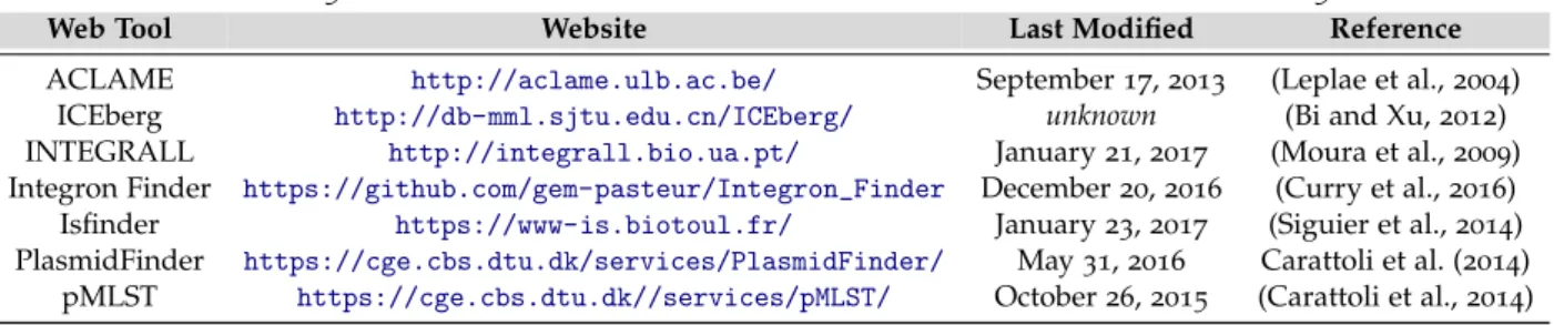
Table 5.: Websites and references for the web tools described in section 2.5
Web Tool Website Last Modified Reference
ACLAME http://aclame.ulb.ac.be/ September 17, 2013 (Leplae et al., 2004) ICEberg http://db-mml.sjtu.edu.cn/ICEberg/ unknown (Bi and Xu, 2012) INTEGRALL http://integrall.bio.ua.pt/ January 21, 2017 (Moura et al., 2009) Integron Finder https://github.com/gem-pasteur/Integron_Finder December 20, 2016 (Curry et al., 2016) Isfinder https://www-is.biotoul.fr/ January 23, 2017 (Siguier et al., 2014) PlasmidFinder https://cge.cbs.dtu.dk/services/PlasmidFinder/ May 31, 2016 Carattoli et al. (2014)
3
D E V E L O P M E N T
In this chapter, the stages of the development of this thesis are described. Namely, the information used to create and populate the database and the adopted approach for the classification into Incompatibility and MOB groups and in terms of Putative Transferability are presented. In the last section, the features and functionality of the created Shiny appli-cation – named PlasmidClassifier: The Bacterial Plasmids Database – and Classifiappli-cation tool are characterized, the used functions from the shiny package are explained and various prints are shown.
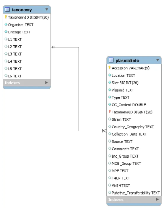
3.1 c o n s t r u c t i o n a n d s t r u c t u r e o f t h e p l a s m i d d ata b a s e
In order to construct the plasmid database, MySQL, an open-source relational database management system (RDBMS), was used. The database was firstly designed using the Enhanced Entity-Relationship (EER) diagram shown in Figure 3.
The plasmid database, as illustrated in the diagram, is organized in two tables:
• plasmidinfo: this is the main table, containing information about the plasmids’ acces-sion number, location, size, name, type (circular or linear), GC content of its genomic sequence, taxonomy id, strain, geographical data (the country and/or region in which the plasmid was found), collection date (year of isolation), isolation source and any additional information (if the plasmid is found in extreme conditions or if it is a symbiont, for example). Their Inc and MOB groups (see subsection 3.2.3) are also included. For the MOB groups, the type of Mating Pair Formation (Mpf), Type 4 Coupling Protein (T4CP), VirB4 and their putative transferability, that is, if the plas-mid is conjugative, mobilizable or non-mobilizable (see section 2.2 for details) are also comprised.
• taxonomy: linked to the plasmidinfo table by the plasmid’s taxonomy id, that is, an identification number which, when used in NCBI’s Taxonomy database (available at
https://www.ncbi.nlm.nih.gov/taxonomy), returns the classification and
3.1. Construction and Structure of the Plasmid Database 23
ture of the plasmid. This table includes the organism and its lineage (columns L1 to L6 refer to the detailed information).
Figure 3.: Enhanced Entity-Relationship (EER) diagram depicting the structure of the database. The two tables have a multiplicity relationship of N:1, since a plasmid can only have one Taxonomy ID, but the same Taxonomy ID can be present in multiple plasmids: the pIL105 (NC 000906) and pMRC01 (NC 001949) plasmids, for example, share the Taxonomy ID 1358, which means that they were both found in the Streptococcus lactis species.
3.2. Populating the Database 24
3.2 p o p u l at i n g t h e d ata b a s e
3.2.1 Obtaining the Plasmid Files
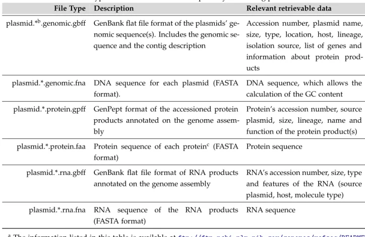
One of the purposes of this thesis, as mentioned throughout the previous sections, was to provide a database gathering as much information about bacterial plasmids as possible. To achieve this goal, genome files containing information about plasmid reference sequences (RefSeq) were downloaded from the NCBI FTP repository (ftp://ftp.ncbi.nlm.nih.gov/ genomes/refseq/plasmid/), which contains several file types (see Table 6).
Table 6.: File types found at the NCBI FTP repository concerning plasmid dataa.
File Type Description Relevant retrievable data
plasmid.*b.genomic.gbff GenBank flat file format of the plasmids’
ge-nomic sequence(s). Includes the gege-nomic se-quence and the contig description
Accession number, plasmid name, size, type, location, host, lineage, isolation source, list of genes and information about protein prod-ucts
plasmid.*.genomic.fna DNA sequence for each plasmid (FASTA format).
DNA sequence, which allows the calculation of the GC content plasmid.*.protein.gpff GenPept format of the accessioned protein
products annotated on the genome assem-bly
Protein’s accession number, source plasmid, size, lineage, name and function of the protein product(s) plasmid.*.protein.faa Protein sequence of each proteinc (FASTA
format)
Protein sequence
plasmid.*.rna.gbff GenBank flat file format of RNA products annotated on the genome assembly
RNA’s accession number, size, type and features of the RNA (source plasmid, host, molecule type) plasmid.*.rna.fna RNA sequence of the RNA products
(FASTA format)
RNA sequence
aThe information listed in this table is available atftp://ftp.ncbi.nlm.nih.gov/genomes/refseq/README. txt.
bThe symbol * corresponds to the number of the file. cNo reference to which plasmid it belongs.
From the file types listed in Table 6, only the .genomic.gbff and .genomic.fna files were used. These compiled files (containing information about multiple plasmids) were then separated into 8935 files, each one containing the GenBank information or DNA sequence, respectively, of a single plasmid, corresponding to a specific accession number (used to name each record). From these, only the files corresponding to complete genomic molecules