ANTITUMOR ACTIVITY OF XANTHONE DERIVATIVES: EFFECTS
ON THE IMMUNE MICROENVIRONMENT
Dissertação de Candidatura ao grau de Mestre em
Oncologia
(Especialização
em
Oncologia
Molecular)
submetida ao Instituto de Ciências Biomédicas Abel Salazar
da Universidade do Porto.
Orientador:
Doutor Rui Medeiros
Professor Associado Convidado de Ciências Médicas
Instituto de Ciências Biomédicas Abel Salazar, Universidade
do Porto, Porto, Portugal.
Co-orientadores:
Doutora Fátima Cerqueira
Professora Associada de Ciências Médicas
Faculdade de Ciências da Saúde, Universidade Fernando
Pessoa, Porto, Portugal.
Doutora Madalena M.M. Pinto
Professora
Catedrática
em
Química
Orgânica
e
Farmacêutica
Faculdade de Farmácia, Universidade do Porto, Porto,
Portugal
This work was developed in the Center of Investigation in Biomedicine of Fernando Pessoa – Energy, environment and health research unit (CEBIMED of FP-ENAS), in Laboratório de Química Orgânica e Farmacêutica, Departamento de Ciências Químicas, Faculdade de Farmácia da Universidade do Porto, and in the Centro de Química Medicinal da Universidade do Porto – CEQUIMED-UP and was funded through national funds from Fernando Pessoa Fundation, FCT – Fundação para a Ciência e a Tecnologia under the project CEQUIMED – PEst-OE/SAU/UI4040/2014, FEDER, POCI, POPH/FSE/QREN.
Não posso deixar de agradecer a quem direta ou indiretamente colaborou para a realização deste trabalho e que, assim, contribuiu para o meu crescimento tanto profissional como pessoal:
Ao Instituto de Ciências Biomédicas Abel Salazar, na pessoa do Digníssimo Diretor Professor Doutor António Sousa Pereira, e à coordenação do mestrado de Oncologia, na pessoa da Professora Doutora Berta Silva, pela oportunidade de aprofundar os meus conhecimentos ao longo destes dois anos de mestrado.
À Universidade Fernando Pessoa, na pessoa do Digníssimo Reitor Professor Doutor Salvato Trigo, pelas condições de trabalho que me proporcionou.
Ao Professor Doutor Rui Medeiros, agradeço a orientação e a oportunidade que me proporcionou, a simpatia com que sempre me recebeu e todos os conhecimentos que me transmitiu.
À Professora Doutora Fátima Cerqueira, minha coorientadora, o meu sincero agradecimento pelo apoio constante, pela disponibilidade, pela amabilidade e pelos indispensáveis conselhos que forma determinantes na realização deste trabalho.
À Professora Doutora Madalena Pinto, pela coorientação, porque me permitiu integrar este projeto, obrigada por toda a sabedoria que me transmitiu e pela enorme simpatia.
Aos meus colegas de trabalho, nomeadamente à Jani Silva, Nair Campos, Liliana e Carla Gabriel pelos conselhos, pelo apoio e principalmente pelos bons momentos.
Aos restantes membros do FP-ENAS/CEBIMED, aos professores, técnicos e auxiliares o meu muito obrigado pela simpatia e pela disponibilidade.
À Mariana Santos, porque a tua amizade é indispensável, assim como os teus conselhos, obrigada por toda a ajuda e por, apesar de seres a que está mais longe, estares sempre presente.
Ao Raphael Costa, pela enorme paciência, por me ajudares em tudo que podes, por estares ao meu lado em todos os momentos e porque o teu amor me ajudou a superar os piores momentos.
Aos meus pais, porque as palavras não são suficientes para vos agradecer por tudo que fizeram e fazem por mim, por serem um exemplo de humildade e determinação, porque sempre me apoiaram incondicionalmente e porque me dão força para nunca desistir. Agradeço do fundo do coração todo o esforço que fazem diariamente para me proporcionarem uma vida melhor.
Nomenclature ... i
Resumo ... iii
Abstract ... v
I. General Introduction ...1
Chemistry of xanthones: General considerations ...2
Biological activities of xanthones ...3
Melanoma ...5
Immune System ...6
Tumor immunology ...11
References ...14
II. State of art ...23
Xanthones as potential agents in melanoma treatment ...24
Importance of immunomodulation in melanoma treatment ...25
References ...27
III. Objectives and outline ...29
IV. Materials and Methods ...31
Chemicals and reagents ...32
Xanthones ...32
Cell lines ...32
Cell growth assay ...33
Antitumor effect of conditioned macrophages culture medium ...34
NO production assay ...34
NO scavenging assay ...35
Human mononuclear cells MTT-proliferation assay ...35
Cytokine quantification ...35
MTT-viability assay ...36
Statistical analysis ...36
V. Natural Xanthones: alpha-mangostin ...39 Paper 1 (submitted to Fitoterapia in 29-09-2014): Mangosteen extract: “angel or demon”? The role of xanthones of mangosteen in potential adverse effects...40
Paper 2 (draft): “Alpha-mangostin antitumor activity: cytotoxicity and influence on the immune system microenvironment” ...69
VI. Synthetic xanthone: 1,2-dihydroxyxanthone ...89 Paper 3 (draft): “1,2-dihydroxyxanthone antitumor activity: cytotoxicity and influence on the immune system microenvironment” ...90
Figure 1 Xanthone basic skeleton ...2
Figure 2: Structure of the main xanthones from mangosteen...3
Figure 3: Polarization of macrophages and corresponding functions. Legend: GC indicate glucocorticoid; IC, immune complex; IL-1ra, IL-1 receptor antagonist; LPS, lipopolysaccharide; MR, mannose receptor; SR, scavenger receptor - adapted from (Chanmee et al. 2014). ...8
Figure 4 Cytokine network in immune system – adapted from (Zhang and An 2007) ..10
Figure 5: Schematic presentation of the role of immune system in cancer. Legend: APC indicates antigen -presenting cell; CTL, cytotoxic T lymphocyte or CD8+ T cell; NK, natural killer cell; Th, T helper cell; Treg, regulatory T cell and TAA, tumor-associated antigens - adapted from (Lakshmi Narendra et al. 2013)...13
Figure 6 Xanthone basic skeleton ...44
Figure 7 Structure of the main xanthones from mangosteen ...45
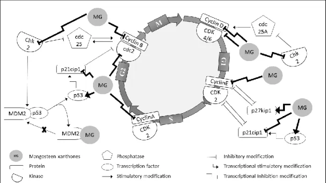
Figure 8 Effect of xanthones of mangosteen at different stages of cell cycle regulation. Legend: cdc indicate cell division cycle protein; CDK, cyclin-dependent kinases; Chk or CHEK2, checkpoint kinase; G1, gap phase; G2, gap phase 2; M, mitosis phase; MDM2, murine doble minute 2; MG, mangosteen xanthones; p21cip1, cyclin-dependent kinase inhibitor 1; p27kip1, cyclin-dependent kinase inhibitor; p53, tumor protein p53; S, DNA synthesis phase. ...49
Figure 9: alpha-Mangostin ...71
Figure 10: Production of IL-1β, IL-10, TGF-β1 and TNF-α by THP-1 macrophages. Cytokine production was evaluated on unstimulated macrophages (basal), LPS-stimulated macrophages and macrophages treated with 3 and 6 µM of alpha-mangostin. Data are the mean ± SEM from tree independent experiments performed in duplicate. * p < 0.001; ┼ p > 0.05. ...76
mononuclear cells. Cytokines production was evaluated on unstimulated lymphocutes (basal), PHA-stimulated lymphocytes and lymphocytes treated with 5, 10 and 20 µM of α-mangostin. Data are the mean ± SEM from tree independent experiments, performed in duplicate. * p < 0.001. ...77
Figure 12: Antitumor effect of mangostin, macrophages supernatants and alpha-mangostin conditioned macrophage culture medium on A375-C5 melanoma cell line. THP-1 PMA-differentiated macrophages were treated with xanthone and supernatants were added to melanoma cells. Results show mean values ± SEM (n = 3). * p < 0.001 ...78
Figure 13: 1,2-Dihydroxyxanthone ...91
Figure 14: Cytotoxic activity of 1,2-Dihydroyixanthone, macrophages supernatants and 1,2-dihydroxyxanthone conditioned macrophages supernatants on A365-C5 melanoma cell line.THP-1 PHA-differentiated macrophages were treated with the compound and supernatants were added to melanoma cells. Results show mean values ± SEM (n = 3). * p < 0.001 ...95
Figure 15: Effect of 1,2-DHX on IL-1β, IL-10, TGF-β1 and TNF-α production by THP-1 macrophages. Cytokines production was evaluated on unstimulates macrophages (basal), LPS-stimulated macrophages (positive control) and macrophages treated with 6 and 3 µM 1,2-dihydroxyxanthone. Data are the mean ± SEM from one experiment, performed with duplicate cultures, and it representative of tree experiments carried out independently. * p < 0.001; ┼ p > 0.05 ...96
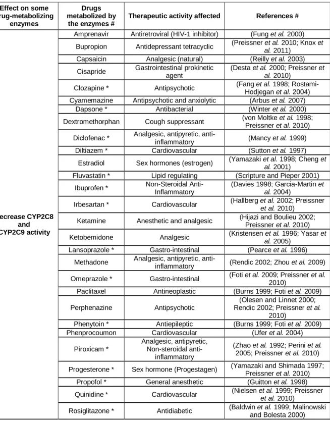
Table 1. Potential effects of xanthones derivatives on drugs metabolism. ...52
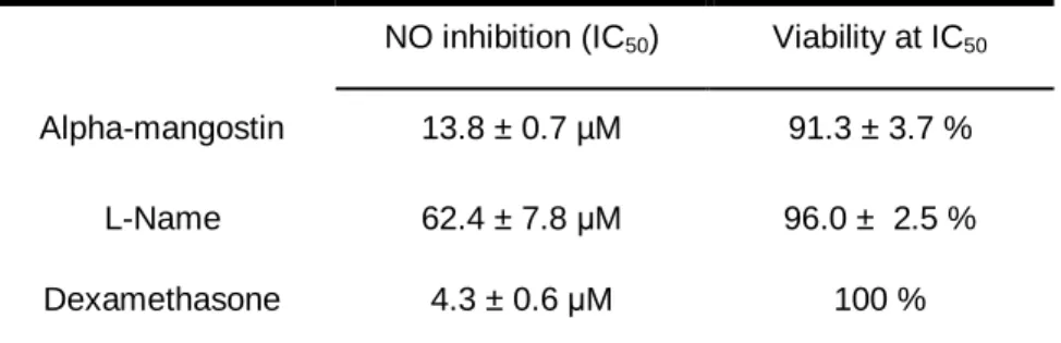
Table 2: Effect of α-MG on NO production by LPS-stimulated RAW 264.7 macrophages. ...74
Table 3: Inhibitory effect of α-MG on NO production by RAW264.7. ...75
Table 4: Effect of α-MG on the PHA-induced proliferation on human lymphocytes...76
Table 5: Effects of α-mangostin on the growth of A375-C5 human melanoma cell line. ...77
Table 6: Effect of 1,2-dihydroxyxanthone on the growth of A375-C5 human melanoma cell line...95
Table 7: Effect of 1,2-DHX on NO production by LPS-stimulated RAW 264.7 macrophages ...97
i 1,2-DHX: 1,2-Dihydroxyxanthone
AP-1: activator protein 1 APC: antigen-presenting cell
cdc2: cell division cycle protein 2 homolog CDK: cyclin-dependent kinases
CHEK2 – checkpoint kinase 2
CTL or CD8+ T: cytotoxic T cell CYP: cytochrome P450;
DMF: N.N-Dimetilformamida DMSO: Dimethyl sulfoxide
DMXAA or Vadimezan: 5,6-dimethylxanthenone-4-acetic acid; ELISA: Enzyme-Linked Immunosorbent Assay
NOS: nitric oxide synthases FBS: fetal bovine serum
GI50: half maximal growth-inhibitory concentrations GST: glutathione-S-transferase
IC50: half maximal inhibitory concentration IL: interleukin;
INF-γ: interferon gamma
L-NAME: N-nitro-L-arginine methyl ester LPS: lipopolysaccharides
M1: classic polarization of macrophages M2: alternative polarization of macrophages; MAPK: mitogen-activated protein kinase MDM2: murine doble minute 2
ii
MTT: 3-(4,5-Dimethylthiazol-2-yl)-2,5-Diphenyltetrazolium Bromide NF-κB: nuclear factor kappa B
NK: natural killer cell NO: nitric oxide
p21cip1: cyclin-dependent kinase inhibitor 1 p27: cyclin-dependent kinase inhibitor p53: tumor protein p53
PBS: Phosphate buffered saline PHA: phytohemaglutinin
PMA: phorbol-12-myristate-13-acetate; SEM: standard error of the mean SRB: sulphorrodamine B
TAM: tumor-associated macrophages TGF: transforming growth factor Th: T helper cell
TNF-α: tumour necrosis factor α Treg: regulatory T cell
iii
O cancro é uma das patologias mais agressivas e letais, permanecendo um desafio para investigadores e médicos, mesmo após décadas de investigação e investimento nesta área. A deteção tardia e as limitações das terapias atuais, associadas a quimio-resistência e efeitos adversos, são os principais problemas.
O melanoma é um tumor melanocítico altamente imunogénico e com elevada predisposição para metastizar. A sua incidência tem vindo a aumentar em todo o mundo e as taxas de mortalidade associadas a esta doença são muito elevadas. Deste modo, é necessário estabelecer terapias alternativas que superem as atuais limitações do tratamento, nomeadamente aumentando o espectro e a eficácia e diminuindo a toxicidade dos medicamentos usados atualmente. Uma boa estratégia para evitar a resistência tumoral exige moléculas com múltiplos alvos.
As xantonas são naturalmente obtidas em plantas de grande porte e também em microrganismos. Notáveis propriedades farmacêuticas foram descritas para estes compostos, principalmente o seu potencial anti-tumoral, que contribuiu para a comercialização de suplementos alimentares, para o isolamento e para a síntese de numerosos derivados xantónicos. No cancro, o efeito promovido por estes, compromete várias vias fisiológicas, interferindo com múltiplas características do cancro, incluindo a apoptose, ciclo celular, angiogénese, a inflamação e a vigilância imunitária, tornando-os um potencial candidato a fármaco.
O objetivo principal deste estudo foi avaliar os efeitos de duas xantonas, alfa-mangostina e 1,2-dihidroxixantona, no crescimento da linha celular humana de melanoma A375-C5 e a sua capacidade para modular o micro-ambiente imunológico dependente de macrófagos. Foi avaliada a capacidade dos compostos para interagir com os diferentes parâmetros imunológicos que podem interferir com o tratamento de tumores, incluindo a produção de óxido nítrico e a expressão de citocinas, como a interleucina1 beta e o fator de necrose tumoral alfa (características do fenótipo M1 em macrófagos) e a interleucina 10 e o fator transformador do crescimento beta 1 (características do fenótipo M2). A alfa-mangostina também foi testada em linfócitos, isto é, na proliferação celular e produção de citoquinas. Esta atividade foi previamente descrita para a 1,2-dihidroxixantona. Em todos os ensaios, foram usados sistemas celulares in vitro, maioritariamente de origem humana.
As xantonas causaram uma diminuição na produção de óxido nítrico e interferiram com a expressão de citocinas por macrófagos e / ou linfócitos estimulados. A alfa-mangostina inibiu a expressão da interleucina 1 beta e do fator transformador do crescimento beta 1, e estimulou o fator de necrose tumoral alfa. A 1,2-dihidroxixantona
iv
crescimento beta 1 e o fator de necrose tumoral alfa em macrófagos. A alfa-mangostina também interferiu com a proliferação de linfócitos e a expressão da interleucina 10 e do fator de necrose tumoral alfa por estas células.
Quanto ao impacto direto dessa modulação no crescimento de melanoma, os compostos apresentam uma melhoria de efeito citotóxico. No entanto, só no tratamento com 1,2-dihidroxixantona, esta melhoria pode ser associada com a modulação de macrófagos pela xantona, uma vez que o efeito da alfa-mangostina em doses baixas é semelhante aos macrófagos não tratados na linha celular em estudo.
Em conclusão, estes resultados permitiram inferir um impacto potencial da alfa-mangostina e da 1,2-dihidroxixantona no tratamento do melanoma, devido à sua atividade citotóxica e ao sugestivo efeito imunoterapêutico em tumores.
v
Cancer is one of the most aggressive and lethal diseases, remaining a challenge for researchers and physicians, in spite of decades of research and investment in this area. Later detection and constrains of current therapies, associated with chemoresistance and toxic side effects, are the main problems.
Melanoma is a melanocytic tumor highly immunogenic and with a great predisposition to metastasize. Its incidence is increasing worldwide and the mortality rates associated to the disease are very high. Therefore, it is imperative to find therapeutic alternatives that overcome the actual limitations of the treatment, namely improving the spectrum and efficacy of actuation and decrease the toxicity of currently used drugs. A good strategy to avoid the tumor resistance needs a drug that targets multiple pathways.
Xanthones are naturally obtained from higher plants and microorganisms. Remarkable pharmacologic proprieties have been described for these compounds, mainly their antitumor potential, which contributed to their commercialization as dietary supplement, isolation and synthesis of numerous derivatives of xanthones. In cancer, their effect comprises several physiologic pathways, interfering with multiple hallmarks of cancer, including apoptosis, cell cycle, angiogenesis, inflammation and immune surveillance making them a prospective drug candidate.
The main gold of this study was the evaluation of the effects of two xanthones, alpha-mangostin and 1,2-dihydroxyxanthone, on A375-C5 melanoma cell growth and their capacity to modulate the immune macrophages-dependent microenvironment. The ability for the compounds to interact with different immunologic parameters that may interfere with tumor treatment were evaluated, including production of nitric oxide and expression of cytokines interleukin-1β and tumor necrosis factor-α (characteristics of M1 phenotype) and interleukin-10 and transforming growth factor-β1 (characteristic of M2 phenotype). alpha-Mangostin was also tested in lymphocytes, namely in cell proliferation and cytokines production. This activity was previously described for 1,2-dihydroxyxanthone. In all assays, in vitro cellular systems were used, mostly of human origin.
Xanthones caused a decrease in nitric oxide production and interfere with the expression of cytokines by stimulated macrophages and/or lymphocytes. Alpha-mangostin suppressed expression of interleukin-1β and transforming growth factor-β1, and stimulated tumor necrosis factor-α. 1,2-Dihydroxyxanthone inhibited interleukin-1β and interleukin-10, stimulated transforming growth factor-β1 and tumor necrosis factor-α in macrophages. Alpha-mangostin also interferes with lymphocytes proliferation and the expression of interleukin-10 and tumor necrosis factor-α by these cells. Concerning the direct impact of this modulation on melanoma growth, the compounds exhibit an
vi
improvement could be associated with modulation of macrophages, since the effect of alpha-mangostin at lower doses is similar to non-treated macrophages in the cell line in study.
In conclusion, these finds allowed to infer a prospective impact of alpha-mangostin and 1,2-dihydroxyxanthone in melanoma treatment, due to their cytotoxic activity and suggestive effect in cancer immunotherapy.
I.
General Introduction
Xanthones, melanoma and immune system2 Chemistry of xanthones: General considerations
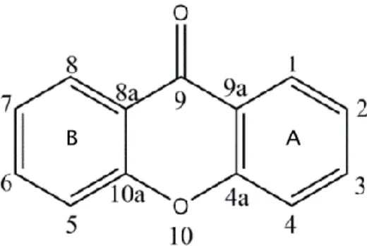
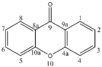
Xanthones are biologically active tricyclic molecules characterized by a dibenzo-γ-pyrone nucleus or 9H-xanthen-9-one (Figure 1) (Pinto et al. 2005; Mazimba et al. 2013).
The diversity of xanthone derivatives is possible due to the variation of the nature and position of substituents on the A and B rings. According to that, natural xanthones may be categorized into: simple oxygenated, glycosylated, prenylated and their derivatives (xanthone dimers, xanthonolignoids, and miscellaneous). On the other hand, the synthetic xanthones can have simple groups such as hydroxyl, methoxyl, methyl, carboxyl, as well as more complex substituents such as epoxide, azole, methylidenebutyrolactone, aminoalcohol, sulfamoyl, methylthiocarboxylic acid, and dihydropyridine in their scaffold (Pinto et al. 2005).
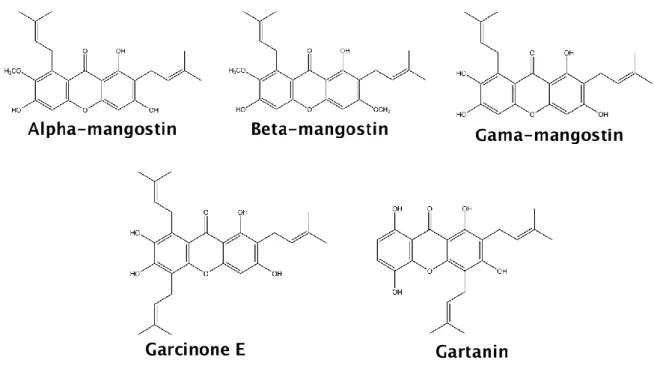
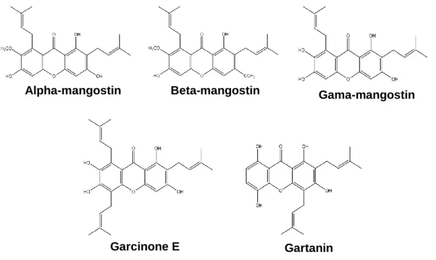
Natural xanthones of higher plants mainly occur in two families, Guttiferae and Gentianaceae, and can also be found in microorganisms as fungi and lichens (Vieira and Kijjoa 2005; Mazimba et al. 2013). The majority of these compounds was obtained from Garcinia mangostana Linn, being the most abundant and frequently studied, the α-mangostin, β-α-mangostin, γ-α-mangostin, garcinone E and gartanin (Figure 2) (Shan et al. 2011).
3
Xanthones have shown remarkable biological/pharmacological activities linked with their tricyclic scaffold, depending on the nature and/or position of the diverse constituents (Mazimba et al. 2013). As xanthones from natural origin are quite limited in type and position of the substituents due to the biosynthetic pathways, the syntheses of new xanthones can attempt to alter or improve their activity by having different nature and positions of the substituents on the nucleus of these compounds (Pedro et al. 2002).
Biological activities of xanthones
In the last decade, the interest for natural or derivative xanthones has been growing as readily confirmed by the increased numbers of scientific reports (Gutierrez-Orozco and Failla 2013). These have allowed to find a great variety of biological/pharmacological activities associated with xanthone derivatives including analgesic (Bianco et al. 1989; Garrido et al. 2001; Cui et al. 2010), antioxidant (Madan et al. 2002; Jung et al. 2006), anti-inflammatory (Lin et al. 1996; Madan et al. 2002; Teixeira et al. 2005; Chen et al. 2008), antitumor (Pinto et al. 2005; Pedraza-Chaverri et al. 2008; Shan et al. 2011; Gutierrez-Orozco and Failla 2013), antiallergic (Pfister et al. 1972; Nakatani et al. 2002), antimicrobial (Pinto et al. 2005; Pedraza-Chaverri et al. 2008), neuroprotective (Li and Ohizumi 2004; Weecharangsan et al. 2006) and immunomodulatory (Makare et al. 2001; Tang et al. 2009).
In spite of all promising proprieties in the improvement of treatment of a number of pathologies as cancer (Pinto et al. 2005; Shan et al. 2011), diabetes (Ichiki et al. 1998;
4
Bumrungpert et al. 2009), cardiac (Jiang et al. 2004; Devi Sampath and Vijayaraghavan 2007), psychiatric (Chairungsrilerd et al. 1996), autoimmune (Madan et al. 2002; Yusuf-Makagiansar et al. 2002; Leiro et al. 2004) and neurodegenerative pathologies (Weecharangsan et al. 2006; El-Seedi et al. 2010), it is important to note that the majority of the compounds were only evaluated in cell lines or animal models. According to that the safety and efficacy of these products cannot be completely assure.
Vadimezan (5,6-dimethylxanthenone-4-acetic acid, DMXAA), a promising anticancer xanthone that presented important proprieties as vascular disrupting-agent is an example of the need of appropriated clinical trial. This compound was tested until phase II trial suggesting potential application in combination with paclitaxel and carboplatin for non-small-cell lung cancer (McKeage et al. 2009). Although, the phase III revealed a lack of utility to human use against this cancer due to a specie-specific role (Baguley and Ching; Lara et al. 2011).
Antitumor activity
Among all physiological activities mediated by xanthone compounds, the antitumor capacity seemed to be quite remarkable since they exert their inhibitory effect in a significant range of tumors. This activity was demonstrated in vitro and/or in vivo on breast (Pedro et al. 2002; Moongkarndi et al. 2004; García-Rivera et al. 2011), colorectal (Gobbi et al. 2002; Nakagawa et al. 2007; Watanapokasin et al. 2010), prostate (Johnson et al. 2012), colon (Chitchumroonchokchai et al. 2013), lung (Kostakis et al. 2002; Suksamrarn et al. 2006; Rajendran et al. 2008), glioma (Chao et al. 2011), hepatoma (Ho et al. 2002; Zou et al. 2004), leukemia (Kostakis et al. 2002; Seo et al. 2002; Matsumoto et al. 2003; Yao et al. 2010) and melanoma (Joseph et al. 1999; Pedro et al. 2002; Wang et al. 2011), among others (Pinto et al. 2005; Gutierrez-Orozco and Failla 2013). Analyze analyse
The chemotherapeutic and chemopreventive potential owing their inhibitory effect on every steps in the process of tumorigenesis (initiation, promotion, and progression) and on multiple signaling targets (Sun et al. 2002; Liu et al. 2013). Indeed, these compounds are able to modulate a considerable number of hallmarks of cancer by induce cell cycle arrest, suppression of tumor cell proliferation, induction of apoptosis, differentiation, reduction of inflammation, and inhibition of adhesion, invasion, and metastasis (Pinto et al. 2005; Akao et al. 2008; Pedraza-Chaverri et al. 2008; Shan et al. 2011; Gutierrez-Orozco and Failla 2013).
The induction of apoptosis may be possible, among other factors, due activation of caspase cascade and disruption of mitochondrial membrane and consequent release of cytochrome c (Matsumoto et al. 2004). Antiproliferative effects of xanthones were demonstrated as result of cell cycle arrest at G1 and S phases (Matsumoto et al. 2005).
5
The inhibition of metastatic process is likely associated to inhibition of matrix metalloproteinase (MMPs; particularly MMP-2 and MMP-9) and u-PA (urokinase - plasminogen activator) expression by JNK1/2 (Jun N-terminal kinase), NF-κB (nuclear factor kappa-B) and AP-1 (activator protein 1) activity suppression (Hung et al. 2009).
Therefore, xanthones could be agents of an emerging antitumor therapy capable of alter multiple signaling targets, affect various traits of cancer and consequently prevent adaptive resistance (Hanahan and Weinberg 2011; Liu et al. 2013).
Anti-inflammatory activity
There is a range of evidences from the involvement of inflammatory pathways in tumorigenesis (Kundu and Surh 2008). Xanthones have demonstrated both, antitumor and anti-inflammatory potential.
Several xanthone derivatives, mainly the natural ones, attenuated the expression of inflammatory mediators as TNF-α (tumor necrosis factor-α) and interleukins 6 (IL-6) in cell lines of macrophages and adipocytes and decreased the activation of signaling pathways including IL-1, mitogen-activated protein kinase (MEK), Jun N-terminal kinase (JNK), extracellular signal-regulated kinase (ERK), signal transducer and activator of transcription 1 (STAT-1), NF-κB, AP-1 in these cells (Kumar et al. 2003; Leiro et al. 2004; Bumrungpert and Kalpravidh 2010; Liu et al. 2012). Immunomodulatory effects were also observed in murine models (Jang et al. 2012).
Reduction of inducible NO synthase (iNOS) mRNA was reported for several xanthones in murine macrophages cell lines (Garrido et al. 2004; Teixeira et al. 2005; Chen et al. 2008; Tewtrakul et al. 2009) or murine models (Leiro et al. 2003). Decreased levels of cyclooxygenase-2 (COX-2) were also associated with activity of xanthones (Leiro et al. 2004; Chen et al. 2008; Tewtrakul et al. 2009).
This represents a small part of all the reported anti-inflammatory effects of xanthones, many others could be added to those as demonstrated in some published reviews (Pinto et al. 2005; Shan et al. 2011; Gutierrez-Orozco et al. 2013).
Melanoma
Melanoma is a malignancy of melanocytes or their precursors, the melanoblasts (Asnaghi et al. 2012). This is primarily located in the skin, but can also be found in ears, gastrointestinal tract, eyes, oral and genital mucosa and leptomeninges (McCourt et al. 2014). The main risk factor associated to this cancer is the excess of sun exposition as repeatedly referred in several prevention campaigns. In spite of this attempt to control
6
emerging cases, its incidence rate is still rising worldwide at highest levels than other tumors (Maio 2012; Liu et al. 2014).
Melanocytic tumors presented a highly predisposition to metastasize, however, when early detected frequently means a successful treatment and increasing of survival. Contrariwise, metastatic tumors represent poor prognostic with high associated mortality (Korn et al. 2008; Gast et al. 2011).
Melanoma tumorigenesis requires a multistep process, however, a great variety of evidences referred the crucial involvement of immune system in tumor progression (Hussein 2004). The character highly immunogenic of melanocytic tumors and the influence of host immune response and microenvironment inflammatory cells in cancer growth were verified (Hussein 2004; Dranoff 2009; Chen et al. 2011). Indeed, primary melanomas undergo spontaneous regression much more frequently than any other cancer that may be a sign of immunosurveillance or, by other way, it may be due melanoma are easier to visualize (Printz 2001; Kalialis et al. 2009). Nevertheless, other evidences corroborate the involvement of immune system, namely the relatively amount of tumor-infiltrating lymphocytes in melanoma microenvironment compared with other cancers and associated with favorable prognostic (Maio 2012; Kushnir and Merimsky 2013) and the appearance of autoimmune condition as vitiligo in melanoma patients or patients treated with immunotherapy which normally means a better disease outcome (Le Gal et al. 2001; Phan et al. 2001; Boasberg et al. 2006).
In more advanced stages, this cancer became capable to avoid immunosurveillance (Reiman et al. 2007; Speeckaert et al. 2011) and its progression and metastatic potential may be supported by immune cells present in tumor microenvironment, as the tumor-associated macrophages (TAMs) since they are the most abundant leucocytes in melanoma and represent a poor prognostic (Brocker et al. 1988; Bernengo et al. 2000; Makitie et al. 2001; Varney et al. 2005; Porta et al. 2007; Solinas et al. 2009; Mantovani and Sica 2010; Qian and Pollard 2010).
Immune System
Immune system is a complex network of cells, tissues and organ that plays an important role in defense against multiple microorganisms and toxins and it is essential to organism homeostasis by respond not only to exogenous, but also to endogenous signals (Matzinger 2002). As is known, this system could be divided in two components, the innate and adaptive immunity.
7
Innate immunity is the immediate response to a “danger” and comprises a variety of cells, including dendritic cells (DC), macrophages, neutrophils and natural killer (NK) cells (Vesely et al. 2011; Lakshmi Narendra et al. 2013).
Adaptive immunity is antigen specific due to somatic rearrangement on genes that codify each receptor of lymphocytes, i.e. the T cell receptor (TCR) for T lymphocytes and immunoglobin for B lymphocytes. Beyond T and B lymphocytes, adaptive immune system comprises humoral mediators including cytokines and antibodies (Vesely et al. 2011; Lakshmi Narendra et al. 2013).
Macrophages
Macrophages are essential cells of innate immune system that play an important role in host defense and tissue homeostasis maintenance (Gordon and Martinez 2010). They derived from circulating monocytes originates from monocytic precursors of bone marrow. Monocytes are attracted to target tissues where differentiate into mature macrophages (Murray and Wynn 2011; Davies et al. 2013).
Macrophages are heterogeneous cells able to polarize in different subtypes among the classic phenotype (M1) and the alternatives (M2-like), according to the received stimuli which influence their immune response (Figure 3) (Mantovani et al. 2002; Martinez et al. 2009). The M1 phenotype is triggered by Th1 (T-helper 1 lymphocyte) cytokine interferon-γ (IFN-interferon-γ
)
, bacteria constituents (like LPS) or Toll-like receptors (TLRs) agonists. Once activated, M1 macrophages produce pro-inflammatory cytokines such as TNF-α, 1β, IL-6 and IL-23, great quantities of MHC (major histocompatibility complex) class I and II (essential for antigen presentation), nitric oxide (NO) and pro-inflammatory chemokines. They promote the recruitment of Th1 cells, CD8+ CTL (cytotoxic T cells) and NK cells leading to an inflammatory response and antitumor immunity. (Mantovani et al. 2002; Mantovani et al. 2004; Fairweather and Cihakova 2009; Hao et al. 2012; Sica and Mantovani 2012).Conversely, M2 macrophages are activated by Th2 cytokines, including IL-4 and IL-13, or only by IL-10. They express high levels of IL-10 and TGF-β and immunosuppressive chemokines. M2-like phenotype favors the recruitment and development of Treg (regulatory T cells) and Th2, leading to a response that supports tumor growth through immunosuppression (Coffelt et al. 2009; Martinez et al. 2009; Siveen and Kuttan 2009; Hao et al. 2012; Sica and Mantovani 2012). However, is difficult to establish a linear effect of M1 and M2 macrophages in immune system.
8
Figure 3: Polarization of macrophages and corresponding functions. Legend: GC indicate glucocorticoid; IC,
immune complex; IL-1ra, IL-1 receptor antagonist; LPS, lipopolysaccharide; MR, mannose receptor; SR, scavenger receptor - adapted from (Chanmee et al. 2014).
T lymphocytes
T lymphocytes or T cells derived from lymphoid precursors and develop in the thymus. They express specific receptors (TCRs) capable to recognize peptides (antigens) presented by APCs (antigen-presenting cells, namely dendritic cells) and direct or indirectly by macrophages. T cells may be classified into cytotoxic T lymphocytes (CD8+
CTL) when recognize peptides presented by MHC class I or T helper cells (CD4+
Th) if they detect antigens from MHC class II (Fauci et al. 2009; Lakshmi Narendra
et al. 2013).
Naïve CD8+ T could proliferate and differentiate into effectors (or CTLs) and
memory CD8+ T cells after appropriate activation signals. CTLs produce cytokines
(as INF-γ) and effector molecules (perforin and granzyme-B) with cytotoxic effect
on the targets including infected or cancer cells (Lakshmi Narendra et al. 2013).
CD4+ T cells are a much heterogeneous group: they can be subdivided into
Th1 (expressing IFN-γ, TNF-α and 2), Th2 (expressing 4-, 5, 10 and
IL-13), Th17 (producing IL-17), T follicular helper cells (TFH), and Treg depending of
stimuli (Munk and Emoto 1995; Wilson et al. 2009; Zhou et al. 2009).
Th1 collaborate with CTLs and regulate the duration and magnitude of
pro-inflammatory or anti-tumor CTLs actions (Romagnani et al. 1997).
9
In contrast with Th1, Th2 induce T cells anergy, suppressing CTLs response
and promoting protumor humoral immune response (Parker 1993; Lakshmi
Narendra et al. 2013).
Regulatory T cells (Treg) may be separated into natutal (nTreg) and inducible
(iTreg or Tr1). Natural Treg mediate immunossupressive via cell
contact-dependent mechanisms (as granzyme-B/perforin or Fas/Fas ligand pathways) and
maintain the immune tolerance (Raimondi et al. 2007). Tr1 is induced by
microenvironment signals as antigens, IL-2, TGF-β and IL-10, and exert an
immunosuppressive effect by produce anti-inflammatory cytokines (Roncarolo et
al. 2001; Roncarolo et al. 2006; Bergmann et al. 2007).
Nitric Oxide
Nitric oxide (NO) is an endogenous gas produced by nitric oxide synthases (NOS) by L-arginine. It is an important cellular signaling molecule involved either in physiologic (including maintenance of blood pressure, neuronal mediation and inflammation) or pathological process (as vascular shock, stroke, diabetes, arthritis, chronic inflammation and cancer) (Gross and Wolin 1995; Kuo and Schroeder 1995; Hou et al. 1999). In mammalians, the main isoforms of the NOS enzyme are the endothelial (eNOS or NOS3), neuronal (nNOS or NOS1) and inducible (iNOS or NOS2). NOS1 and NOS3 are expressed constitutively in neuronal and endothelial cells, respectively, depending on calcium concentration. NOS2 is an inducible calcium-independent isoform expressed after immunologic stimuli in, theoretically, all cells (Moncada et al. 1991; Gross and Wolin 1995; U. 2012).
In cancer, NO effect seemed to be biphasic. Below a critical concentration of NO, it causes DNA mutations (Wink et al. 1998), inhibits apoptosis (Choi et al. 2002), promotes angiogenesis (Ziche and Morbidelli 2000), limits immune response against cancer (Wink et al. 1991) and promotes metastasis (Lala and Orucevic 1998). When it exceeds the critical concentration, NO induce apoptosis and suppress the growth of the tumor (Choudhari et al. 2013).
10 Cytokines
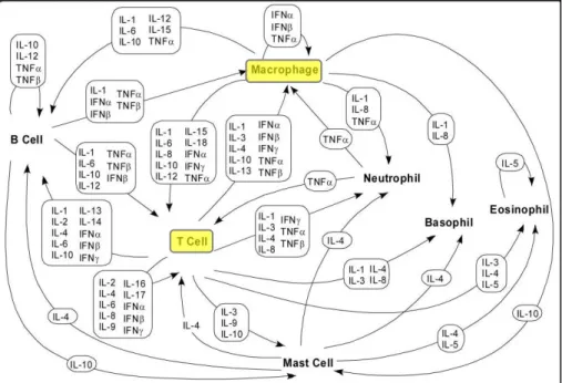
Cytokines are small proteins essentials to the interaction and communications between cells. They interact in a complex pathway in an autocrine, paracrine or endocrine manner with synergic or antagonist outcome, since a single cytokine may act on several different cell types (Figure 4) (Zhang and An 2007).
Monokines, interleukins and lymphokines are different types of cytokines. They are produced by a variety of immune or endothelial cells; however helper T cells (Th) and macrophages are the predominant producers (Zhang and An 2007).
In spite of their importance in several physiologic processes, they have been also associated to pathologic condition. Tumor cell and cells from tumor microenvironment, for example, showed aberrant production of cytokine (Kurzrock 2001; Jin et al. 2004).
Cytokines are commonly divided in pro-inflammatory and anti-inflammatory. Interleukin-1β (IL-1β) and tumor necrosis factor-α (TNF-α) are pro-inflammatory chemokines frequently associated either to immunosurveillance against cancer, or tumor progression. Anti-inflammatory cytokines, as transforming growth factor- β (TGF-β) and IL-10, also show complex effects on tumor development.
IL-1β is only produced after stimulation by inflammatory signals. It has been associated with all steps of malignancy (carcinogenesis, progression, invasion and metastasis) (Apte and Voronov 2008) and was related with metastasis promotion in melanoma models in vivo (Giavazzi et al. 1990; Meyer et al. 2011). In contrast, this interleukin is highly produced by M1 macrophages, important promoters of immune response against
11
malignant cells, which means that IL-1β may facilitate immunosurveilance, limiting tumor growth and progression (Fairweather and Cihakova 2009).
TNF-α can induce different signaling pathways, including a pro-apoptotic and an anti-apoptotic, among others (Chen and Goeddel 2002). According to that, its effect is a double-edge sword in carcinogenesis. Higher concentration of this cytokine can induce an antitumor response (Wiemann and Starnes 1994; Herman et al. 2013). In agreement, inhibitors of TNF appear to increase the risk of skin cancer, including melanoma (Mariette et al. 2011; Kouklakis et al. 2013). In contrast, low levels of it can induce tumor phenotypes by promote reactive oxygen/nitrogen species generation, causing DNA damage and promoting tumorigenesis (Woo et al. 2000; Hussain et al. 2003; Balkwill 2006).
TGF-β presents immunosuppressive and anti-inflammatory properties, involved in multiple physiologic pathways including embryogenesis, proliferation and differentiation (Santibanez et al. 2011). Their role in cancer is controversial. In early stages of cancer it promotes cell cycle arrest and apoptosis, acting as tumor suppressor. In later stages TGF-β stimulated the invasion and metastasis, inducing epithelial-mesenchymal transition (Akhurst and Derynck 2001; Morrison et al. 2013). In melanoma, TGF-β isoforms (TGF-β1/2/3) are highly expressed and increase in parallel with tumor progression (Krasagakis et al. 1998; Javelaud et al. 2008). According to that, this cytokine is commonly produced by M2-like macrophages leads to a Th2 response, i.e., promotes an immunosuppressive microenvironment that allows the tumor immune escape (Coffelt et al. 2009; Martinez et al. 2009; Siveen and Kuttan 2009; Hao et al. 2012; Sica and Mantovani 2012).
IL-10 is produced by almost all immune cells (as T and B cell, macrophages and monocytes, mast cells, granulocytes, dendritic cells and keratinocytes) (Sabat et al. 2010; Costa et al. 2013) and can also be produced by tumor cells (Gastl et al. 1993). In cancer, its effect is, once again, paradoxical. IL-10 can exert antitumor activity in glioma, melanomas, breast and ovarian carcinomas (Lin and Karin 2007) may be through downregulation of MHC I and consequent induction of NK-mediated tumor cell lysis (Kundu and Fulton 1997). However, IL-10 can allow the immune escape by tumor and reduce antigen presentation, cell maturation, differentiation and apoptosis (Zeng et al. 2010; Hamidullah et al. 2012).
Tumor immunology
In case of malignant transformation, characteristic mutations of cancer produce tumor-associated antigens (TAAs) that may allow immune activation and tumor control (Spurrell and Lockley 2014).
12
However, the persistence of tumors implies immune scape by several mechanisms. Same tumors lose essential functions to immune activation as the failure in expression of MHC molecules (responsible to antigen presentation and consequent T cell activation). Others express immunosuppressive cytokines or create physical barriers to avoid immunosurveillance (Spurrell and Lockley 2014).
From all inflammatory cells, macrophages or tumor-associated macrophages (TAMs) seemed to have the most important functions in malignancies (Chen et al. 2011). In fact, during tumor development macrophages have mostly M1 phenotypes and promote an immune response against cancer, however, in later-stages of tumor, TAMs frequently exhibit M2-like phenotypes (Sica and Mantovani 2012). M2-like TAMs represent a worst prognostic, since they favor tumor growth and survival through induction of angiogenesis and suppression of cytotoxic activity of T lymphocytes (Tsutsui et al. 2005; Zijlmans et al. 2006; Mantovani and Sica 2010).
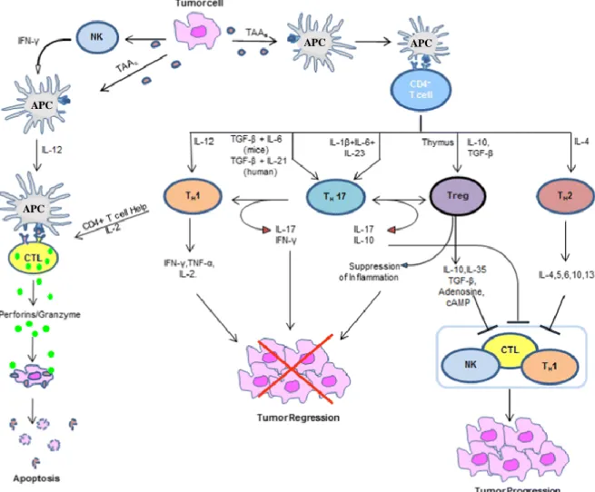
Figure 5 represents the interaction between innate and adaptive immunity and consequent effects on tumors. It reinforces the controversial effect of immune system, i.e. a set of immune cell (as M1 macrophages, NK, CTLs and Th1 cells) and specifics cytokines detect and control tumor progression, while other cells (like M2-macrophages, Th2 and Tregs) and derived cytokines, frequently present at later stages of cancer, contribute to their progression and immune scape.
Nevertheless, the role of immune system in cancer is not so linear because, this is influenced by the tumor microenvironment. Some immune cells are heterogeneous and present high plasticity, altering their phenotypes in response to cancer which explains the frequent inconsistent effects verified. In fact, the protumor or antitumor effects of each immune cell depend on cytokines signaling present in tumor microenvironment and it will determine their mode of differentiation (Lakshmi Narendra et al. 2013).
13
Figure 5: Schematic presentation of the role of immune system in cancer. Legend: APC indicates antigen
-presenting cell; CTL, cytotoxic T lymphocyte or CD8+ T cell; NK, natural killer cell; Th, T helper cell; Treg, regulatory T cell and TAA, tumor-associated antigens - adapted from (Lakshmi Narendra et al. 2013).
APC
APC
APC
14 References
Akao Y, Nakagawa Y, Iinuma M et al (2008) Anti-cancer effects of xanthones from pericarps of mangosteen. Int J Mol Sci 9: 355-70.
Akhurst RJ & Derynck R (2001) TGF-β signaling in cancer – a double-edged sword. Trends in Cell Biology 11: S44-S51.
Apte RN & Voronov E (2008) Is interleukin-1 a good or bad ‘guy’ in tumor immunobiology and immunotherapy? Immunological Reviews 222: 222-241.
Asnaghi L, Ebrahimi KB, Schreck KC et al (2012) Notch signaling promotes growth and invasion in uveal melanoma. Clin Cancer Res 18: 654-65.
Baguley BC & Ching L-M DMXAA: An antivascular agent with multiple host responses. International Journal of Radiation Oncology • Biology • Physics 54: 1503-1511.
Balkwill F (2006) TNF-alpha in promotion and progression of cancer. Cancer Metastasis Rev 25: 409-16.
Bergmann C, Strauss L, Zeidler R et al (2007) Expansion of human T regulatory type 1 cells in the microenvironment of cyclooxygenase 2 overexpressing head and neck squamous cell carcinoma. Cancer Res 67: 8865-73.
Bernengo MG, Quaglino P, Cappello N et al (2000) Macrophage-mediated immunostimulation modulates therapeutic efficacy of interleukin-2 based chemoimmunotherapy in advanced metastatic melanoma patients. Melanoma Res 10: 55-65.
Bianco A, Passacantilli P, Righi G et al (1989) Synthesis of 2-hydroxyacetyl-7-acetyl-xanthone, a new xanthone derivative endowed with antianaphylactic, analgesic, and antiinflammatory activities. Farmaco 44: 547-54.
Boasberg PD, Hoon DS, Piro LD et al (2006) Enhanced survival associated with vitiligo expression during maintenance biotherapy for metastatic melanoma. J Invest Dermatol 126: 2658-63.
Brocker EB, Zwadlo G, Holzmann B et al (1988) Inflammatory cell infiltrates in human melanoma at different stages of tumor progression. Int J Cancer 41: 562-7.
Bumrungpert A, Kalpravidh R, Chitchumroonchokchai C et al (2009) Xanthones from mangosteen prevent lipopolysaccharide-mediated inflammation and insulin resistance in primary cultures of human adipocytes. The Journal of Nutrition: 1185-1191.
Bumrungpert A & Kalpravidh RW (2010) Xanthones from mangosteen inhibit inflammation in human macrophages and in human adipocytes exposed to macrophage-conditioned media. The Journal of …: 1-6.
Chairungsrilerd N, Furukawa K, Ohta T et al (1996) Histaminergic and serotonergic receptor blocking substances from the medicinal plant Garcinia mangostana. Planta Med 62: 471-2.
Chanmee T, Ontong P, Konno K et al (2014) Tumor-associated macrophages as major players in the tumor microenvironment. Cancers (Basel) 6: 1670-90.
Chao AC, Hsu YL, Liu CK et al (2011) alpha-Mangostin, a dietary xanthone, induces autophagic cell death by activating the AMP-activated protein kinase pathway in glioblastoma cells. J Agric Food Chem 59: 2086-96.
Chen G & Goeddel DV (2002) TNF-R1 signaling: a beautiful pathway. Science 296: 1634-5.
Chen LG, Yang LL & Wang CC (2008) Anti-inflammatory activity of mangostins from Garcinia mangostana. Food Chem Toxicol 46: 688-93.
Chen P, Huang Y, Bong R et al (2011) Tumor-associated macrophages promote angiogenesis and melanoma growth via adrenomedullin in a paracrine and autocrine manner. Clinical cancer research : an official journal of the American Association for Cancer Research 17: 7230-9.
Chitchumroonchokchai C, Thomas-Ahner JM, Li J et al (2013) Anti-tumorigenicity of dietary alpha-mangostin in an HT-29 colon cell xenograft model and the tissue distribution of xanthones and their phase II metabolites. Mol Nutr Food Res 57: 203-11.
15
Choi BM, Pae HO, Jang SI et al (2002) Nitric oxide as a pro-apoptotic as well as anti-apoptotic modulator. J Biochem Mol Biol 35: 116-26.
Choudhari SK, Chaudhary M, Bagde S et al (2013) Nitric oxide and cancer: a review. World J Surg Oncol 11: 118.
Coffelt SB, Hughes R & Lewis CE (2009) Tumor-associated macrophages: effectors of angiogenesis and tumor progression. Biochim Biophys Acta 1796: 11-8.
Costa NL, Valadares MC, Souza PP et al (2013) Tumor-associated macrophages and the profile of inflammatory cytokines in oral squamous cell carcinoma. Oral Oncol 49: 216-23.
Cui J, Hu W, Cai Z et al (2010) New medicinal properties of mangostins: analgesic activity and pharmacological characterization of active ingredients from the fruit hull of Garcinia mangostana L. Pharmacol Biochem Behav 95: 166-72.
Davies LC, Jenkins SJ, Allen JE et al (2013) Tissue-resident macrophages. Nat Immunol 14: 986-95.
Devi Sampath P & Vijayaraghavan K (2007) Cardioprotective effect of alpha-mangostin, a xanthone derivative from mangosteen on tissue defense system against isoproterenol-induced myocardial infarction in rats. J Biochem Mol Toxicol 21: 336-9.
Dranoff G (2009) Targets of protective tumor immunity. Ann N Y Acad Sci 1174: 74-80. El-Seedi HR, El-Barbary MA, El-Ghorab DM et al (2010) Recent insights into the biosynthesis and biological activities of natural xanthones. Curr Med Chem 17: 854-901.
Fairweather D & Cihakova D (2009) Alternatively activated macrophages in infection and autoimmunity. J Autoimmun 33: 222-30.
Fauci A, Braunwald E, Kasper D et al (2009) Harrison's Manual of Medicine, 17th Edition, McGraw-Hill Education.
García-Rivera D, Delgado R, Bougarne N et al (2011) Gallic acid indanone and mangiferin xanthone are strong determinants of immunosuppressive anti-tumour effects of Mangifera indica L. bark in MDA-MB231 breast cancer cells. Cancer Letters 305: 21-31.
Garrido G, Delgado R, Lemus Y et al (2004) Protection against septic shock and suppression of tumor necrosis factor alpha and nitric oxide production on macrophages and microglia by a standard aqueous extract of Mangifera indica L. (VIMANG). Role of mangiferin isolated from the extract. Pharmacol Res 50: 165-72.
Garrido G, Gonzalez D, Delporte C et al (2001) Analgesic and anti-inflammatory effects of Mangifera indica L. extract (Vimang). Phytother Res 15: 18-21.
Gast A, Bermejo JL, Claus R et al (2011) Association of inherited variation in Toll-like receptor genes with malignant melanoma susceptibility and survival. PLoS One 6: e24370.
Gastl GA, Abrams JS, Nanus DM et al (1993) Interleukin-10 production by human carcinoma cell lines and its relationship to interleukin-6 expression. International Journal of Cancer 55: 96-101.
Giavazzi R, Garofalo A, Bani MR et al (1990) Interleukin 1-induced augmentation of experimental metastases from a human melanoma in nude mice. Cancer Res 50: 4771-5.
Gobbi S, Rampa A, Bisi A et al (2002) Synthesis and antitumor activity of new derivatives of xanthen-9-one-4-acetic acid. J Med Chem 45: 4931-9.
Gordon S & Martinez FO (2010) Alternative activation of macrophages: mechanism and functions. Immunity 32: 593-604.
Gross SS & Wolin MS (1995) Nitric oxide: pathophysiological mechanisms. Annu Rev Physiol 57: 737-69.
Gutierrez-Orozco F, Chitchumroonchokchai C, Lesinski GB et al (2013) alpha-Mangostin: anti-inflammatory activity and metabolism by human cells. J Agric Food Chem 61: 3891-900.
Gutierrez-Orozco F & Failla ML (2013) Biological activities and bioavailability of mangosteen xanthones: a critical review of the current evidence. Nutrients 5: 3163-83.
Hamidullah, Changkija B & Konwar R (2012) Role of interleukin-10 in breast cancer. Breast Cancer Res Treat 133: 11-21.
16
Hanahan D & Weinberg RA (2011) Hallmarks of cancer: the next generation. Cell 144: 646-74.
Hao N-B, Lü M-H, Fan Y-H et al (2012) Macrophages in Tumor Microenvironments and the Progression of Tumors. Clinical and Developmental Immunology 2012: 11.
Herman JM, Wild AT, Wang H et al (2013) Randomized phase III multi-institutional study of TNFerade biologic with fluorouracil and radiotherapy for locally advanced pancreatic cancer: final results. J Clin Oncol 31: 886-94.
Ho CK, Huang YL & Chen CC (2002) Garcinone E, a xanthone derivative, has potent cytotoxic effect against hepatocellular carcinoma cell lines. Planta Medica 68: 975-979.
Hou YC, Janczuk A & Wang PG (1999) Current trends in the development of nitric oxide donors. Curr Pharm Des 5: 417-41.
Hung SH, Shen KH, Wu CH et al (2009) Alpha-mangostin suppresses PC-3 human prostate carcinoma cell metastasis by inhibiting matrix metalloproteinase-2/9 and urokinase-plasminogen expression through the JNK signaling pathway. J Agric Food Chem 57: 1291-8.
Hussain SP, Hofseth LJ & Harris CC (2003) Radical causes of cancer. Nat Rev Cancer 3: 276-85.
Hussein MR (2004) Genetic pathways to melanoma tumorigenesis. J Clin Pathol 57: 797-801.
Ichiki H, Miura T, Kubo M et al (1998) New antidiabetic compounds, mangiferin and its glucoside. Biol Pharm Bull 21: 1389-90.
Jang HY, Kwon OK, Oh SR et al (2012) Mangosteen xanthones mitigate ovalbumin-induced airway inflammation in a mouse model of asthma. Food Chem Toxicol 50: 4042-50.
Javelaud D, Alexaki VI & Mauviel A (2008) Transforming growth factor-beta in cutaneous melanoma. Pigment Cell Melanoma Res 21: 123-32.
Jiang DJ, Dai Z & Li YJ (2004) Pharmacological effects of xanthones as cardiovascular protective agents. Cardiovasc Drug Rev 22: 91-102.
Jin P, Panelli MC, Marincola FM et al (2004) Cytokine polymorphism and its possible impact on cancer. Immunol Res 30: 181-90.
Johnson JJ, Petiwala SM, Syed DN et al (2012) α-Mangostin, a xanthone from mangosteen fruit, promotes cell cycle arrest in prostate cancer and decreases xenograft tumor growth. Carcinogenesis 33: 413-419.
Joseph WR, Cao Z, Mountjoy KG et al (1999) Stimulation of tumors to synthesize tumor necrosis factor-alpha in situ using 5,6-dimethylxanthenone-4-acetic acid: a novel approach to cancer therapy. Cancer Res 59: 633-8.
Jung H-A, Su B-N, Keller WJ et al (2006) Antioxidant Xanthones from the Pericarp of Garcinia mangostana (Mangosteen). Journal of Agricultural and Food Chemistry 54: 2077-2082.
Kalialis LV, Drzewiecki KT & Klyver H (2009) Spontaneous regression of metastases from melanoma: review of the literature. Melanoma Res 19: 275-82.
Korn EL, Liu PY, Lee SJ et al (2008) Meta-analysis of phase II cooperative group trials in metastatic stage IV melanoma to determine progression-free and overall survival benchmarks for future phase II trials. J Clin Oncol 26: 527-34.
Kostakis IK, Magiatis P, Pouli N et al (2002) Design, synthesis, and antiproliferative activity of some new pyrazole-fused amino derivatives of the pyranoxanthenone, pyranothioxanthenone, and pyranoacridone ring systems: a new class of cytotoxic agents. J Med Chem 45: 2599-609.
Kouklakis G, Efremidou EI, Pitiakoudis M et al (2013) Development of primary malignant melanoma during treatment with a TNF-alpha antagonist for severe Crohn's disease: a case report and review of the hypothetical association between TNF-alpha blockers and cancer. Drug Des Devel Ther 7: 195-9.
Krasagakis K, Tholke D, Farthmann B et al (1998) Elevated plasma levels of transforming growth factor (TGF)-beta1 and TGF-beta2 in patients with disseminated malignant melanoma. Br J Cancer 77: 1492-4.
17
Kumar IV, Paul BN, Asthana R et al (2003) Swertia chirayita mediated modulation of interleukin-1beta, interleukin-6, interleukin-10, interferon-gamma, and tumor necrosis factor-alpha in arthritic mice. Immunopharmacol Immunotoxicol 25: 573-83.
Kundu JK & Surh YJ (2008) Cancer chemopreventive and therapeutic potential of resveratrol: mechanistic perspectives. Cancer Lett 269: 243-61.
Kundu N & Fulton AM (1997) Interleukin-10 inhibits tumor metastasis, downregulates MHC class I, and enhances NK lysis. Cell Immunol 180: 55-61.
Kuo PC & Schroeder RA (1995) The emerging multifaceted roles of nitric oxide. Ann Surg 221: 220-35.
Kurzrock R (2001) Cytokine deregulation in cancer. Biomed Pharmacother 55: 543-7. Kushnir I & Merimsky O (2013) The evolution in melanoma treatment as a reflection of precision-oriented medicine. Oncol Lett 5: 424-426.
Lakshmi Narendra B, Eshvendar Reddy K, Shantikumar S et al (2013) Immune system: a double-edged sword in cancer. Inflamm Res 62: 823-34.
Lala PK & Orucevic A (1998) Role of nitric oxide in tumor progression: lessons from experimental tumors. Cancer Metastasis Rev 17: 91-106.
Lara PN, Jr., Douillard JY, Nakagawa K et al (2011) Randomized phase III placebo-controlled trial of carboplatin and paclitaxel with or without the vascular disrupting agent vadimezan (ASA404) in advanced non-small-cell lung cancer. J Clin Oncol 29: 2965-71.
Le Gal FA, Avril MF, Bosq J et al (2001) Direct evidence to support the role of antigen-specific CD8(+) T cells in melanoma-associated vitiligo. J Invest Dermatol 117: 1464-70.
Leiro J, Arranz JA, Yanez M et al (2004) Expression profiles of genes involved in the mouse nuclear factor-kappa B signal transduction pathway are modulated by mangiferin. Int Immunopharmacol 4: 763-78.
Leiro JM, Alvarez E, Arranz JA et al (2003) In vitro effects of mangiferin on superoxide concentrations and expression of the inducible nitric oxide synthase, tumour necrosis factor-alpha and transforming growth factor-beta genes. Biochem Pharmacol 65: 1361-71. Li Y & Ohizumi Y (2004) Search for constituents with neurotrophic factor-potentiating activity from the medicinal plants of paraguay and Thailand. Yakugaku Zasshi 124: 417-24.
Lin CN, Chung MI, Liou SJ et al (1996) Synthesis and anti-inflammatory effects of xanthone derivatives. J Pharm Pharmacol 48: 532-8.
Lin W-W & Karin M (2007) A cytokine-mediated link between innate immunity, inflammation, and cancer. The Journal of Clinical Investigation 117: 1175-1183.
Liu J, Fukunaga-Kalabis M, Li L et al (2014) Developmental pathways activated in melanocytes and melanoma. Arch Biochem Biophys.
Liu SH, Lee LT, Hu NY et al (2012) Effects of alpha-mangostin on the expression of anti-inflammatory genes in U937 cells. Chin Med 7: 19.
Liu Z, Antalek M, Nguyen L et al (2013) The effect of gartanin, a naturally occurring xanthone in mangosteen juice, on the mTOR pathway, autophagy, apoptosis, and the growth of human urinary bladder cancer cell lines. Nutr Cancer 65 Suppl 1: 68-77.
Madan B, Singh I, Kumar A et al (2002) Xanthones as inhibitors of microsomal lipid peroxidation and TNF-alpha induced ICAM-1 expression on human umbilical vein endothelial cells (HUVECs). Bioorg Med Chem 10: 3431-6.
Maio M (2012) Melanoma as a model tumour for immuno-oncology. Ann Oncol 23 Suppl 8: viii10-4.
Makare N, Bodhankar S & Rangari V (2001) Immunomodulatory activity of alcoholic extract of Mangifera indica L. in mice. J Ethnopharmacol 78: 133-7.
Makitie T, Summanen P, Tarkkanen A et al (2001) Tumor-infiltrating macrophages (CD68(+) cells) and prognosis in malignant uveal melanoma. Invest Ophthalmol Vis Sci 42: 1414-21.
Mantovani A & Sica A (2010) Macrophages, innate immunity and cancer: balance, tolerance, and diversity. Curr Opin Immunol 22: 231-7.
Mantovani A, Sica A, Sozzani S et al (2004) The chemokine system in diverse forms of macrophage activation and polarization. Trends Immunol 25: 677-86.
18
Mantovani A, Sozzani S, Locati M et al (2002) Macrophage polarization: tumor-associated macrophages as a paradigm for polarized M2 mononuclear phagocytes. Trends Immunol 23: 549-55.
Mariette X, Matucci-Cerinic M, Pavelka K et al (2011) Malignancies associated with tumour necrosis factor inhibitors in registries and prospective observational studies: a systematic review and meta-analysis. Ann Rheum Dis 70: 1895-904.
Martinez FO, Helming L & Gordon S (2009) Alternative activation of macrophages: an immunologic functional perspective. Annu Rev Immunol 27: 451-83.
Matsumoto K, Akao Y, Kobayashi E et al (2003) Induction of apoptosis by xanthones from mangosteen in human leukemia cell lines. Journal of Natural Products 66: 1124-1127.
Matsumoto K, Akao Y, Ohguchi K et al (2005) Xanthones induce cell-cycle arrest and apoptosis in human colon cancer DLD-1 cells. Bioorg Med Chem 13: 6064-9.
Matsumoto K, Akao Y, Yi H et al (2004) Preferential target is mitochondria in α-mangostin-induced apoptosis in human leukemia HL60 cells. Bioorganic & Medicinal Chemistry 12: 5799-5806.
Matzinger P (2002) The danger model: a renewed sense of self. Science 296: 301-5. Mazimba O, Nana F, Kuete V et al (2013) 11 - Xanthones and Anthranoids from the Medicinal Plants of Africa. In: KUETE, V. (ed.) Medicinal Plant Research in Africa. Oxford: Elsevier.
Mccourt C, Dolan O & Gormley G (2014) Malignant Melanoma: A Pictorial Review. Ulster Med J 83: 103-110.
Mckeage MJ, Reck M, Jameson MB et al (2009) Phase II study of ASA404 (vadimezan, 5,6-dimethylxanthenone-4-acetic acid/DMXAA) 1800mg/m(2) combined with carboplatin and paclitaxel in previously untreated advanced non-small cell lung cancer. Lung Cancer 65: 192-7.
Meyer C, Sevko A, Ramacher M et al (2011) Chronic inflammation promotes myeloid-derived suppressor cell activation blocking antitumor immunity in transgenic mouse melanoma model. Proc Natl Acad Sci U S A 108: 17111-6.
Moncada S, Palmer RM & Higgs EA (1991) Nitric oxide: physiology, pathophysiology, and pharmacology. Pharmacol Rev 43: 109-42.
Moongkarndi P, Kosem N, Kaslungka S et al (2004) Antiproliferation, antioxidation and induction of apoptosis by Garcinia mangostana (mangosteen) on SKBR3 human breast cancer cell line. Journal of Ethnopharmacology 90: 161-166.
Morrison CD, Parvani JG & Schiemann WP (2013) The relevance of the TGF-beta Paradox to EMT-MET programs. Cancer Lett 341: 30-40.
Munk ME & Emoto M (1995) Functions of T-cell subsets and cytokines in mycobacterial infections. Eur Respir J Suppl 20: 668s-675s.
Murray PJ & Wynn TA (2011) Protective and pathogenic functions of macrophage subsets. Nat Rev Immunol 11: 723-37.
Nakagawa Y, Iinuma M, Naoe T et al (2007) Characterized mechanism of α-mangostin-induced cell death: Caspase-independent apoptosis with release of endonuclease-G from mitochondria and increased miR-143 expression in human colorectal cancer DLD-1 cells. Bioorganic and Medicinal Chemistry 15: 5620-5628.
Nakatani K, Atsumi M, Arakawa T et al (2002) Inhibitions of histamine release and prostaglandin E2 synthesis by mangosteen, a Thai medicinal plant. Biol Pharm Bull 25: 1137-41.
Parker DC (1993) T cell-dependent B cell activation. Annu Rev Immunol 11: 331-60. Pedraza-Chaverri J, Cárdenas-Rodríguez N, Orozco-Ibarra M et al (2008) Medicinal properties of mangosteen (Garcinia mangostana). Food and Chemical Toxicology 46: 3227-3239.
Pedro M, Cerqueira F, Sousa ME et al (2002) Xanthones as inhibitors of growth of human cancer cell lines and their effects on the proliferation of human lymphocytes in vitro. Bioorganic & medicinal chemistry 10: 3725-30.
19
Pfister JR, Ferraresi RW, Harrison IT et al (1972) Xanthone-2-carboxylic acids, a new series of antiallergic substances. J Med Chem 15: 1032-5.
Phan GQ, Attia P, Steinberg SM et al (2001) Factors associated with response to high-dose interleukin-2 in patients with metastatic melanoma. J Clin Oncol 19: 3477-82.
Pinto MM, Sousa ME & Nascimento MS (2005) Xanthone derivatives: new insights in biological activities. Curr Med Chem 12: 2517-38.
Porta C, Subhra Kumar B, Larghi P et al (2007) Tumor promotion by tumor-associated macrophages. Adv Exp Med Biol 604: 67-86.
Printz C (2001) Spontaneous regression of melanoma may offer insight into cancer immunology. J Natl Cancer Inst 93: 1047-8.
Qian BZ & Pollard JW (2010) Macrophage diversity enhances tumor progression and metastasis. Cell 141: 39-51.
Raimondi G, Turner MS, Thomson AW et al (2007) Naturally occurring regulatory T cells: recent insights in health and disease. Crit Rev Immunol 27: 61-95.
Rajendran P, Ekambaram G, Magesh V et al (2008) Chemopreventive efficacy of mangiferin against benzo(a)pyrene induced lung carcinogenesis in experimental animals. Environmental Toxicology and Pharmacology 26: 278-282.
Reiman JM, Kmieciak M, Manjili MH et al (2007) Tumor immunoediting and immunosculpting pathways to cancer progression. Semin Cancer Biol 17: 275-87.
Romagnani S, Parronchi P, D'elios MM et al (1997) An update on human Th1 and Th2 cells. Int Arch Allergy Immunol 113: 153-6.
Roncarolo MG, Bacchetta R, Bordignon C et al (2001) Type 1 T regulatory cells. Immunol Rev 182: 68-79.
Roncarolo MG, Gregori S, Battaglia M et al (2006) Interleukin-10-secreting type 1 regulatory T cells in rodents and humans. Immunol Rev 212: 28-50.
Sabat R, Grutz G, Warszawska K et al (2010) Biology of interleukin-10. Cytokine Growth Factor Rev 21: 331-44.
Santibanez JF, Quintanilla M & Bernabeu C (2011) TGF-beta/TGF-beta receptor system and its role in physiological and pathological conditions. Clin Sci (Lond) 121: 233-51.
Seo EK, Kim NC, Wani MC et al (2002) Cytotoxic prenylated xanthones and the unusual compounds anthraquinobenzophenones from Cratoxylum sumatranum. J Nat Prod 65: 299-305.
Shan T, Ma Q, Guo K et al (2011) Xanthones from mangosteen extracts as natural chemopreventive agents: potential anticancer drugs. Curr Mol Med 11: 666-77.
Sica A & Mantovani A (2012) Macrophage plasticity and polarization: in vivo veritas. J Clin Invest 122: 787-95.
Siveen KS & Kuttan G (2009) Role of macrophages in tumour progression. Immunol Lett 123: 97-102.
Solinas G, Germano G, Mantovani A et al (2009) Tumor-associated macrophages (TAM) as major players of the cancer-related inflammation. J Leukoc Biol 86: 1065-73.
Speeckaert R, Van Geel N, Vermaelen KV et al (2011) Immune reactions in benign and malignant melanocytic lesions: lessons for immunotherapy. Pigment Cell Melanoma Res 24: 334-44.
Spurrell EL & Lockley M (2014) Adaptive immunity in cancer immunology and therapeutics. Ecancermedicalscience 8: 441.
Suksamrarn S, Komutiban O, Ratananukul P et al (2006) Cytotoxic prenylated xanthones from the young fruit of Garcinia mangostana. Chemical and Pharmaceutical Bulletin 54: 301-305.
Sun J, Chu YF, Wu X et al (2002) Antioxidant and antiproliferative activities of common fruits. J Agric Food Chem 50: 7449-54.
Tang YP, Li PG, Kondo M et al (2009) Effect of a mangosteen dietary supplement on human immune function: a randomized, double-blind, placebo-controlled trial. J Med Food 12: 755-63.