UNIVERSIDADE DE LISBOA
FACULDADE DE MEDICINA DE LISBOA
ENDOTHELIAL NOTCH LIGANDS IN BONE
MARROW FUNCTION AND IN MALIGNANCY
Inês Sofia Alvarez Martins
Orientador:
Professor Doutor Sérgio Jerónimo Rodrigues Dias
Tese especialmente elaborada para obtenção do grau de Doutor em Ciências Biomédicas, especialidade de Biologia Celular e Molecular
Juri: Doutor Rui Miguel dos Santos Benedito, Assistant Professor, Investigador do Centro Nacional de Investigaciones Cardiovasculares Carlos III, Barcelona;
- Doutor Carlos Filipe Ribeiro Lemos Pereira, Investigador Auxiliar do Centro para Neurociências e Biologia Celular da Universidade de Coimbra;
- Doutro António José de Freitas Duarte, Professor Associado de Faculdade de Medicina Veterinária da Universidade de Lisboa;
- Doutor Cláudio Areias Franco, Especialista de Reconhecido Mérito e Competência,
Investigador, Group Leader do Instituto de Medicina Molecular da Faculdade de Medicina da Universidade de Lisboa;
- Doutor Sérgio Gerónimo Rodrigues Dias, Professor Associado Convidado da Faculdade de Medicina da Universidade de Lisboa (Orientador);
- Doutor Luís António Marques Costa, Professor Associado Convidado da Faculdade de Medicina da Universidade de Lisboa;
- Doutor Domingos Manuel Pinto Henrique, Investigador Auxiliar, da Faculdade de Medicina da Universidade de Lisboa.
Presidente: Doutor José Augusto Gamito Melo Cristino, Professor Catedrático e Presidente do Conselho Científico da Faculdade de Medicina da Universidade de Lisboa.
Fundação para a Ciência e Tecnologia, bolsa SFRH/BD/78249/2011 2017
A impressão desta tese foi aprovada pelo Conselho Científico da Faculdade
de Medicina de Lisboa em reunião de 19 de Abril de 2016
ACKNOWLEDGMENTS
I
ACKNOWLEDGMENTS
Though only my name appears on the cover of this dissertation, a great many people have contributed to its production. I owe my debt of gratitude to all those who made this degree possible, for their scientific and, mostly, moral support throughout these challenging few years.
First of all, I would like to thank my supervisor, Dr. Sérgio Dias, for his support and contributions throughout this process. His patience and encouragement have helped me overcome many setbacks and his mentorship was essential for my success.
The people I’ve crossed paths with during this journey have made this experience one that I will cherish forever. None of this would have been accomplished without the past and present members of the SDias lab. I would like to thank Leonor, for her teaching when I first came to the lab, Ana Magalhães, Sandrina and Hélia, for the scientific discussions, and especially to Ana de Barros, Inês Matias, Clemente and Cabrita for putting up with my morning grumpiness, for the joyful environment in the lab, and for the great friendship. Also, to the iMMers who contributed for this work, especially to the members of the histology, flow cytometry, imaging and rodent facilities, for all the advices and technical support.
I am grateful that I have been surrounded by good friends who have kept me sane and my views in perspective in the process. A heartfelt thank you goes particularly to Isaura, Diogo, Memé, Marta, Alice and Joana who have been the most wonderful supporters in the quest for my PhD.
Most importantly, none of this would have been possible without the love and patience of my family. My wonderful parents, who never stopped trying to understand just what I was doing and who have been a constant source of strength and encouragement all these years. My little brother, Rui, who is the most amazing enthusiast in live. When my confidence wavered, you inspired me. I am deeply grateful.
II
RESUMO
O sistema cardiovascular é o primeiro a ser formado no embrião de organismos vertebrados, sendo composto por uma rede de vasos que permite a circulação do sangue por todo o corpo, entregando oxigénio e nutrientes. As células endoteliais que formam os vasos sanguíneos têm um papel instrutor em diversos processos fisiológicos e patológicos, como a manutenção das células estaminais, a regeneração e reparação de órgãos ou o desenvolvimento de tumores, através da produção e libertação de factores parácrinos, designados factores angiócrinos. Por conseguinte, a fim de compreender de que forma as células endoteliais comunicam com as células circundantes, a identificação destes factores tem recebido muita atenção nos últimos anos.
Nesta Tese usámos ratinhos geneticamente modificados para explorar o envolvimento de dois factores angiócrinos pertencentes à via de sinalização Notch - Jagged 1 (Jag1) e Delta-like 4 (Dll4) - em dois processos distintos. Primeiro explorámos a função do ligando Jag1 especificamente em células endoteliais no recrutamento de macrófagos para tumores da próstata em desenvolvimento. Com esse intuito, bloqueámos Jag1 especificamente em
células endoteliais VE-Caderina+ e sobrexpressámos Jag1 em células endoteliais positivas
para Tie2, em ratinhos TRAMP*Jag1lox/lox*VE-Cadherin-Cre-ERT2 (eJag1KO) e
TRAMP*Tet-O-Jag1*Tie2-rtTA (eJag1OE), respectivamente. Em ratinhos que desenvolvem tumores da
próstata espontâneos (TRAMP), a expressão do ligando Jag1 em células endoteliais induz a proliferação de células tumorais, promove angiogénese e maturação dos novos vasos. No entanto, o envolvimento de Jag1 endotelial (eJag1) no recrutamento e activação de macrófagos para o tumor ainda não era conhecida. O segundo processo que estudámos foi a modulação da hematopoiese e do nicho vascular da medula óssea através da modulação de Dll4 endotelial, usando ratinhos com mutações condicionais e indutíveis que promovem o knock-out ou a sobrexpressão de Dll4 em células endoteliais. Um vasto número de estudos mostrou o envolvimento de Dll4 no desenvolvimento vascular e na diferenciação de células hematopoiéticas, mas o papel que o ligando desempenha no nicho vascular e na comunicação das células endoteliais com células hematopoiéticas na medula óssea não tinha sido estudado.
RESUMO
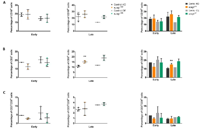
III O primeiro objectivo desta tese foi investigar se o ligando Jag1 em células endoteliais poderia afectar o desenvolvimento de tumores da próstata através do recrutamento de macrófagos para o tumor. Os macrófagos são um grande componente celular do microambiente tumoral e podem ser activados diferencialmente para um fenótipo anti- (M1) ou pro-tumoral (M2). As células endoteliais e os macrófagos estabelecem interacções específicas que modulam as propriedades angiogénicas do tumor e que se pensa serem capazes de afectar a polarização dos macrófagos. Os nossos resultados mostram que eJag1 regula positivamente o recrutamento e polarização de macrófagos para um fenótipo
pro-tumoral em tumores da próstata. De facto, ratinhos eJag1KO têm menos macrófagos
intra-tumorais que os controlos e maior polarização M1 e ratinhos eJag1OE mais macrófagos que
os respectivos controlos, e maior polarização M2. Este fenótipo foi também observado in vitro quando células endoteliais do cordão umbilical (HUVECs) tratadas com um anticorpo neutralizante para Jag1 promoveram uma diminuição do número de macrófagos do tipo M2, sugerindo que esta modulação da activação de macrófagos é um efeito directo da expressão de Jag1 em células endoteliais.
Investigámos a modulação da expressão de genes que codificam para factores angiócrinos e demonstrámos que a modulação dos níveis de Jag1 no endotélio afectou a expressão de moléculas de adesão e de quimioatractores, bem como de outros ligandos da via Notch. Particularmente em ratinhos eJag1OE, a sobrexpressão de Jag1 induziu um aumento nos níveis de Angpt2, Dll4 e Jag2 e uma subexpressão de Cxcl12, Vcam1 e Dll1 nas células endoteliais isoladas do tumor. O aumento de Angpt2, em particular, pode explicar o recrutamento e polarização dos macrófagos para um fenótipo M2.
De forma semelhante, os macrófagos M1 e M2 também mostraram alterações na expressão de alguns genes. Os níveis de eJag1 correlacionavam-se com um aumento da expressão de Il6 e Tnfa e com a subexpressão de Notch2, particularmente em macrófagos M2. Adicionalmente, macrófagos M2 também exibiam uma sobrexpressão de Notch1 e Cxcr4, independentemente da modulação de eJag1. Em conclusão, a expressão de Jag1 em células endoteliais promove o recrutamento de macrófagos para tumores da próstata e polarização para um fenótipo M2, possivelmente através da indução da modificação da expressão de outros factores angiócrinos o que induz uma modulação nos padrões de expressão genética dos macrófagos.
IV
A fim de compreender de que forma a expressão do ligando Dll4 no nicho vascular afectava a hematopoiese, tirámos partido de ratinhos com ganho e perda-de-função de Dll4 especificamente em células endoteliais (eDll4OE e eDll4KO) e analisámo-los sem serem irradiados ou 8 e 26 dias após serem submetidos a uma irradiação sub-letal, que induziu mielossupressão. Em animais não irradiados, a modulação de Dll4 endotelial (eDll4)
perturbou a hematopoiese, como evidenciado pela redução de células mielóides (CD11b+)
no sangue periférico de ratinhos eDll4OE e pelo aumento de células linfóides B (B220+) na medula óssea e sangue periférico de ratinhos eDll4KO. Adicionalmente, ensaios de diferenciação em metilcelulose in vitro demonstraram que ratinhos knock-out para Dll4 endotelial têm um aumento de progenitores mielóides (CFU-G) mas uma diminuição no número de progenitores multipotentes (CFU-GEMM).
Após exposição a radiação, ratinhos com menores níveis de eDll4 (eDll4KO e Control
OE) recuperaram mais rapidamente o número de células na medula que os restantes modelos. Detectámos também uma diminuição no número de plaquetas em ratinhos eDll4OE e um aumento de eritrócitos em circulação em ratinhos eDll4KO, 8 dias após mielossupressão. Vinte e seis dias após irradiação, as proporções relativas das diferentes linhagens hematopoiéticas também tinham sido modificadas em função dos níveis de eDll4. A sobrexpressão de Dll4 induziu diferenciação mielóide e linfóide T, em detrimento
da linhagem linfóide B, e o oposto foi detectado em ratinhos eDll4KO, consistente com o
papel atribuído ao ligando Dll4 na especificação das linhagens hematopoiéticas, quando expresso noutro tipo de células do microambiente. Analisámos ainda a recuperação hematopoiética após mielossupressão no contexto de transplante de medula. Transplante
de medula total de ratinhos eDll4KO para ratinhos controlo letalmente irradiados promoveu
uma recuperação da celularidade da medula mais rápida, e diminuiu o dano na medula óssea, quando comparado com um transplante de medula de ratinhos controlo para ratinhos eDll4KO.
Esta modificação no compartimento hematopoiético levou-nos a questionar se o nicho vascular da medula óssea também seria modificado pela modulação de eDll4. Verificámos que o nicho vascular não estava alterado em ratinhos que não tinham sido expostos a radiação ou que foram sacrificados 26 dias após mielossupressão, mas que 8 dias após
RESUMO
V irradiação, ratinhos eDll4KO tinham um aumento de vasos positivos para VE-Caderina e
VEGFR2 e ratinhos eDll4OE exibiam uma diminuição de vasos VEGFR2-positivos. Esta
modulação da identidade dos vasos na medula óssea, sem afectar o número total de vasos
(CD105+), pode explicar a recuperação mais rápida de ratinhos eDll4KO quando expostos a
radiação. Adicionalmente, tal modulação foi acompanhada de uma modificação na localização de células B e de megacariócitos relativamente aos sinusoides da medula óssea. Oito dias após mielossupressão, ratinhos com menores níveis de eDll4 tinham menos
células B e megacariócitos em contacto com vasos VE-Caderina+, o que se correlacionou
com menos células B dentro dos vasos ou em circulação 26 dias após irradiação. Investigámos ainda a modulação da expressão de genes que codificam factores angiócrinos que poderiam modular a hematopoiese. A sobrexpressão de eDll4 induziu uma diminuição da expressão de Cxcl12, Vcam1 e Thpo e o knock-out de eDll4 levou a uma subexpressão de Cxcl12. Particularmente em ratinhos eDll4KO, a diminuição do número de células B e megacariócitos em contacto com os sinusoides pode ser explicada pela redução dos níveis de Cxcl12, uma vez que o tratamento de HUVECs com um anticorpo neutralizante para Dll4 levou a uma redução dos níveis de CXCL12 e uma consequente diminuição na migração de células CD34+. A diminuição do número de plaquetas em ratinhos eDll4OE, por sua vez, pode ser explicado pela redução dos níveis de Thpo.
O trabalho realizado neste projecto de doutoramento revelou os efeitos da modulação de eJag1 e eDll4 na progressão de tumores da próstata e na recuperação da medula óssea após mielossupressão, respectivamente. Em ambos os casos, a expressão dos ligandos da via Notch em células endoteliais revelou ser prejudicial. eJag1 promove directa e indirectamente a progressão de tumores e eDll4 bloqueia a normal recuperação da medula após irradiação sub-letal ou num cenário de transplante de medula, indicando que estes ligandos devem ser considerados como possíveis alvos terapêuticos.
Palavras-chave
Tumor da próstata; Jagged 1; Macrófagos; Polarização de macrófagos; M1; M2; Medula Óssea; Nicho vascular; Delta-like 4; Hematopoiese; Sinalização Notch
VI
SUMMARY
Endothelial cells have emerged as instructive players in distinct physiological and pathological tasks, maintaining resident stem cell homeostasis, orchestrating tissue regeneration, and inducing tumor growth through the release of paracrine factors, known as angiocrine factors. In this Thesis, we explored the role of two particular angiocrine genes, Jagged 1 (Jag1) and Delta-like 4 (Dll4), in the development of prostate tumors through the recruitment of macrophages and in bone marrow (BM) regeneration following myeloablation, respectively.
To address endothelial Jag1 (eJag1) function in prostate tumor progression, we used genetically engineered mouse models, in which mice that develop spontaneous prostate tumors (TRAMP) were crossed with endothelial-specific Jag1 loss- or gain-of-function mice (eJag1KO and eJag1OE). We showed that eJag1 induces macrophage recruitment into the tumors and polarization into a pro-tumoral M2 phenotype, both in vivo and in vitro. This
was accompanied by a modulation in angiocrine gene expression, particularly in eJag1OE
mice, and in the macrophage expression pattern. Our preliminary data thus suggest that eJag1 modulates tumor growth and angiogenesis indirectly by promoting macrophage recruitment and polarization into an M2 state.
To understand how endothelial Dll4 modulation affected the BM vascular niche and hematopoiesis, we used two conditional mouse models with endothelial-specific Delta-like 4 (Dll4) loss- or gain-of-function (eDll4KO and eDll4OE) and analyzed their hematopoietic and vascular compartments with and without sub-lethal irradiation. Although the BM vascular niche was not affected by eDll4 modulation at steady state or 26 days after irradiation, by day 8 post-irradiation eDll4 induced changes in BM vessel identity, without affecting the overall BM vessel content. This modulation of the BM vascular niche was accompanied by a modulation in the angiocrine gene expression pattern and induced changes in hematopoiesis. Particularly, eDll4 levels correlated with increased erythropoiesis and decreased megakaryopoiesis and B lymphopoiesis, although it promoted the migration of B cells and megakaryocytes to the vicinity of BM sinusoids. These modifications resulted in
an improvement of hematopoietic recovery both in sub-lethally irradiated eDll4KO and in
SUMMARY
VII eDll4 impairs BM recovery following myeloablation and may be relevant in a setting of BM transplantation or in patients receiving chemotherapy.
Taken together, our data suggests that the Notch ligands Jag1 and Dll4 may be possible targets in tumor progression and BM recovery, respectively, as their expression in endothelial cells is unfavorable in a clinical setting.
Keywords
Prostate tumor; Jagged 1; Macrophage; Macrophage polarization; M1; M2; Bone marrow; Vascular niche; Delta-like 4; Hematopoiesis; Notch signaling
VIII
ABBREVIATIONS
ADAM A desintegrin and metalloprotease
Angpt Angiopoietin
BC Before Christ
BM Bone marrow
CAR CXCL12 abundant reticular cells
CD Cluster of differentiation
CDC42 Cell division cycle 42
CFU Colony forming unit
CFU-E CFU-erythrocyte
CFU-G CFU-granulocyte
CFU-GEEM CFU-granulocyte-erythrocyte-macrophage-megakaryocyte
CFU-M CFU-macrophage
CLP Common lymphoid progenitor
COUP-TFII Chicken ovalbumin upstream promoter transcription factor II
Cox Cyclooxygenase CSF Colony-stimulating factor CXCL C-X-C chemokine ligand CXCR C-X-C chemokine receptor Dll Delta-like E Embryonic day EC Endothelial cell
ECM Extracellular matrix
eDll4 Endothelial Dll4
eDll4KO Endothelial Dll4 knockout mice (Dll4lox/lox*VE-Cadherin-Cre-ERT2)
eDll4OE Endothelial Dll4 overexpressing mice (Dll4-Tet-O7*Tie2-rtTA)
EGF Epidermal growth factor
eJag1 Endothelial Jag1
eJag1KO Endothelial Jag1 knockout mice (TRAMP*Jag1lox/lox*VE-Cadherin-Cre-ERT2)
eJag1OE Endothelial Jag1 overexpressing mice (TRAMP*Tet-O-Jag1*Tie2-rtTA)
EPC Endothelial progenitor cell
ER Endoplasmic reticulum
ERK Extracellular signal-regulated kinases
FBS Fetal bovine serum
ABBREVIATIONS
IX
FGFR Fibroblast growth factor receptor
G-CSF Granulocyte colony-stimulating factor
GPCR G-protein-coupled receptor
GTPase Guanosine triphosphatase
HES Hairy/enhancer of split
HEY HES-related protein
HIF Hypoxia inducible factor
HUVEC Human umbilical cord vein endothelial cells
HSC Hematopoietic stem cell
HSPC Hematopoietic stem and progenitor cell
ICAM1 Intercellular adhesion molecule 1
IFNγ Interferon gamma
IGF Insulin-like growth factor
IL Interleukin
IP Intraperitoneally
Jag Jagged
JAM Junctional adhesion molecules
LepR Leptin receptor
LPS Lipopolysaccharide
LSEC Liver sinusoidal endothelial cells
LT-HSC Long term hematopoietic stem cell
Maml Mastermind-like
MAPK Mitogen-activated protein kinase
MCP1 Monocyte chemoattractant protein 1
MHC II Major histocompatibility complex class II
MK Megakaryocyte
MMP Matrix metalloprotease
MSC Mesenchymal stromal cell
MT1-MMP Membrane-type 1 matrix metalloprotease
N-Cadherin Neuronal cadherin
Nes Nestin
NFκB Nuclear factor of kappa light polypeptide gene enhancer in B-cells 1
NICD Notch intracellular domain
NO Nitric oxide
NOS3 Nitric oxide synthase 3
X
Osx Osterix
PB Peripheral Blood
PBS Phosphate Buffered Saline
PlGF Placental growth factor
Pre-pro-B Early B lymphocyte progenitor
Pre-B Late B lymphocyte progenitor
Prx1 Paired related homeobox protein 1
RBPjk Recombining binding protein suppressor of hairless kappa
Robo4 Roundabout 4
Sca1 Stem cell antigen 1
SCF Stem cell factor
SDF1 Stromal derived factor 1
SEC Sinusoidal endothelial cell
SMA Smooth muscle actin
SMC Smooth muscle cell
TAMs Tumor-associated macrophages
TEM Tie2-expressing monocyte/macrophage
TGF Transforming growth factor
Thpo Thrombopoietin
Tie Tyrosine kinase with immunoglobulin-like EGF-like domains
TNFα Tumor necrosis factor
TRAMP Transgenic adenocarcinoma mouse prostate
Unc5b Unc-5 homolog b
VCAM1 Vascular cell adhesion molecule 1
VE-Cadherin Vascular endothelial cadherin
VEGF Vascular endothelial growth factor
VEGFR Vascular endothelial growth factor receptor
VHL von Hippel-Lindau protein
vWF Von Willebrand Factor
CONTENTS XI
CONTENTS
ACKNOWLEDGMENTS ... I RESUMO ... II SUMMARY ... VI ABBREVIATIONS ... VIII CONTENTS ... XI FIGURES INDEX ... XV TABLES INDEX... XVIIIC
HAPTER1
-
I
NTRODUCTION…………..………
1
1.1. THE VASCULAR SYSTEM ... 2
1.2. BLOOD VESSEL FORMATION: VASCULOGENESIS AND ANGIOGENESIS ... 3
1.2.1. Sprouting angiogenesis ... 5
1.2.2. Signaling pathways involved in sprouting angiogenesis ... 8
1.2.3. Vessel maturation ... 20
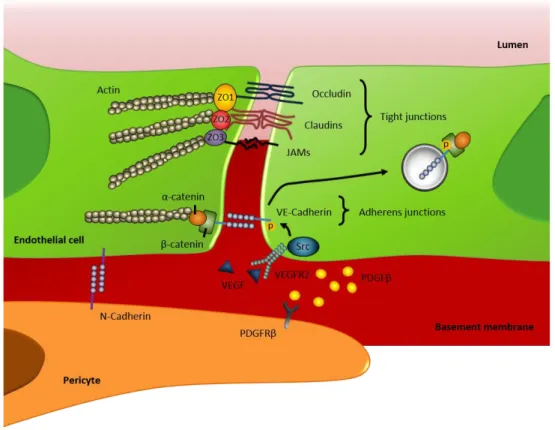
1.3.ENDOTHELIALCELLGENERALPROPERTIES ... 23
1.3.1. Endothelial cell junctions ... 23
1.3.2. Endothelial-pericyte interactions ... 28
1.3.3. Extracellular matrix ... 29
1.4. DISRUPTION OF ENDOTHELIAL CELL PROPERTIES ... 30
1.4.1. Endothelial cells in inflammation ... 31
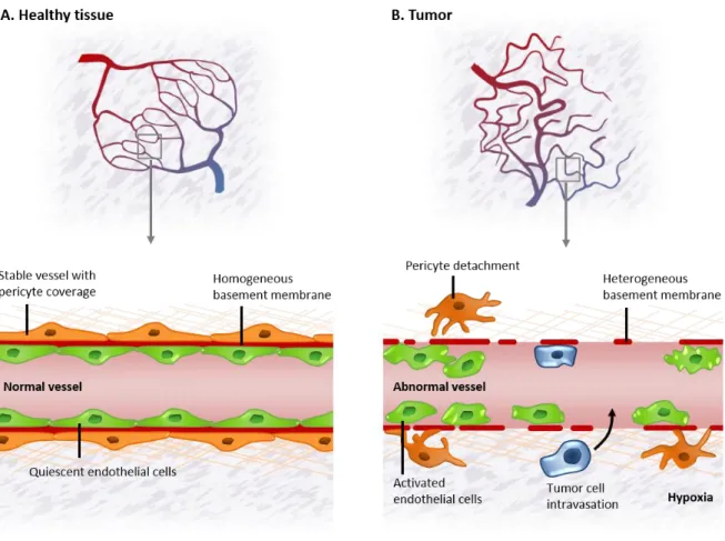
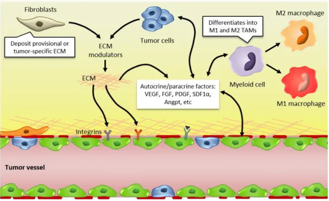
1.4.2. Endothelial cells in cancer... 38
1.5. ENDOTHELIAL CELLS HAVE AN INSTRUCTIVE ROLE IN ORGAN REGENERATION ... 45
1.5.1. Regulation of hematopoiesis by the bone marrow niches ... 47
1.6. AIMS OF THE THESIS ... 52
1.7. REFERENCES ... 53
C
HAPTER2
-
E
NDOTHELIALJ
AGGED1
ENHANCES MACROPHAGE RECRUITMENT AND ALTERNATIVE ACTIVATION IN A SETTING OF PROSTATE ADENOCARCINOMA...
69
XII
2.1. ABSTRACT ... 70
2.2. INTRODUCTION ... 71
2.3. METHODS ... 72
2.3.1. Animal experiments ... 72
2.3.2. Flow cytometry and cell sorting... 73
2.3.3. Cell culture and macrophage polarization assay ... 74
2.3.4. RNA isolation and quantitative PCR ... 75
2.3.5. Statistical analysis ... 75
2.4. RESULTS ... 76
2.4.1. Modulation of endothelial Jag1 affects the recruitment of immune cells to the tumor site ... 76
2.4.2. Expression of the Notch ligands Jag1 and Dll4 in endothelial cells directly affects macrophage polarization in vitro ... 80
2.4.3. eJag1 modulation affects the transcription profile of “angiocrine genes” in endothelial tumor-associated cells ... 81
2.4.4. Modulation of eJag1 influences the transcription profile of tumor associated macrophages. ... 83
2.5. DISCUSSION ... 84
2.6. REFERENCES ... 90
C
HAPTER3
-
C
HAPTER3
-
E
NDOTHELIALD
ELTA-
LIKE4
MODULATES THE HEMATOPOIETIC SYSTEM AND HINDERS BONE MARROW RECOVERY FOLLOWING MYELOABLATION...
97
3.1. ABSTRACT ... 98 3.2. INTRODUCTION ... 98 3.3. METHODS ... 100 3.3.1. Animal genotyping ... 100 3.3.2. Animal experiments ... 101 3.3.3. Sample collection ... 1023.3.4. In vitro colony forming assay ... 102
3.3.5. Flow cytometry ... 103
CONTENTS
XIII
3.3.7. Statistical analysis ... 104
3.4. RESULTS ... 104
3.4.1. Endothelial Dll4 affects bone marrow cell content and the colony forming potential of eDll4KO hematopoietic stem and progenitor cells ... 104
3.4.2. Endothelial specific Dll4 modulates bone marrow and peripheral blood hematopoietic content ... 105
3.4.3. eDll4 modulation affects BM recovery and hematopoiesis after myeloablation ... 108
3.4.4. Endothelial Dll4 depletion affects BM recovery and B cell content after BM transplantation. ... 117
3.5. DISCUSSION ... 119
3.6. REFERENCES ... 123
C
HAPTER4
-
E
NDOTHELIALD
ELTA-
LIKE4
IS REQUIRED FOR NORMAL BONE MARROW VASCULAR NICHE RECOVERY AND HEMATOPOIETIC CELL MIGRATION FOLLOWING MYELOABLATION. ...
127
4.1. ABSTRACT ... 128 4.2. INTRODUCTION ... 129 4.3. METHODS ... 130 4.3.1. Animal genotyping ... 130 4.3.2. Animal experiments ... 131 4.3.3. Flow cytometry ... 132
4.3.4. Immunostaining and imaging ... 132
4.3.5. RNA isolation and quantitative PCR ... 134
4.3.6. Cell culture and treatment... 134
4.3.7. Chemotaxis assay ... 135
4.3.8. Statistical analysis ... 135
4.4. RESULTS ... 136
4.4.1. At steady state, endothelial-specific Delta-like 4 modulation does not affect the BM vascular niche. ... 136
4.4.2. Sub-lethal irradiation causes a modulation in BM vessel identity in early stages of recovery ... 138
XIV
4.4.3. eDll4 affects B cell and megakaryocyte localization within the BM vascular niche
following myelosuppression ... 141
4.4.4. eDll4 modulates the expression of “angiocrine genes” throughout recovery .. 146
4.4.5. In vitro neutralization of Dll4 in HUVECs inhibits HSPC migration ... 147
4.5. DISCUSSION ... 148
4.6. REFERENCES ... 152
C
HAPTER5
-
D
ISCUSSION. ... 159
5.1. THE ROLE OF ENDOTHELIAL JAGGED 1 IN MACROPHAGE RECRUITMENT IN A PROSTATE TUMOR MOUSE MODEL ... 161
5.2. THE IMPACT OF ENDOTHELIAL DELTA-LIKE 4 MODULATION IN THE BM MICROENVIRONMENT AND HEMATOPOIESIS ... 165
5.3. CONCLUDING REMARKS ... 170
5.4. REFERENCES ... 172
FIGURES INDEX
XV
FIGURES INDEX
Figure 1.1. Mechanisms of blood vessel formation ... 4
Figure 1.2. Cellular mechanisms of angiogenic sprouting ... 7
Figure 1.3. Canonical Notch signaling pathway ... 12
Figure 1.4. Notch signaling regulates endothelial tip/stalk cell specification ... 17
Figure 1.5. Angpt-Tie system regulates mural cell recruitment and vascular permeability ... 19
Figure 1.6. Adhesive proteins involved in the establishment of endothelial cell-cell and endothelial-pericyte interactions ... 27
Figure 1.7. Endothelial cell activation ... 33
Figure 1.8. Tumor vessels are structurally and functionally abnormal ... 40
Figure 1.9. Interactions between tumor cells and their microenvironment modulate tumor and angiogenic responses ... 43
Figure 1.10. The bone marrow HSC niche is composed by distinct stromal cell types and differentiated hematopoietic cells ... 49
Figure 2.1. eJag1 modulates the percentage of lymphoid cells that are found in the tumor site ... 77
Figure 2.2. Endothelial Jag1 affects macrophage polarization towards an M2-like phenotype... 79
Figure 2.3. In vitro neutralization of the Notch ligands Jag1 and Dll4 affects macrophage polarization into the M2 state ... 80
Figure 2.4. eJag1 modulates angiocrine gene expression in tumor-associated endothelial cells ... 82
Figure 2.5. The expression profile of tumor associated macrophages is modified upon endothelial Jag1 modulation ... 84
Figure 3.1. Experimental setup ... 102
Figure 3.2. Endothelial Dll4 knockout affects BM cellularity and the CFU potential of HSPCs ... 105
Figure 3.3. eDll4 modulation affects BM and PB lymphoid and myeloid content ... 106
Figure 3.4. eDll4 does not affect the levels of erythrocytes, leukocytes or platelets in circulation ... 107
XVI
Figure 3.6. Endothelial Dll4 modulates the BM lymphoid content 8 days after irradiation
... 110
Figure 3.7. eDll4 modulates erythrocyte and platelet levels and the percentage of
peripheral blood myeloid and lymphoid cells 8 days after myeloablation ... 111
Figure 3.8. Modulation of eDll4 induces differential BM hematopoietic recovery by day 26
post-irradiation ... 112
Figure 3.9. eDll4 modulation does not affect complete blood counts but modifies the
peripheral blood hematopoietic composition 26 days after irradiation. ... 113
Figure 3.10. Notch ligand Dll4 expression in endothelial cells modulates BM recovery
following sub-lethal irradiation ... 115
Figure 3.11. The recovery of PB hematopoietic lineages following irradiation is affected by
endothelial Dll4... 116
Figure 3.12. Knocking out endothelial Dll4 affects BM recovery after BM transplantation
... 117
Figure 3.13. Endothelial Dll4 knockout in donor mice modulates myeloid and B lymphoid
percentages after BM transplantation ... 118
Figure 4.1. Experimental setup ... 132 Figure 4.2. Modulating endothelial Dll4 does not affect the BM vascular niche at steady
state ... 137
Figure 4.3. Endothelial Dll4 modulation induces changes in VEGFR2 and VE-Cadherin vessel
number 8 days after irradiation ... 139
Figure 4.4. Normal vessel number is restored by 26 days after irradiation ... 140 Figure 4.5. The number of Dll4-positive vessels correlates with eDll4 knockout and
overexpression ... 141
Figure 4.6. B lymphocyte localization relative to the BM vascular niche is affected by eDll4
levels ... 142
Figure 4.7. eDll4 knockout decreases megakaryocyte number and localization near BM
sinusoids upon myeloablation ... 144
Figure 4.8. Megakaryocyte number is increased by both eDll4 knockout and overexpression
in later stages of recovery, but MK localization relative to BM sinusoids is not affected 145
Figure 4.9. Endothelial-specific Dll4 modulates angiocrine gene expression in the BM
microenvironment throughout recovery ... 146
Figure 4.10. Dll4 neutralization in HUVECs downregulates CXCL12 and decreases HSPC
FIGURES INDEX
XVII
Figure 5.1. Our proposed model for the role of endothelial Jag1 in prostate tumor
progression ... 164
Figure 5.2. Delta-like 4 expression in the bone marrow ... 166 Figure 5.3. Major findings included in Chapters 3 and 4 ... 170
XVIII
TABLES INDEX
Table 1.I. Vascular defects associated with Notch-pathway mutants. ... 13 Table 2.I. Primers list. ... 75 Table 3.I. Primers used for genotyping. ... 101 Table 4.I. Primers used for genotyping. ... 131 Table 4.II. Antibodies list. ... 133 Table 4.III. Primers for qPCR. ... 134
I
NTRODUCTION
CONTENTS
1.1. THE VASCULAR SYSTEM ... 2
1.2. BLOOD VESSEL FORMATION: VASCULOGENESIS AND ANGIOGENESIS ... 3 1.2.1. Sprouting angiogenesis ... 5 1.2.2. Signaling pathways involved in sprouting angiogenesis ... 8 1.2.3. Vessel maturation ... 20
1.3.ENDOTHELIALCELLGENERALPROPERTIES ... 23 1.3.1. Endothelial cell junctions ... 23 1.3.2. Endothelial-pericyte interactions ... 28 1.3.3. Extracellular matrix ... 29
1.4. DISRUPTION OF ENDOTHELIAL CELL PROPERTIES ... 30 1.4.1. Endothelial cells in inflammation ... 31 1.4.2. Endothelial cells in cancer... 38
1.5. ENDOTHELIAL CELLS HAVE AN INSTRUCTIVE ROLE IN ORGAN REGENERATION ... 45 1.5.1. Regulation of hematopoiesis by the bone marrow niches ... 47
1.6. AIMS OF THE THESIS ... 52
2
1.1. THE VASCULAR SYSTEM
The human body can be described as a series of biological systems that function together to sustain life, each serving a particular function. One of these systems, the circulatory or cardiovascular system, is comprised of a complex network of hollow tubes that allow blood to circulate throughout the entire body, delivering oxygen and nutrients to all tissues. One of the earliest and most accurate descriptions of the cardiovascular system was made by Aristoteles in 350 BC, who placed the heart at the center of the vascular system and understood that both arteries and veins originated from it (Praagh & Praagh 1983; Shoja et al. 2008). Later, in the second century, Galen demonstrated that arteries, and not just veins, contained blood and not air and verified that the systems of arteries and veins were completely distinct, differing in their location, their capacity of pulsating, the thickness of their tunic and the type of blood they carried (Aird 2011; Khan et al. 2005). However, he wrongly believed that the arterial and venous systems were closed and separated, communicating only through extremely small and invisible pores in the septum that separated both ventricles. In his model, the blood was not recycled but constantly formed in the liver from the ingested food and then consumed by the organs. It
was only in the 17th century that William Harvey postulated that the blood flows through
the body in a circular motion, pumped by the heart (Aird 2011; Khan et al. 2005). He explained that blood pumps with ventricular contraction through the lungs, then back to the heart, and then through the entire body. In the periphery it passes through “pores in the flesh” and returns to the heart through veins that increase in size as they approach the heart (Garber et al. 2008; Androutsos et al. 2012). Although he did not have the means to visualize it, he inferred that blood passed from the arteries to the veins through a small network of vessels, an hypothesis that was later proven by Marcelo Malpighi in 1661, who used microscopy to clearly prove a direct continuity between arteries and veins through capillaries (Motta 1998; Stapleton 2009). It took the scientific community 200 more years to establish that capillaries were lined by an unique epithelial cell type, which was termed endothelium by Wilhelm His in 1865 (Eliseyeva 2013).
For a long time, endothelial cells (ECs) were seen as a homogeneous population of cells only responsible for the formation of an inert barrier separating the vascular space from
BLOOD VESSEL FORMATION: VASCULOGENESIS AND ANGIOGENESIS
3 the interstitium. However, studies performed over the last 40 years have clearly identified the endothelium as more than a barrier between blood and tissues. In 1977, Moncada and his colleagues published the first report indicating that the endothelium plays a central role in the control of vascular tone via the production of vasoactive substances (Moncada et al. 1977; Sandoo et al. 2010). Later, it was shown that it has a crucial role in regulating thrombosis, maintaining the adequate blood fluidity in different organs and parts of the vascular tree (Van Hinsbergh 2012; Yau et al. 2015) and in regulating coagulation by cross-talk with platelets (Marcus et al. 2003; Marcus et al. 1991; Galley 2004). This, together with the observations that activated endothelial cells play major roles in the pathophysiology of conditions such as inflammation and cancer (Ribatti 2008) has led to an exponential increase in studies focusing on endothelial cells and on their participation both in physiological and pathological conditions (Nachman 2012; Nachman & Jaffe 2004; Cines et al. 1998). In particular, the relation between endothelial cells and hematopoietic cells and their role as instructive players in the immune response to tumors has received increasing attention in recent years and will be the focus of this thesis.
1.2. BLOOD VESSEL FORMATION: VASCULOGENESIS AND
ANGIOGENESIS
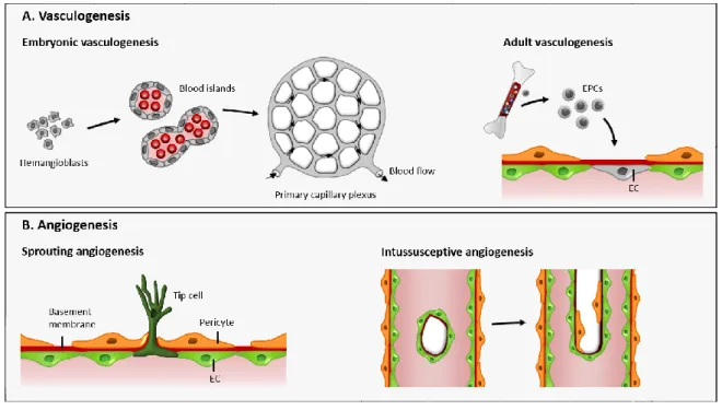
The cardiovascular system is the first functional organ system to develop in the vertebrate embryo in a process called vasculogenesis. The blood islands found in the extraembryonic yolk sac are the earliest vascular structures observed during development. These blood islands are believed to derive from the hemangioblasts which are commonly defined as precursors of endothelial and hematopoietic cells (Choi et al. 1998; Cao & Yao 2011). While the central cells within the blood islands give rise to embryonic hematopoietic cells, the peripheral cells differentiate into endothelial cells that will connect to form a primitive vascular plexus with lumen (Figure 1.1) (Risau & Flamme 1995; Weinstein 1999). Although vasculogenesis is a term usually employed to describe the formation of the primitive blood vessels inside the embryo and its surrounding membranes, it actually defines the de novo blood vessel formation. Several reports have shown that such phenomenon is not restricted to early embryogenesis, and instead occurs throughout adult
4
life, both in physiological and pathological conditions, in a process that is dependent on the recruitment of bone marrow-derived endothelial progenitor cells (EPCs) (Shi et al. 1998; Tepper et al. 2005; Asahara et al. 1999; Ribatti et al. 2001; Drake 2003).
After the primary vascular plexus is formed, it undergoes extensive remodeling where the specification of vessels to arteries and veins by distinct signals and underlying genetic programming takes place. The vascular wall of primitive blood vessels becomes structurally stabilized by mural cells that include vascular smooth muscle cells (SMC) for larger vessels and single pericytes around smaller vessels (Coultas et al. 2005; Adams & Alitalo 2007). Simultaneously, the primary plexus significantly expands in a process where new blood vessels arise from preexisting ones, called angiogenesis.
Angiogenesis can occur through intussusception or sprouting (Figure 1.1). Intussusceptive angiogenesis is the term used when a preexisting capillary is internally divided giving rise to daughter vessels (Djonov et al. 2000; Burri et al. 2004). It was first
Figure 1.1. Mechanisms of blood vessel formation
(A) Mesodermal cells in the early embryo differentiate into endothelial and hematopoietic precursors (hemangioblasts)
and aggregate to form blood islands. Fusion of blood islands leads to the vasculogenic formation of a honeycomb-shaped primary capillary plexi that presents arterial and venous specification of the endothelial cells. In the adult, endothelial progenitor cells (EPCs) are recruited from the bone marrow and differentiate into endothelial cells (ECs), contributing to vessel growth. (B) The expansion and remodeling of the vascular network occurs during angiogenesis by formation of new vessels from preexisting ones in a process called sprouting angiogenesis, or by internally splitting a vessel into two daughter vessels, which is called intussusceptive angiogenesis.
BLOOD VESSEL FORMATION: VASCULOGENESIS AND ANGIOGENESIS
5 described in 1986 by Caduff et al. who showed that the postnatal transformation of the capillary network in the lungs was dependent on the insertion of new transcapillary pillars (Caduff et al. 1986). Such pillars are formed by the protrusion of opposing capillary walls into the lumen of a vessel with subsequent formation of an inter-endothelial zone of contact that, with the invasion of growth factors or cells, such as fibroblasts, leads to the formation of a channel in the vessel that eventually enlarges and splits the vessel into two (Burri et al. 2004). However, the predominant form of angiogenesis involves sprouting of new vessels from preexisting ones, by migration and proliferation of endothelial cells towards a pro-angiogenic stimulus.
1.2.1. Sprouting angiogenesis
Sprouting angiogenesis is a complex process, involving the expression of numerous genes by different cell types, all contributing to an integrated sequence of events (Conway et al. 2001; Geudens & Gerhardt 2011). It is induced by an inadequate supply of oxygen in tissues and organs. In response to hypoxia, the oxygen-sensible hypoxia-inducible factor 1 alpha (HIF1α) is no longer targeted for degradation by the von Hippel-Lindau (VHL) E3 ubiquitin ligase and accumulates inside the cells, leading to the subsequent expression and secretion of pro-angiogenic molecules coded by hypoxia-inducible genes. Examples include the vascular endothelial growth factor (VEGF), angiopoietin 2 (Angpt2), basic fibroblast growth factor (FGF2) and placental growth factor (PlGF) (Pugh & Ratcliffe 2003).
Sprouting endothelial cells
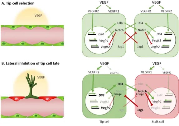
Sprouting angiogenesis is triggered by a pro-angiogenic stimulus, usually VEGF (described later in this Chapter), that induces the activation of quiescent endothelial cells (Figure 1.2A and 1.2B). Depending on each cell responsiveness to VEGF, different types of endothelial cells with unique morphologies are formed within a sprout (Gerhardt et al. 2003). The tip of the vascular sprout comprises a single endothelial cell, usually characterized by being the most responsive to VEGF due to its higher levels of VEGF receptor 2 (VEGFR2), that extends multiple long filopodia in a polarized manner (Figure
1.2B) (Wacker & Gerhardt 2011; Blanco & Gerhardt 2013). This cell, known as the tip cell,
6
(MT1-MMP) (Yana et al. 2007), that favors the degradation of the surrounding basal lamina and presents a motile and invasive behavior that allows it to grow towards attractive cues (Ribatti & Crivellato 2012; Geudens & Gerhardt 2011). The endothelial cells that trail the tip cell and that are responsible for the elongation of the sprout are called stalk cells. Contrasting with the tip cells, stalk cells are highly proliferative and elongate the sprout as the tip cell migrates. In addition, stalk cells undergo morphological and positional rearrangements to form the lumen of the nascent sprout (Figure 1.2C) (Adams & Alitalo 2007; Iruela-Arispe & Davis 2009; Wacker & Gerhardt 2011).
Consistent with the distinct functions of each cell type, endothelial tip and stalk cells also differ in their expression profile. Although a tip cell-specific marker has not been identified, tip cells express high levels of Delta-like 4 (Dll4), VEGFR2, VEGFR3, Angpt2, platelet derived growth factor b (PDGFb) and unc-5 homolog b (Unc5b) and have low levels of Notch signaling activity. Contrastingly, stalk cells express Jagged1, VEGFR1 and Robo4 more strongly than tip cells (Gerhardt et al. 2003; Phng & Gerhardt 2009; Claxton & Fruttiger 2004; Lu et al. 2004; Tammela et al. 2008; Siekmann & Lawson 2007; Ribatti & Crivellato 2012).
Sprout fusion and lumen formation
To ensure that the newly formed sprouts are perfused, they need to have a functional lumen. As the sprouts elongate, lumen formation occurs either by fusion of growing vacuoles of one stalk cell with the vacuoles of adjacent stalk cells or by basal polarization and subsequent repulsion of the apical side of opposing stalk cells (Strilić et al. 2009; Lammert & Axnick 2012; Iruela-Arispe & Davis 2009).
Sprout formation and elongation is followed by a second crucial step towards the formation of a functional vascular network, the vessel anastomosis (Figure 1.2C). When a tip cell encounters the tips of other sprouts or existing capillaries, they suppress their motile and explorative behavior and fuse to create a new circuit that expands the vascular network (Figure 1.2D).
The fusion of migrating tip cells is mediated by Tie2- and Neurophilin-1-positive macrophages that act as chaperones by bridging neighboring tip cells (Figure 1.2C) (Fantin et al. 2010). VEGF-C expression by the tissue macrophages stimulates the VEGFR3- positive
BLOOD VESSEL FORMATION: VASCULOGENESIS AND ANGIOGENESIS
7
Figure 1.2. Cellular mechanisms of angiogenic sprouting
(A) In the absence of pro-angiogenic stimuli, endothelial cells (green) are retained in a quiescent state. (B) During
angiogenesis, high levels of pro-angiogenic factors (such as VEGF, FGF, Angpt2 and PlGF) induce the selection of tip cells (dark green) for sprouting. Sprouting requires the induction of motile and invasive activity, modulation of cell-cell contacts and matrix metalloprotease-mediated degradation of extracellular matrix (ECM). The selected tip cells inhibit adjacent endothelial cells from responding to the pro-angiogenic signals and becoming tip cells. However, adjacent endothelial cells constantly compete for the tip cell position and they may shuffle and exchange positions with tip cells during angiogenic sprouting. (C) During sprout elongation, tip cells are followed by stalk cells (light green), which maintain connectivity with parental vessels and initiate vascular lumen formation through vacuole fusion. Upon contact with other sprouts, tip cell behavior is repressed and vessels fuse in a process called anastomosis, which is mediated by macrophages (purple) that act as chaperones by bridging the tip cells together. (D) Fusion processes establish a continuous and perfused lumen and subsequent maturation processes occur, such as the stabilization of endothelial cell-cell contacts, recruitment of pericytes and establishment of pericyte-endothelial cell contacts and matrix deposition that together re-establish a quiescent endothelial phenotype.
8
tip cells to turn on Notch target genes (VEGF and Notch signaling interaction will be addressed in Section 1.2.2), which decreases VEGF sensitivity in these cells and thus converts them into stalk cells, facilitating the assembly of functional microcirculatory loops (Tammela et al. 2011).
Once lumenised connections have been established and the previously poorly perfused tissues have a suitable oxygen delivery, paracrine VEGF expression is downregulated. This induces the endothelial cells to adopt a quiescent, immotile and non-proliferative phenotype, becoming phalanx cells (Mazzone et al. 2009). Although both the proliferative stalk cells and the quiescent phalanx cells are covered by supporting pericytes (Geudens & Gerhardt 2011), phalanx cells organize in a more regular, “cobblestone” appearance, show an increased expression of junctional molecules, such as ZO-1 and VE-Cadherin, and are surrounded by a more stable basement membrane, which improves tissue perfusion and oxygenation (De Bock et al. 2009; Mazzone et al. 2009).
1.2.2. Signaling pathways involved in sprouting angiogenesis
During the angiogenic process, the responses to the pro- and anti-angiogenic factors, and subsequent modulation of the phenotypic characteristics of the tip, stalk and phalanx cells, are tightly regulated. It depends on the interplay between several signaling pathways, including the VEGF and VEGFRs-, the Delta-Notch-, Angiopoietin/Tie receptor-, FGF- and PDGF-signaling pathways, that will be described in this section, with particular focus on the Delta-Notch signaling pathway.
VEGF signaling
The VEGF signaling pathway has been established as the key regulator of sprouting angiogenesis, both in physiological conditions and in disease. The VEGF family currently comprises seven members: VEGF-A, VEGF-B, VEGF-C, VEGF-D, VEGF-E, VEGF-F and PlGF (Hoeben et al. 2004), each interacting differentially with specific tyrosine kinase receptors, VEGFR1, VEGFR2 and VEGFR3 and the non-signaling co-receptors neuropilin 1 (Nrp1) and Nrp2 (Roy et al. 2006; Hoeben et al. 2004). VEGF-A, often referred to simply as VEGF, is thought to be of singular importance in sprouting angiogenesis as, among other functions,
BLOOD VESSEL FORMATION: VASCULOGENESIS AND ANGIOGENESIS
9 it promotes endothelial cell migration and proliferation and controls endothelial cell-cell junctions (Ferrara 2001; Phng & Gerhardt 2009). In fact, VEGF importance during vascularization is emphasized by the observations that both VEGF null and VEGF heterozygous mice are embryonically lethal (Carmeliet et al. 1996; Ferrara et al. 1996).
Most of the endothelial responses to VEGF are mediated through VEGFR2 (Waltenberger et al. 1994), which positively drives the mitogenic and chemotactic responses of endothelial cells to VEGF (Bernatchez et al. 2002). VRGFR-1 is a high-affinity receptor for VEGF, but its weak tyrosine-kinase activity turn it into a trap for VEGF by suppressing its availability to bind VEGFR2 (Shibuya 2001; Park et al. 1994; Kappas et al. 2008). However, genetic inactivation of either of the receptors causes embryonic lethality (Fong et al. 1995; Shalaby et al. 1995). Consistent with VEGFR1 function as a negative
regulator of VEGF, Vegfr1-/- mice die due to severe vessel overgrowth and disorganization
(Park et al. 1994), a phenotype that is rescued by the expression of the soluble form (that lacks the tyrosine kinase domain) of VEGFR-1 (Hiratsuka et al. 1998). On the other hand, VEGFR2 null mutation disturbs the vasculogenesis process, inhibiting the differentiation of endothelial cells and hematopoietic progenitor cells (Shalaby et al. 1995), suggesting it plays a key role in both vasculogenesis and angiogenesis.
The selection of the tip cell in the sprouting process depends mainly on the levels of VEGFR1 and VEGFR2 present on each cell (Jakobsson et al. 2010). Mosaic analysis have demonstrated that Vegfr2+/- cells have a disadvantage in adopting the tip cell position,
whereas Vegfr1+/- cells have an advantage for the tip cell position. Moreover, as the levels
of VEGF receptors are continuously altered in consequence of Notch activation (Williams et al. 2006), tip cell specification is highly transient, and the cells with higher VEGFR2 and lower VEGFR1 expression dynamically compete with and overtake their neighboring tip cells (Jakobsson et al. 2010).
Two other VEGF receptors are also involved in the tip cell selection and guidance process by interacting with VEGFR2: VEGFR3 and Nrp1. Similar to VEGFR1 and VEGFR2, VEGFR3 null mice also present an embryonic lethal phenotype and die at embryonic day (E) 10.5 due to defects in the primary vessel remodeling (Dumont et al. 1998). VEGFR3 is activated by the VEGF homologues VEGF-C and VEGF-D and is most strongly expressed in the leading tip cells during both mouse and zebrafish angiogenesis (Shawber et al. 2007;
10
Siekmann & Lawson 2007). Loss of VEGFR3 function in retinal endothelial cells results in hypersprouting (Zarkada et al. 2015; Tammela et al. 2011), probably through the loss of VEGFR2/VEGFR3 heterodimers, that negatively modulate VEGFR2 activity (Nilsson et al. 2010; Dixelius et al. 2003; Tammela et al. 2011; Zhang et al. 2010).
Nrp1 is expressed in the angiogenic vasculature, including tip cells, and enhances VEGF-mediated signaling by establishing interactions with VEGFR2. However, consistent with the observations that Nrp1 mutant mice exhibit defects in the heart, vasculature and nervous system and die at E10.5-12.5 (Kawasaki et al. 1999; Gu et al. 2003), this gene has been implicated in tip cell function and guidance in the embryo (Fantin et al. 2013; Jones et al. 2008; Gerhardt et al. 2004), in a process that is independent on the VEGFR2 co-receptor function (Aspalter et al. 2015).
Once tip cells are selected, they polarize and start moving towards the stimulus through the formation of filopodia and lamelopodia. Simultaneously, new sprouts are formed because of the proliferation and migration of adjacent stalk cells. Both the migration of tip cells and the proliferation of stalk cells is mediated by VEGF-induced VEGFR2 signaling. However, whereas tip cell migration depends on a gradient of VEGF, proliferation is regulated by its concentration, showing that VEGF distribution can regulate distinct cellular responses in defined populations of endothelial cells (Gerhardt et al. 2003; Geudens & Gerhardt 2011). Indeed, VEGFR2 activation in endothelial cells leads to activation of intracellular signaling cascades such as the mitogen-activated protein kinase (MAPK) and phosphatidyl-inositol-3 kinase (PI3K) (Karar & Maity 2011; Mavria et al. 2006). While the remodeling of the actin cytoskeleton required for the filopodia and lamelopodia formation seems to be induced by PI3K/Akt activation of Rho small GTPases, particularly CDC42 (Lamalice et al. 2004; Lamalice et al. 2007; Abraham et al. 2015), MAPK signaling has been implicated in endothelial cell proliferation (Meadows et al. 2001) and survival (Gupta et al. 1999; Berra et al. 2000). A balance in the activation of these two pathways might be what regulates the differential response to VEGF in tip and stalk cells.
Notch signaling
The Notch pathway is an evolutionarily conserved contact-dependent signaling machinery that is required for embryonic development, regulation of tissue homeostasis
BLOOD VESSEL FORMATION: VASCULOGENESIS AND ANGIOGENESIS
11 and maintenance of the stem cell pool in adults. In fact, the Notch signaling pathway is a critical regulator of multiple cell fate decisions, tissue patterning and morphogenesis by coordinating proliferation, differentiation and survival in a broad range of cell types in a single organism and at different steps during cell lineage progression (J. Liu et al. 2010; Andersson et al. 2011).
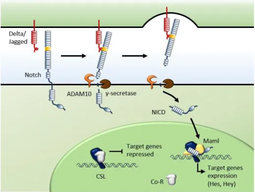
In mammals, five canonical ligands, Delta-like 1 (Dll1), Delta-like 3 (Dll3), Delta-like 4 (Dll4), Jagged 1 (Jag1) and Jagged 2 (Jag2), interact with four Notch receptors (Notch1-Notch4). Both the Notch receptors and ligands are transmembrane proteins with large extracellular domains containing multiple epidermal growth factor (EGF)-like repeats. When receptor-ligand interactions are established between neighboring cells, two proteolytic cleavage events are triggered in the Notch receptor. The first cleavage is catalyzed by ADAM-family metalloproteases within the juxtamembrane region, whereas the second one is mediated by the γ-secretase complex within the single transmembrane domain of Notch receptors. The final cleavage releases the Notch intracellular domain (NICD) from the cell membrane which subsequently translocates to the nucleus where it interacts directly with the transcription factor RBPjk, also known as CSL (CBF1, Su(H), Lag2, after its mammalian, Drosophila and Caenorhabditis elegans orthologues).
In the absence of NICD, CSL functions as a transcriptional repressor by establishing interactions with a transcriptional corepressor complex. Following NICD binding to CSL, as a result of Notch activation, the corepressors are displaced, and the transcriptional coactivator Mastermind-like (Maml) is recruited. This NICD/CSL/Maml complex recruits additional coactivators to activate transcription of downstream target genes, such as hairy/enhancer of split (HES) and HES-related proteins (HEY/HRT/HERP), which in turn act as transcriptional regulators of further downstream genes (Figure 1.3) (Bray 2006; Blanco & Gerhardt 2013; Andersson et al. 2011; Kopan & Ilagan 2009; Holderfield & Hughes 2008; Phng & Gerhardt 2009). Several observations indicate that the Notch signaling pathway plays a key role at different stages of vascular development. The Notch ligands Dll1, Dll4 and Jag1 and the Notch receptors 1 and 4 are expressed in the vasculature (Hofmann & Iruela-Arispe 2007). Furthermore, deletion of genes involved in Notch signaling transduction, including receptors, ligands, transcription factors, downstream targets, and molecules that are involved in Notch processing lead to embryonic lethality in mice due to
12
severe vascular defects (Table 1.I). Knockout mice for Notch1, Notch1/Notch4 or the Notch ligand Jag1 die in utero between E9.5 and E10.5 of gestation, exhibiting major vascular abnormalities due to defects in the primary vascular plexus remodeling into a functional and organized vascular network (Krebs et al. 2000; Xue et al. 1999; Krebs et al. 2004; Gale et al. 2004; Duarte et al. 2004). Such defects seem to be caused particularly by the absence of those genes in endothelial cells because endothelial-specific knockout of Notch1 or Jag1, as well as expression of a constitutively active form of Notch4 (Notch4/int3) and Jag1 overexpression in an endothelial-specific manner, all cause embryonic lethality due to severe vascular defects similar to the ones found in Notch1-deficient mice (Limbourg 2005; Uyttendaele et al. 2001; Benedito et al. 2009; High et al. 2008). Moreover, although they are not found in the vasculature, Notch2 and Notch2/Notch3 knockout also induce vascular deficiencies that seem to relate with the lack of smooth muscle cell coverage, highlighting
Figure 1.3. Canonical Notch signaling pathway
Delta/Jagged binding to the Notch receptor on an adjacent cell induces two proteolytic cleavages of the receptor catalyzed by ADAM10 and γ-secretase complex. This proteolytic processing mediates the release of Notch intracellular domain (NICD), which translocates to the nucleus and physically associates with DNA-binding CSL protein. In the absence of Notch activation, CSL recruits corepressors and silences transcription of Notch target genes. When bound to NICD, CSL recruits coactivators, such as Mastermind-like (Maml), and functions as transcriptional activator of Notch downstream target genes encoding various Hey and Hes family proteins.
BLOOD VESSEL FORMATION: VASCULOGENESIS AND ANGIOGENESIS
13 the role of the Notch signaling pathway in several steps of vascular development (Hamada et al. 1999; Wang et al. 2012; McCright et al. 2001). Consistent with these findings, knockout mice for Adam10, components of the γ-secretase complex (Ps1-/-/Ps2-/-) or
downstream targets of the Notch pathway (Hey-/-/Hey2-/-) all die during embryonic
development with defects in the primary vascular plexus remodeling and in arteriovenous specification (Hartmann et al. 2002; Fischer et al. 2004; Kokubo et al. 2005; Herreman et al. 1999; Donoviel et al. 1999).
The Notch ligand Dll4 seems to be of particular importance in vascular development, which is highlighted by reports showing it is the only Notch pathway component causing haploinsufficiency in most genetic backgrounds (Duarte et al. 2004; Gale et al. 2004; Krebs et al. 2004), a penetrance only comparable to VEGF (Carmeliet et al. 1996). Knocking out one allele of Dll4 causes vascular remodeling defects and arteriovenous malformations such as the ones found in Notch1-/- mutants (Gale et al. 2004; Duarte et al. 2004; Krebs et
al. 2004). Moreover, Dll4+/- mice have an increased number of vascular sprouts and vessel
branches in the growing front of some vascular beds, such as the yolk sac (Gale et al. 2004; Suchting et al. 2007). The opposite phenotype is found in both ubiquitous and endothelial-specific Dll4 overexpression in mice, which cause decreased sprouting and increased arterialization (Trindade et al. 2008).
Table 1.I. Vascular defects associated with Notch-pathway mutants.
Mutated Gene Phenotype
Receptors
Notch1-/-
Lethal at E10.5-11. Failure in remodeling the primary vascular plexus and disorganized embryonic vasculature. Impaired generation of HSCs from the hemangioblast and long-term definitive hematopoiesis (Krebs et al. 2000; Kumano et al. 2003; Hadland et al. 2004)
Notch2-/- Embryonic lethal at around E11.5. Reduced coverage of vascular smooth muscle cells
(Hamada et al. 1999; Wang et al. 2012)
Notch2 hypomorphic allele (Notch2del1/del1)
Embryonic lethality prior to E16.5. Defects in glomerular development in the kidney and in the development of the eye vasculature. Myocardial hypoplasia, hemorrhaging, and edema (McCright et al. 2001)
Jagged1–/+/Notch2del/+ Mimics human Alagille syndrome defects. Heart and kidney glomerular defects (McCright et al. 2002)
Notch3
-/-Viable and fertile. Decreased retinal vascularization at early stages; Defective arterial specification and maturation of arterial vascular smooth muscle. (Krebs et al. 2003; Domenga et al. 2004; H. Liu et al. 2010)
14
Mutated Gene Phenotype
Notch1-/-/Notch4-/-
Lethal around E9.5. Failure in remodeling the primary vascular plexus and disorganized embryonic vasculature. Vascular defects were more severe than in Notch1-/- embryos
(Krebs et al. 2000)
Notch2-/-/Notch3-/- Lethal at E11.5. Enlarged vessels and thin vessel walls with reduced coverage of vascular smooth muscle cells (Wang et al. 2012)
EC-specific Notch1 knockout
(Tie2-Cre/Notch1lox/lox)
Embryonic lethal between E10.5-E11.5. Phenocopies Notch1-deficient mice, with absence of primary vascular plexus remodeling to form large and small blood vessels of the mature yolk sac (Limbourg 2005)
Constitutively active form of Notch4 (int3) regulated by the VEGFR-2 (Flk1) locus (Flk1/Int3)
Embryonic lethality between E9.5-10.5. Displayed disorganized vascular networks and dilated vessels. Failure in vascular remodeling and stabilization both in embryos and the yolk sac (Uyttendaele et al. 2001)
Notch4(int3)-inducible
inactivation in ECs
(Tie2-tTA/TRE-Int3)
Embryonic lethality. Expression in the adult causes blood vessel enlargement, defective arterialization and increased vascular smooth muscle cells. These phenotypes were reversed upon repression of int3 expression (Carlson et al. 2005)
Ligands
Jag1–/– Lethal between E9.5–11.5. Defective remodeling of the embryonic and yolk sac vasculature (Xue et al. 1999)
Dll1lacZ/Dll1 kineo (combined Dll1 hypomorphic and null alleles)
Viable and fertile with loss of arterial identity (Sörensen et al. 2009)
Dll4+/–
Embryonic lethality at E9.5-10.5. Phenocopies Notch1-/-/Notch4-/- mutants. Absent remodeling of the yolk sac vasculature and defective arterial branching from the aorta. The number of vascular sprouts and vessel branches is increased in the growth front of some vascular beds, such as the yolk sac (Krebs et al. 2004; Gale et al. 2004; Duarte et al. 2004)
Inducible Dll4 overexpression
(chicken beta-actin
(CAG)-Cre/TetO7-Dll4)
Lethal at around E9.0-9.5. Failure in remodeling the primary vascular plexus; arterial cell identity is established in the venous compartment. Defects in vascular sprouting (Trindade et al. 2008)
EC-specific Dll4
overexpression (Tie2-rtTA-M2/TetO7-Dll4)
Lethal at around E10.5 presenting the same defects as ubiquitous Dll4 overexpression (Trindade et al. 2008)
EC-specific Jag1 knockout (Tie1-Cre/Jag1lox/lox or
Tie2-Cre/Jag1lox/lox)
Embryonic lethal at about E10.5. Defects in smooth muscle cell development in both embryonic and yolk sac blood vessels, accompanied with loss of arterial specification (Benedito et al. 2009; High et al. 2008)
EC-specific Jag1 overexpression
(VE-Cadherin-tTA/TetO-Jag1)
Lethal before E16.5. Extensive hemorrhaging in the skin. (Benedito et al. 2009) Regulators
Rbpsuh–/– Embryonic lethal before E9.5. Vascular defects similar to Notch1-/-/Notch4-/- double
mutants. Loss of arterial specification (Oka et al. 1995; Krebs et al. 2004)
Adam10–/– Lethal at around E9.5. Vascular defects resemble the ones found in Notch1-/-/Notch4
-/-mice (Hartmann et al. 2002) EC-specific Adam10
knockout
(Tie2-Cre/Adam10lox/lox)
Embryonic lethal at E11.5. Exhibit large-caliber vessels on the liver surface and myocardium. Augmented expansion of erythroid precursors in the BM, but increased hemolysis (Glomski et al. 2011)
Ps1–/– (component of the
γ-secretase complex)
Lethal at late gestation or after birth. Vascular remodeling failure in stomach and skin; reduction of cerebral sprouting in the brain with increased diameter of sprouting capillaries and brain hemorrhages (Nakajima et al. 2003)
Ps2–/– (component of the
γ-secretase complex)
Viable and fertile. Hemorrhages and pulmonary fibrosis found in adult mice (Herreman et al. 1999)
Ps1–/–/Ps2–/– Embryonic lethality after E9.5. Failure in remodeling the yolk sac vasculature and
BLOOD VESSEL FORMATION: VASCULOGENESIS AND ANGIOGENESIS
15
Mutated Gene Phenotype
Target genes
Hey1 –/– Viable and fertile. No obvious phenotypic anomaly (Fischer et al. 2004)
Hey2 –/– Mice die within 10 days after birth. Ventricular septal defects (Sakata et al. 2002; Gessler
et al. 2002; Donovan et al. 2002)
Hey1–/–/Hey2–/–
Lethal between E9.5-11.5. Failure in remodeling the primitive vascular plexus in the yolk sac, massive hemorrhages and absence of large embryonic blood vessels (Fischer et al. 2004; Kokubo et al. 2005)
In the last decade, studies in the mouse retina, in zebrafish intersegmental vessels and in 3D endothelial cell culture sprouting assays have demonstrated that the Dll4/Notch signaling is the major regulator of the tip and stalk cell specification process (Hellström et al. 2007; Lobov et al. 2011; Siekmann & Lawson 2007; Suchting et al. 2007; Leslie et al. 2007; Patel 2005). The mechanism by which Notch imposes differential behavior in endothelial cells that are exposed to similar doses of a pro-angiogenic stimulus is directly connected to VEGF signaling, and a negative regulatory loop is established between these two pathways. VEGF signaling in endothelial cells regulates Dll4 expression both in vivo and in vitro. Studies in retina where VEGF is blocked, either with VEGF-Trap or with a soluble form of VEGFR1, both decreased Dll4 mRNA expression (Suchting et al. 2007; Lobov et al. 2007). Conversely, VEGF stimulation in human umbilical cord vein endothelial cells (HUVECs) increases Dll4 expression (Ridgway et al. 2006), a phenotype that was also observed in human tumor samples where VEGF and Dll4 expression were found to be directly correlated (Patel 2005).
In addition to VEGF acting upstream of Dll4, it has become clear that Dll4-induced Notch signaling activation negatively regulates VEGF signaling by regulating the expression of the different VEGF receptors (VEGFR1, VEGFR2, VEGFR3 and Nrp1). Heterozygous mice for Dll4 (Dll4+/-) were shown to have a downregulation in VEGFR2 accompanied with
VEGFR1 upregulation in retinal vessels, which correlated with increased sprouting (Suchting et al. 2007; Jakobsson et al. 2010). In vitro studies have also helped to shed some light on the Notch-dependent VEGF signaling regulation. Notch activation in HUVECs decreases both VEGFR2 and Nrp1 mRNA expression (Williams et al. 2006; Ridgway et al. 2006) and simultaneously upregulates of VEGFR1 and VEGFR3 (Harrington et al. 2008; Funahashi et al. 2010; Shawber et al. 2007), which altogether renders the cells less responsive to VEGF.