Rev Bras Hematol Hemoter. 2011;33(1) 73
Simultaneous lymph node involvement by Castleman disease and
Kaposi sarcoma
Both multicentric Castleman disease and Kaposi sarcoma are more frequently observed in HIV infected patients. The coexistence of these Human herpesvirus 8 related lesions, in the same tissue, has been observed, but literature reports are scant. On the other hand, the expression of HHV-8-LANA-1 is easily demonstrable by immunohistochemistry. This has been shown to be a powerful tool for the diagnosis of these entities. The aim of this report is to communicate our experience with a case of multicentric Castleman disease occurring in the setting of HIV infection, which demonstrated microscopic Kaposi sarcoma in the same lymph node during the pathological work-up.
Keywords: Sarcoma, Kaposi; Herpesvirus 8, human; Giant lymph node hyperplasia; Herpesviridae infections; Virus latency; Human; Male; Adult; Case reports
Introduction
Castleman disease (CD) is a rare lymphoproliferative disease, initially observed in patients with localized disease, who are asymptomatic or mildly symptomatic, and were cured with surgical resection. Subsequent observations, based on studies of case series and histopathologic reviews, broadened the knowledge about the disease, which is traditionally grouped into the following histologic forms: hyaline-vascular variant, plasma cell variant, and those with mixed characteristics. It can present as localized lymphadenopathy (localized form) or as disseminated disease (multicentric form).(1)
The symptoms associated with morphologic aspects, suggest the participation of a virus in the pathogenesis of the disease. The observation that approximately 13% of patients with multicentric Castleman Disease (MCD) develop Kaposi sarcoma (KS), led to an interest in investigating the presence of human herpesvirus 8 (HHV-8) in the lymphoid tissues of patients with MCD.(2) Currently it is known that HHV-8 is present in 100% of the cases of
MCD in patients infected with the human immunodeficiency virus (HIV) and in 40% to 50% of HIV-negative cases.(3)
Although the pathogenesis of CD is still not fully understood, the first step in the development of the disease is the production of interleukin-6 (IL-6) – a cytokine with lymphoproliferative properties – by B cells in the mantle zone of lymph nodes. The production of IL-6 is stimulated by HHV-8 infection and it is believed that some as yet unidentified endogenous or exogenous factor must be involved in the cases not associated with the vírus.(4) In addition to the increase in IL-6, it has been observed that
vascular endothelial growth factor (VEGF) levels are increased in the lymph nodes of patients with CD.(1)
MCD has a poor prognosis and a clinical course that is rapidly fatal. It occurs more frequently in HIV-infected patients that present constitutional symptoms, generalized lymphadenopathy, hepatosplenomegaly, polyclonal hypergamma-globulinemia, anemia, and elevated serum levels of IL-6.(5) The histopathologic
examination of the lymph nodes shows lymphoid follicles largely devoid of germinal centers, some degree of hyalinization and expansion of the mantle zones, where one observes variable quantities of plasmablasts with large and vesicular nuclei, with prominent nucleoli, expressing the latency-associated nuclear antigen 1 (LANA-1) of HHV-8. The interfollicular areas appear expanded and infiltrated by mature plasma cells. The plasmablasts can coalesce and form microlymphomas or even HHV-8 positive plasmablastic lymphoma, an entity recently included in the WHO classification for tumors of hematopoietic and lymphoid tissues.(3,6)
KS, similar to MCD, is observed with increased frequency in HIV-positive patients, with the microscopic involvement of lymph nodes and spleen by KS common in patients
Conflict-of-interest disclosure: The authors declare no competing financial interest
Submitted: 10/13/2010 Accepted: 12/24/2010
Correspondence: Luciana Wernersbach Pinto
Instituto de Pesquisa Clínica Evandro Chagas – Fiocruz
Av Brasil , 4365 – Manguinhos 21340-360 – Rio de Janeiro (RJ), Brazil [email protected]
www.rbhh.org or www.scielo.br/rbhh
DOI: 10.5581/1516-8484.20110018
Instituto de Pesquisa Clínica Evandro Chagas (IPE), Fiocruz Rio de Janeiro (RJ), Brazil
Luciana Wernersbach Pinto Estevão Portela Nunes
74 Rev Bras Hematol Hemoter. 2011;33(1)
Pinto LW, Nunes EP
with acquired immunodeficiency syndrome (AIDS).(7) The
coexistence of these two diseases related to HHV-8 in the same tissue has been observed, however there are few reports in the literature and only one recent well-documented case series evaluating the presence of the two lesions in the same tissue specimen.(5,8) On the other hand, the expression of
HHV-8-LANA-1 is easily demonstrated by immunohistochemistry. The objective of this case report is to share our experience with one case of MCD in an HIV-positive patient, which demonstrated the coexistence of microscopic KS in the same lymph node specimen, during the immunohistochemical analysis for the detection of HHV-8.
Case report
The patient is a 40-year-old male who reports daily fever (39º-40ºC), enlarged lymph nodes, a 6 kg weight loss in the past three months, and productive cough for two weeks. Antibiotic therapy was initiated, without improvement. Serologic testing during this period was positive for HIV-1. At the time of hospitalization he presented with exertional dyspnea, cough, fever, cervical and axillary lymphadenomegaly, and hepatosplenomegaly. Laboratory tests revealed anemia and thrombocytopenia. Blood and sputum cultures were negative for mycobacterium. Chest tomography revealed a ground glass infiltrate at the left base and enlarged mediastinal and axillary lymph nodes. Direct examination of sputum did not reveal acid-fast resistant bacilli. Nevertheless, treatment with anti-tuberculous and antifungal regimens was started. The patient, however, developed shortness of breath and worsening thrombocytopenia and renal function, and died ten days after admission. During the hospitalization a cervical lymph node biopsy was performed.
The lymph node submitted to histopathologic examination underwent routine processing and immuno-histochemical studies, using antibodies to assess the expression of the following antigens: CD20 (1:800, DAKO), CD3 (1:400, DAKO), CD138 (1:200, Cell Marque), Bcl-2 (1:200, Novocastra) and HHV-8 (LANA-1) (1:200, Novocastra).
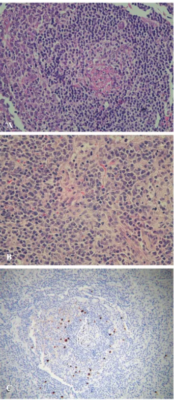
Microscopic evaluation of the lymph node revealed atrophic or fibrotic lymphoid follicles with increased vascularization, surrounded by expanded mantle zones, with a concentric appearance, interspersed with scattered plasmablasts. The interfollicular areas also were expanded – containing a large quantity of mature plasma cells – and highly vascularized (Figures 1A and 1B). Focal areas of capsular thickening in the routine H & E stain were also observed (Figure 2A).
The immunohistochemical study with HHV-8-LANA-1 revealed plasmablasts with nuclear positivity within the mantle zones, as well as the positivity in scattered endothelial cells in the lymphoid follicles (Figure 1C). This pattern of expression associated with histological features led to the diagnosis of HHV-8-associated CD (plasmablastic).
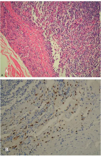
The microscopic involvement of the lymph node by KS was demonstrated by nuclear positivity for the HHV-8-LANA-1 antigen in spindle cells and in endothelial cells lining poorly formed vascular channels in the regions of capsular thickening (Figure 2B).
Figure 1 – Microscopic aspects of Castleman's disease
A – Atrophic lymphoid follicle, with expanded mantle zone with a concentric appearance (H & E x 200)
B – Plasma cells in interfollicular areas (H & E x 200)
C – Immunohistochemistry showing plasmablasts positive for HHV8-LANA1
A
B
Rev Bras Hematol Hemoter. 2011;33(1) 75
Discussion
Although the frequency of CD as a cause of lymphadenopathy in the HIV-infected population is not known, it is not rare. CD in these patients has been described as a new variant, the HHV-8-associated CD (plasmablastic). It can occur with any CD4 T lymphocyte count and it appears that antiretroviral therapy has no effect on the course of the disease.(1,6)
The cells infected by HHV-8 in MCD related to HIV have been localized and characterized by studies using immunohistochemistry. There is a consensus that they are B cells with plasmablastic morphology or plasma cell differentiation, localized in the mantle zone of lymphoid follicles and interfollicular areas. Brousset et al., (9) in a study
that used double staining to evaluate the expression of LANA-1 and v-IL-6 in cases of MCD and KS, observed that in MCD cases, about one-third of the lymphoid cells were
positive for both of the markers, indicating the expression of genes of the lytic phase and of the latency in the plasmablasts. These authors further showed that in some of the lymph nodes in MCD there was expression of LANA-1 in spindle cells (some clearly endothelial cells and others resembling fibroblasts), as well as in lymphoid cells. In one of the cases studied, spindle cells were abundant, with areas that recalled KS. (9)
Abe et al.(8) reported a case of KS and MCD in a single
lymph node from a HIV-positive patient, in which they demonstrated, using immunohistochemistry, the differential expression of the viral proteins in the two lesions. In MCD, proteins of the lytic phase and of the latency of HHV-8 are observed in the cells of the mantle zone, while in KS neoplastic cells are positive for LANA-1, with rare expression of lytic proteins.
Naresh et al.(5) recently investigated the coexistence of
these two entities related to HHV-8 (KS and MCD) in 24 lymph nodes and five spleens from patients with MCD associated with HIV. Among the lymph node specimens, 63% had microscopic involvement by KS involving the capsule, trabeculae, or hilum. This group was compared with 20 lymph nodes of HIV-positive patients without evidence of lymphoproliferative disorders. In this second group, 25% of the lymph nodes had microscopic involvement by KS, a percentage significantly lower than in the group with MCD. The hypothesis of the authors was that the association of the two diseases is due to lytic infection of the B lymphoid cells by HHV-8, exposing susceptible cells in the vulnerable sites of the lymph node to very high levels of HHV-8, which results in the formation of KS “tumorlets”. This study also called attention to the fact that the microscopic foci of KS spindle cells in the capsule, trabeculae and hilum of lymph nodes are subtle and can be easily missed in routine histological evaluation or even after immunohistochemistry.(5)
In the study of the present case, the microscopic focus of KS was not detected with routine H & E staining, and initially we only diagnosed Castleman Disease. With the availability of the anti-HHV-8-LANA-1 antibody for routine use, it was possible to demonstrate the presence of HHV-8 positive plasmablasts and to detect the slight proliferation of spindle cells and immunopositive vascular channels close to the lymph node capsule.
In patients with MCD and HIV infection, in addition to antiretroviral therapy, systemic therapy should be instituted. This can be done with chemotherapy, immunomodulatory agents or monoclonal antibodies against the receptor for IL-6 or anti-CD20 (rituximab). Rituximab has shown promise in inducing a durable remission, but it has been observed that some patients experience adverse effects such as the reactivation of Kaposi sarcoma.(10) This finding emphasizes the importance of the
detection by the pathologist of microscopic foci of KS in lymph nodes of patients with MCD.
Given the findings of this case study and the data from the case series of Naresh et al.,(5) we suggest that the
Figure 2 – Areas with nodal Kaposi's sarcoma.
A – Note the capsular thickening and ill-formed vascular channels (H & E x 100)
B – Immunohistochemistry with anti-HHV8-LANA1 showing nuclear positivity in spindle cells and in the endothelial cells lining the vascular channels
A
B
76 Rev Bras Hematol Hemoter. 2011;33(1)
assessment by immunohistochemistry for HHV-8 (LANA-1) should always be considered in lymph nodes of HIV-positive patients with CD or those with ill-defined areas of capsular/ trabecular thickening and vascular proliferation on microscopic examination.
Agradecimentos
The authors thank Leigh Passman for editing the text.
References
1. Casper C. The aetiology and management of Castleman disease at 50 years: translating pathophysiology to patient care. Br J Haematol. 2005;129(1):3-17. Review.
2. Soulier J, Grollet L, Oksenhendler E, Cacoub P, Cazals-Hatem D, Babinet P, et al. Kaposis sarcoma-associated herpesvirus-like DNA sequences in multicentric Castleman's disease. Blood. 1995;86(4): 1276-80. Comment in: Blood. 1996;87(1):414-6.
3. Du MQ, Bacon CM, Isaacson PG. Kaposi sarcoma-associated herpesvirus/human herpesvirus 8 and lymphoproliferative disorders. J Clin Pathol. 2007;60(12):1350-7. Review.
4. Aoki Y, Jaffe ES, Chang Y, Jones K, Teruya-Feldstein J, Moore PS, Tosato G. Angiogenesis and hematopoiesis induced by
Kaposis sarcoma-associated herpesvirus-encoded interleukin-6. Blood. 1999;93(12):4034-43.Comment in: Blood. 1999;93 (12):4031-3.
5. Naresh KN, Rice AJ, Bower M. Lymph nodes involved by multicentric Castleman disease among HIV-positive individuals are often involved by Kaposi sarcoma. Am J Surg Pathol. 2008;32 (7):1006-12.
6. Cronin DM, Warnke RA. Castleman disease: an update on classification and the spectrum of associated lesions. Adv Anat Pathol. 2009;16(4):236-46. Review.
7. Moskowitz LB, Hensley GT, Gould EW, Weiss SD. Frequency and anatomic distribution of lymphadenopathic Kaposis sarcoma in the acquired immunodeficiency syndrome: an autopsy series. Hum Pathol. 1985;16(5):447-56.
8. Abe Y, Matsubara D, Gatanaga H, Oka S, Kimura S, Sasao Y, et al. Distinct expression of Kaposis sarcoma-associated herpesvirus-encoded proteins in Kaposis sarcoma and multicentric Castleman's disease. Pathol Int. 2006;56(10):617-24.
9. Brousset P, Cesarman E, Meggetto F, Lamant L, Delsol G. Colocalization of the viral interleukin-6 with latent nuclear antigen-1 of human herpesvirus-8 in endothelial spindle cells of Kaposis sarcoma and lymphoid cells of multicentric Castleman's disease. Hum Pathol. 2001;32(1):95-100.
10. Casper C. New approaches to the treatment of human herpesvirus 8-associated disease. Rev Med Virol. 2008;18 (5): 321-9. Review.
xxx