UNIVERSIDADE DE LISBOA
FACULDADE DE FARMÁCIA
Development of three-dimensional umbilical cord-derived
mesenchymal stem cell cultures for differentiation into
hepatocyte-like cells: a potential breakthrough in toxicological
drug screening
Madalena Zincke dos Reis Fernandes Cipriano
Orientadora:
Doutora Joana Paiva Gomes Miranda
Co-orientadores: Doutor Jorge Miguel Silva Santos
Doutora Katrin Zeilinger
Tese especialmente elaborada para a obtenção do grau de Doutor em Farmácia,
especialidade de Toxicologia
UNIVERSIDADE DE LISBOA
FACULDADE DE FARMÁCIA
Development of three-dimensional umbilical cord-derived mesenchymal stem
cell cultures for differentiation into hepatocyte-like cells: a potential
breakthrough in toxicological drug screening
Madalena Zincke dos Reis Fernandes Cipriano
Orientadora: Doutora Joana Paiva Gomes Miranda Co-orientadores: Doutor Jorge Miguel Silva Santos
Doutora Katrin Zeilinger
Tese especialmente elaborada para a obtenção do grau de Doutor em Farmácia, especialidade de Toxicologia
Júri:
Presidente: Doutora Matilde da Luz dos Santos Duque da Fonseca e Castro, Professora Catedrática e Directora da Faculdade de Farmácia da Universidade de Lisboa
Vogais: Doutor Jorge Manuel Lira Gonçalves Ruas, Associate Professor, Karolinska Institutet, Sweden
Doutor Félix Dias Carvalho, Professor Catedrático, Faculdade de Farmácia da Universidade do Porto
Doutora Diana Esperança dos Santos Nascimento, Investigadora Auxiliar, Instituto de Engenharia Biomédica da Universidade do Porto
Doutora Sofia de Azeredo Pereira Costa, Professora Auxiliar, Faculdade de Ciências Médicas da Universidade Nova de Lisboa
Doutora Cecília Maria Pereira Rodrigues, Professora Catedrática, Faculdade de Farmácia da Universidade de Lisboa
Doutor Nuno Filipe da Rocha Guerreiro de Oliveira, Professor Auxiliar com Agregação, Faculdade de Farmácia da Universidade de Lisboa
Doutora Joana Paiva Gomes Miranda, Investigadora Auxiliar, Faculdade de Farmácia da Universidade de Lisboa
iMed.ULisboa / Fundação para a Ciência e Tecnologia, Portugal
2017
This work has been performed at:
Chemical Biology and Toxicology Group, Research Institute for Medicines (CBT, iMed.ULisboa) in the Faculty of Pharmacy of University of Lisbon, Portugal; ECBio, S.A., Portugal; and Berlin-Brandenburg Center for Regenerative Therapies (BCRT) in the Charité Universitätsmedizin in Berlin, Germany; under the scientific supervision of Joana Paiva Gomes Miranda, PhD, and co-supervision of Miguel Silva Santos, PhD, and Dr. med. vet. Katrin Zeilinger.
Madalena Cipriano is a recipient of a PhD fellowship (SFRH/BD/87508/2012) from the Fundação para a Ciência e Tecnologia (FCT), Lisboa, Portugal and this work was developed within the framework of the FCT funded project “Development of 3D culture models for human UCX® cell-derived hepatocytes for predictive toxicology and cell therapy”, reference PTDC/SAU-TOX/110457/2009.
ACKNOWLEDGEMENTS
First and foremost I would like to express my special appreciation to my advisors.
To Joana Miranda, my main advisor, special thanks for believing in me. While I was still an undergraduate student, you gave me the opportunity to start a small research project and patiently teach me all about the basic laboratory and cell culture rules, techniques and tricks. You also lighted up the science bug on me and tought me to run for what I wanted. You always pushed me to grow as a researcher. I always felt constant support and friendship, especially in the most difficult times. Thank you for the opportunity to be part of our team, for creating the conditions of a complete education at several levels, scientifically, socially and humanly. With you I learned how important it is to be part of a team, support other team members and to live in different environments to strength your capacities.
A special acknowledgement is also due to Miguel Santos. Thank you for the relentless support and availability, as well as critical thinking and scientific discussions, crucial for the great course of this work.
To Katrin Zeillinger my co-advisor, for all the support and trust, as well as for welcoming me into her research group, in the Charité – Universitätsmedizin Berlin (Germany). Without your cooperation and support a significant part of this work would not have been possible.
An acknowledgement is also due to the Faculdade de Farmácia, Universidade de Lisboa, in particular to the Departamento de Ciências Toxicológicas e Bromatológicas, and the Research Institute for Medicines (iMed.ULisboa) for the facilities and conditions that allowed me to develop my PhD work, as well as to the Charité – Universitätsmedizin and ECBio, SA,. To the Portuguese government (Fundação para a Ciência e a Tecnologia) and FEDER for the funding, namely my PhD grant (SFRH/BD/87508/2012) and to the project funding (PTDC/SAU-TOX/110457/2009), as well as the strategic projects PEst-OE/SAU/ UI4013/2011 and UID/DTP/04138/2013.
To all my colleagues from the Chemical Biology and Toxicology Group, special thanks for all the help and constant support. I would like to thank Professor Matilde Castro, the CBT coordinator at the time I joined CBT, for all support during this thesis.
To my first lab colleagues Ana Sofia Coelho and Joana Marques. Joana, you are much more than a CBT colleague but a friend for life in and outside of science. I would also acknowledge Elysse Filipe, Patrícia Guerreiro, Pedro Pinheiro, Ana and João. To Inês Ferreira for receiving me in her home in Berlin, and for being always there for scientific and political discussions. I
last period of this thesis, for growing together as a team.
To Professor Nuno Oliveira for his support, scientific discussions and for being always present. To ECBio, SA, in particular to Miguel and also to Rita Barcia, Pedro Cruz and Hélder Cruz, for supporting my work, for providing the stem cells, for the scientific discussions and contribution for paper manuscripts. I am also grateful for being always welcome to run experiments there. To AG Zeillinger, Bioreactor Group, the Berlin-Brandenburg Center for Regenerative Therapies (BCRT) in the Charité Universitätsmedizin in Berlin, Germany. Words cannot express how thankful I am for the warm welcome and the way you took me into your group and made me feel at home (even when home was quite far away). To Fanny, for being always there to solve bioreactor problems and to arrange social activities. A special acknowledgement to Nora Freyer, for being always there for scientific, political and cultural discussions. You were my support during a new chapter of my life that made me grow so much in those short 6 months. I would also acknowledge the support of Christine, Marco, Thomas and Jasmina that were also part of these great times.
To Ruas Jorge Group, Molecular and Cellular Exercise Physiology, Department of Physiology and Pharmacology, Karolinska Institutet in Stockholm, Sweden. In particular to Jorge Ruas and Jorge Correia for welcoming me in their lab and for the scientific input on a crucial moment of the work here presented.
To Sofia Pereira and her CEDOC team, namely Catarina, Nádia and Nuno for the precious collaboration and support in the last year of this project in the HPLC methods and data analysis as well as for being so kind and helpful to me.
Finally, I need to acknowledge my always supporting family, my friends and my team for being there and believing in me.
ABSTRACT
A standard truly predictive in vitro hepatic model has been sought for reducing animal testing, increasing drug development efficiency and improving prediction of adverse drug reactions. The liver is the main organ responsible for xenobiotic metabolism, being particularly exposed to chemicals and its metabolites. The currently available hepatic in vitro models are insufficient. Those present low biotransformation activity, quickly lose its differentiated phenotype in vitro and/or have relevant interspecies differences. Therefore, the differentiation of stem cells (SC) into hepatocyte-like cells (HLC) has been suggested as an alternative approach to provide a representative human hepatic model. However, a functional hepatocyte-like phenotype has not yet been achieved.
In this thesis, we intended to demonstrate that by (i) using a human neonatal mesenchymal stem cell (hnMSC) source, UCX®, (ii) designing a protocol to differentiate this hnMSC into HLC using the cytokines and growth factors present during liver embryogenesis and (iii) resorting to 3D culture conditions would provide an environment closer to the in vivo, and allow a more representative cellular morphology, gene expression and biological behaviour of the hepatocyte.
Umbilical cord matrix hnMSC were selected for the hepatic differentiation due to its high availability, expansion capacity and genomic stability upon expansion, and, due to its more primordial origin, its higher differentiation capacity relative to other MSC sources. The hepatic differentiation protocol was step-wise developed under 2D cell culture conditions. FGF-2 at 4 ng/mL and the absence of FGF-4 resulted in an improved endoderm commitment and foregut induction during the first step of the differentiation protocol. The epigenetic modifiers trichostatin A (TSA), 5-azacytidine (5-AZA) and dimethyl sulfoxide (DMSO) were tested for an improved hepatoblast formation and maturation to HLC. The 21 day-long protocol was extended up to day 34, when a global gene expression analysis was performed. Transcriptomic analyses placed HLC between the HepG2 cell line and hpHep and distant from hnMSC and allowed us to understand how close we were from the hepatocyte transcriptome. HLC were also characterized up to day 34 at protein and functional level. DMSO at the concentration of 1 % and the cell passage day 17 of the differentiation with 24h of 5-AZA treatment, resulted in the best tested protocol. HLC were able to store glycogen, produce albumin and urea and express genes encoding for key hepatic enzymes. Most importantly, HLCs displayed stable UGTs, EROD (CY1A1/2), ECOD (CYP2B6, 1A2 and 2E1), CYP1A1, CYP2C9 and CYP3A4-dependent activities for 13 days at levels comparable to those observed in cultured primary rat hepatocytes. The presence of the hepatic drug transporters
enzymes was observed for the first time on hnMSC derived HLCs.
To further mimic the in vivo developing liver and thus achieve a more mature hepatic phenotype we established the 3D cultures. hnMSC 3D cultures aimed at evaluating hnMSC self-assembling capacity as well as the effect of this cell culture condition on its proliferation and/or differentiation potential. Viable and low proliferative hnMSC spheroids were obtained by resorting to (i) dynamic cultures using spinner flask suspension cultures (SFSC); and (ii) static cultures using ultra-low attachment (ULA) plates. In addition, 3D culture primed the hnMSC to produce a rich ECM and was successfully adapted to serum free conditions, which is particularly relevant for the hepatic differentiation.
3D culture of HLCs was established as spheroids, formed in ULA plates, and into a hollow-fiber perfused bioreactor designed for high density hepatocyte culture. 3D-HLC showed a partial hepatic zonation, observed by immunofluorescence and glycogen storage staining, and an improved hepatic functionality relative to 2D-HLC, namely by converting drugs into its specific metabolites and by producing higher amounts of glutathione. A thorough evaluation of 3D-HLCs biotransformation competency was confirmed using the model drug nevirapine (NVP). 3D HLCs distinguished from 2D-HLC by producing all phase I and II metabolites and by showing the modulation of phase I, II and III enzymes expression upon 10 days of NVP treatment. HLC thilomic profile was also presented for the first time. NVP resulted in an increased glutathione synthesis and oxidation in 3D HLC indicating a higher susceptibility to NVP toxic metabolites relative to 2D-HLC.
The thesis that hnMSC were capable of differentiation into HLC was here proved. By resorting to 3D culture conditions a more representative hMSC-based in vitro model of the liver physiological conditions was developed, extensively characterized and reinforced by the NVP study. This work highlights the relevance of stem cell derived HLC as an alternative model to study drug metabolism and unveil toxicity alerts associated with drug metabolism and bioactivation. The cellular system here developed is a step towards the 3 R´s implementation in terms of mechanistic studies as well as high-throughput drug screening.
Keywords:
Umbilical-cord matrix mesenchymal stem cells; Hepatic differentiation; Three-dimensional cell cultures; In vitro alternative models; In vitro toxicology
RESUMO
O processo de desenvolvimento de um novo fármaco é dispendioso, longo e tem uma taxa de sucesso de apenas 11 %. A falta de eficácia ou segurança das moléculas em desenvolvimento correspondem a cerca de 30 % da taxa de insucesso, que ocorre maioritariamente nas fases IIb e III. Pelo que, é essencial compreender melhor e o mais precocemente possível os mecanismos de ação e toxicidade das moléculas em desenvolvimento. Para isso, são necessários modelos celulares mais relevantes. que utilizem células humanas para evitar as diferenças entre espécies, e que sejam fisiologicamente mais representativos, permitindo assim reduzir os custos e a experimentação animal.
O fígado é um órgão essencial para o processo de avaliação da segurança e eficácia de novas moléculas devido ao seu papel na biotransformação. O processo de biotransformação regula, de certa forma, a disponibilidade de um fármaco e seus metabolitos em circulação, em particular no que respeita aos fármacos de administração oral. Quer a molécula mãe quer os seus produtos de biotransformação podem ser responsáveis pelos efeitos tóxicos dos xenóbioticos diretamente no fígado ou em outros órgãos. Assim, o estudo da biotransformação e da hepatotoxicidade é particularmente importante no desenvolvimento não clínico. As linhas celulares atualmente utilizadas em estudos in vitro, como as células HepG2 (de origem tumoral), apresentam uma capacidade de biotransformação baixa e pouco representativa, enquanto os hepatócitos primários (isolados de tecidos animais ou de biopsias humanas), perdem a sua capacidade de biotransformação com o tempo em cultura. Os hepatócitos primários humanos apresentam ainda uma disponibilidade muito limitada. Para tentar colmatar as falhas dos atuais modelos in vitro, a diferenciação de células estaminais (CE) em células tipo-hepatócito (CTH) tem sido sugerida como uma fonte alternativa de células humanas. No entanto, ainda não foi possível originar com sucesso uma população de CTH madura derivada de CE.
Neste sentido, a tese aqui apresentada pretendeu demonstrar que (i) utilizando células estaminais mesenquimais humanas isoladas da matriz do cordão umbilical (UCX®), (ii) desenvolvendo um protocolo de diferenciação baseado no conhecimento das citoquinas e fatores de crescimento envolvidos no desenvolvimento embrionário do fígado e (iii) recorrendo a sistemas de cultura tridimensionais (3D) resultaria num ambiente in vitro mais semelhante ao dos tecidos in vivo, proporcionando uma morfologia celular, expressão génica e comportamento biológico mais representativo do hepatócito. Como tal, permitindo a obtenção de CTH funcionais e competentes.
(CEMnh) neste trabalho prende-se com a sua elevada capacidade de proliferação e estabilidade cromossómica, elevada disponibilidade e reduzidas questões éticas. A origem neonatal desta população de CEMnh surge também como uma vantagem destas por apresentarem uma menor marca epigenética do estilo de vida e idade do dador, uma capacidade de diferenciação superior em comparação com outras células estaminais mesenquimais, e numa reduzida imunogenicidade, que permite uma potencial aplicação clínica das células diferenciadas.
O protocolo de diferenciação hepática foi primeiramente desenvolvido em culturas em monocamada (2D). Os três passos do protocolo de diferenciação hepática in vitro compreendem o comprometimento com a linhagem endodérmica, a diferenciação hepática em hepatoblastos e a sua maturação em CTH. Neste processo, o comprometimento com a linhagem endodérmica e a indução da parte anterior do trato gastrointestinal foram melhorados pela presença de FGF-2 a 4 ng/mL e pela ausência de FGF-4. Os modificadores epigenéticos tricostatina A (TSA), 5-azacitidina (5-AZA) e dimetil sulfóxido (DMSO) foram também testados nas etapas de formação de hepatoblastos e de maturação em CTH. O protocolo de diferenciação tem a duração de 21 dias sendo que a cultura de CTH foi mantida e monitorizada até ao dia 34. O protocolo com DMSO à concentração de 1 %, uma passagem celular ao dia 17 e a adição de 5-AZA a este dia resultou no melhor protocolo testado. As CTH demonstraram capacidade de armazenar glicogénio, produzir albumina e ureia e expressar genes essenciais para as funções dos hepatócitos. As CTH mostram também atividade de UGT, EROD (CY1A1/2), ECOD (CYP2B6, 1A2 e 2E1), CYP1A1, CYP2C9 e CYP3A4 que se manteve ao longo de 13 dias em cultura, com níveis comparáveis aos observados em culturas de hepatócitos primários isolados de rato. A avaliação da atividade e indução de uma vasta gama de enzimas do CYP450 assim como atividade de enzimas de fase II e a presença dos transportadores hepáticos OATP-C e MRP-2 foi observada pela primeira vez em CTH derivadas CEMhn. Finalmente, a análise global de expressão génica (transcriptómica) ao dia 34 da diferenciação mostrou que as CTH se posicionam entre a linha celular HepG2 e os hepatócitos primários humanos e distantes das células indiferenciadas. Esta análise permitiu-nos avaliar a proximidade entre o transcriptoma das CTH e o do hepatócito humano.
No sentido de obter um fenótipo de CTH mais relevante as CEMnh foram cultivadas sob a forma de esferoides com o objetivo de avaliar a sua viabilidade em 3D e qual o efeito deste tipo de cultura no seu potencial de diferenciação e proliferação. Os esferoides foram obtidos
(ii) culturas estáticas usando placas de cultura de baixa aderência (PCBA). As culturas 3D de CEMnh foram caracterizadas de acordo a dimensão dos esferoides, a viabilidade celular e a capacidade de produzir matriz extracelular. Nestas condições de cultura, os esferoides mostraram uma reduzida capacidade de proliferação, como observado pela marcação com Ki-67 e pela quantificação da biomassa, e conduziram à secreção de uma matriz extracelular rica, constituída por colagénio tipo I e IV, fibronectina e laminina. As CEMnh, mantidas sob a forma de esferoides, também mantiveram as suas características de células estaminais mesenquimais durante pelo menos 3 dias em cultura, nomeadamente a capacidade de diferenciação em adipócitos, osteoblastos e condroblastos. Desta forma, assegurando que a diferenciação seria iniciada em CEMnh indiferenciadas. Estas culturas foram ainda adaptadas a condições de cultura sem soro, essencial para as suas futuras aplicações clínicas e na diferenciação hepática.
A diferenciação em CTH foi então implementada em 3D (i) sob a forma de esferoides (PCBA) e (ii) num bioreactor de perfusão de fibra oca, desenvolvido para a cultura de hepatócitos. As culturas 3D mostraram uma zonação hepática parcial, observada pela coloração de glicogénio e pela deteção dos transportadores hepáticos OATP-C e MRP-2 por imunofluorescência. O fenótipo das CTH nas culturas tridimensionais também se distinguiu das culturas em monocamada pela maior capacidade de converter fármacos como o bupropion e o diclofenac nos seus metabolitos de fase I e de sintetizar glutationa. Uma avaliação mais profunda da capacidade de biotransformação das CTH foi avaliada utilizando o fármaco nevirapina (NVP) como modelo. O tratamento com NVP, pelo período de 10 dias, resultou na produção de todos os metabolitos de fase I e II de NVP nas culturas 3D por oposição às culturas 2D onde os metabolitos de fase II foram detetados em muito baixas quantidades. As CTH em 3D também mostraram modulação da expressão génica de enzimas de fase I, II e III enquanto as culturas 2D apenas mostraram modulação de enzimas de fase I. O perfil tiolómico foi determinado pela primeira vez em CTH. O tratamento com NVP alterou particularmente o perfil tiolómico da cultura 3D, tendo as CTH mostrado um aumento da síntese de glutationa assim como um aumento do seu estado de oxidação, indicando que as CTH em 3D apresentam uma maior suscetibilidade à NVP e aos seus metabolitos tóxicos. Em suma, a utilização destas CEMnh e o protocolo de diferenciação desenvolvido nesta tese permitiram obter células do tipo hepatócito. A tese de que as condições de cultura tridimensionais, na presença de citocinas e factores de crescimento envolvidos no desenvolvimento embrionário do fígado resultaria num modelo hepático in vitro mais representativo foi aqui comprovada, e reforçada pelo estudo da biotransformação da nevirapina. Assim, o sistema celular aqui desenvolvido destaca a importância da
metabolismo e toxicidade de fármacos, particularmente dos que dependem de bioactivação. Desta forma, este trabalho apresenta-se como mais um passo no sentido da implementação da política dos 3 Rs indo também ao encontro das necessidades da indústria farmacêutica em termos da sua aplicação para estudos de mecanismos farmacológicos e toxicológicos bem como o estudo inicial de um elevado número de moléculas (high-throughput screening).
Palavras-chave:
Células estaminais da matriz do cordão umbilical; Diferenciação hepática; Culturas celulares tridimensionais; Modelos alternativos; Toxicologia in vitro
LIST OF PUBLICATIONS AND COMUNICATIONS
The scientific content of the present thesis has been included in the following publications:
Cipriano M, Pinheiro PFP, Sequeira C, Santos JM, Oliveira NG, Marques MM, Castro M,
Pereira AS, Miranda JP. The integrated assessment of nevirapine biotransformation unveils
the biocompetence of a 3D in vitro model of human hepatocyte-like cells for liver function studies (submitted for publication)
Cipriano M, Oliveira NG, Miranda JP. Revisiting 3D liver models for drug metabolism and
toxicology studies: A critical perspective. (submitted for publication)
Cipriano M, Miranda JP. Strategies for deriving stem cells into hepatocyte-like cells and its
drug screening potential applications. (in preparation)
Cipriano M, Correia JC, Camões SP, Oliveira NG, Cruz P, Cruz H, Castro M, Ruas JL, Santos
JM, Miranda JP. The role of epigenetic modifiers on extended cultures of functional
hepatocyte-like cells derived from human neonatal mesenchymal stem cells. Arch Toxicol.
2017; 91:2469-89. DOI 10.1007/s00204-016-1838-0.
Cipriano M, Freyer N, Knöspel F, Oliveira NG, Barcia R, Cruz P, Cruz H, Santos JM, Zeilinger
K, Miranda JP. 2016. Self-Assembled 3D Spheroids and Hollow-fibre Bioreactors improve
MSC-Derived Hepatocyte-Like Cell Maturation In Vitro. Arch Toxicol. 2017; 91:1815-32. DOI
10.1007/s00204-016-1838-0
Santos JM, Camões SP, Filipe E, Cipriano M, Barcia R, Filipe M, Teixeira M, Mosqueira D, Nascimento DS, Simões S, Gaspar M, Pinto-do-Ó P, Cruz P, Cruz H, Castro M, Miranda JP.
3D spheroid cell culture of umbilical cord tissue-derived MSCs (UCX®) leads to enhanced paracrine induction of wound healing. Stem Cell Res Ther. 2015; 6(1): 90-109. DOI:
10.1186/s13287-015-0082-5
Other publications containing methods described in this thesis:
Grilo NM, Correia MJ, Miranda JP, Cipriano M, Serpa J, Marques MM, Monteiro EC, Antunes AM, Diogo LL, Pereira SA. Unmasking efavirenz neurotoxicity: Time matters to the underlying
mechanisms. Eur J Pharm Sci. 2017; 105:47-54
Pinheiro PF, Harjivan SG, Martins IL, Marinho AT, Cipriano M, Camões S, Antunes AMM, Pereira SA, Oliveira, C, Castro M, Marques MM, Miranda JP. Stirred cultures of hepatocyte
spheroids as an alternative in vitro system for drug biotransformation studies: Nevirapine bioactivation. Arch Toxicol. 2017; 91: 1199. DOI 10.1007/s00204-016-1792-x.
communications:
Cipriano M, Pinheiro PFP, Santos JM, Oliveira NG, Marques MM, Antunes AM, Castro M,
Pereira AS, Miranda JP JP. 3D in vitro cultures of human Hepatocyte-Like Cells as an
alternative model for drug biotransformation studies: nevirapine case study. Abstract in
Abstract book. SPF 2017 (XLVII Reunião Anual da Sociedade Portuguesa de Farmacologia/XXXVI Reunião de Farmacologia Clínica/XVII Reunião de Toxicologia), Coimbra, Portugal. February 2-4, 2017.
Cipriano M, Correia JC, Camões SP, Oliveira NG, Castro M, Ruas JL, Santos JM, Miranda
JP. Global gene expression analysis of extended cultures of functional hepatocyte-like cells
derived from human neonatal mesenchymal stem cells. Abstract in Abstract book. SPF 2017
(XLVII Reunião Anual da Sociedade Portuguesa de Farmacologia/XXXVI Reunião de Farmacologia Clínica/XVII Reunião de Toxicologia), Coimbra, Portugal. February 2-4, 2017.
Cipriano M, Freyer N, Knöspel F, Camões SP, Santos JM, Cruz H, Oliveira NG, Castro M,
Zeilinger K, Miranda JP. 3D culture models for the maturation of stem cell derived
hepatocyte-like cells applied to drug metabolism studies. Abstract in Abstract book. SPF 2016 (XLVI
Reunião Anual da Sociedade Portuguesa de Farmacologia/XXXV Reunião de Farmacologia Clínica/XVI Reunião de Toxicologia), Oporto, Portugal. February 4-6, 2016.
Cipriano M, Freyer N, Knöspel F, Camões SP, Santos JM, Cruz H, Oliveira NG, Castro M,
Zeilinger K, Miranda JP. 3D culture models for the maturation of stem cell derived
hepatocyte-like cells: spheroid culture and hollow-fiber bioreactor as potential in vitro alternatives for drug metabolism studies. Abstract in Abstract book. HeMiBio International Symposium: Biology
meets technology for liver toxicity testing, Leuven, Belgium. December 2-3, 2015. (Award for best Abstract, Selection for Oral Presentation)
Cipriano M, Camões S, Barcia RN, Cruz P, Cruz H, Santos JM, Oliveira NG, M Castro,
Miranda JP. The effect of epigenetic modifiers on the phenotype of hepatocyte-like cells
derived from human neonatal mesenchymal stem cells. Abstract in Abstract book. 7th
iMed.ULisboa Postgraduate Students Meeting, Lisbon, Portugal. July 15-16, 2015. (Award for Best Oral Presentation)
Cipriano M, Santos JM, Cruz P, Cruz H, Barcia RN, Oliveira NG, Miranda JP. Human
neonatal mesenchymal stem cells undergo hepatic differentiation and present metabolic activity over time. Abstract in Abstract book. SPF 2015 (XLV Reunião Anual da Sociedade
Portuguesa de Farmacologia/XXXIV Reunião de Farmacologia Clínica/XV Reunião de Toxicologia), NOVA Medical School, Lisbon, Portugal. February 5-6, 2015
The scientific content of the present thesis has been presented in the following poster communications:
Cipriano M, Oliveira NG, Cruz P, Cruz H, Castro M, Santos JM, Miranda JP. 3D culture
models improve MSC-derived hepatocyte-like cell maturation in vitro: Usefulness for drug metabolism studies. XIV International Congress of Toxicology and the X Mexican Congress
of Toxicology (ICT2016) Merida, Yucatan, Mexico, 2-6 October, 2016
Cipriano M, Belém B, Rodrigues JS, Cruz P, Cruz H, Oliveira NG, Castro M, Santos JM,
Miranda JP, Off-the-shelf hepatocyte-like cells (HLCs): Characterization of cryopreserved
human mesenchymal stem cell-derived HLCs. 52nd Congress of the European Societies of Toxicology, Eurotox 2016 “Protecting Public and Environmental Health by understanding and communicating toxicology” Seville, Spain, 4-7 September, 2016
Cipriano M, Oliveira NG, Cruz P, Cruz H, Castro M, Santos JM, Miranda JP, Self-Assembled
3D Spheroids of MSC-Derived Hepatocyte-Like Cells for in vitro Toxicity Studies. 17th
EUSAAT 2016 3 Rs Congress, Linz, Austria. August 24-27, 2016.
Cipriano M, Freyer N, Knöspel F, Santos JM, Cruz H, Oliveira NG, Castro M, Zeilinger K,
Miranda JP. 3D culture models for the maturation of stem cell derived hepatocyte-like cells:
spheroid culture and hollow-fiber bioreactor as potential in vitro alternatives for drug metabolism studies. 8th Post-Graduate iMed.UL Students Meeting; Lisbon, Portugal, Jul 15-16, 2016.
Cipriano M, Freyer N, Knöspel F, Camões SP, Santos JM, Cruz H, Oliveira NG, Castro M,
Zeilinger K, Miranda JP. 3D culture models for the maturation of stem cell derived
hepatocyte-like cells: spheroid culture and hollow-fiber bioreactor as potential in vitro alternatives for drug metabolism studies. HeMiBio International Symposium: Biology meets technology for liver
toxicity testing, Leuven, Belgium. December 2-3, 2015.
Cipriano M, Freyer N, Knöspel F, Barcia R, Cruz P, Cruz H, Oliveira NG, Santos JM, Zeilinger
K, Miranda JP. 2015. 3D culture strategies for improved MSCs-derived hepatocyte-like cells:
potential toxicological and clinical applications. The Liver Meeting® 2015, San Francisco, United States of America. November 13-17, 2015.
epigenetic modifiers on human neonatal mesenchymal stem cells differentiation into hepatocyte-like cells. 51st Congress of the European Societies of Toxicology, Eurotox 2015 “Bridging Sciences for Safety”, Oporto, Portugal. September 13-16, 2015.
Cipriano M, Freyer N, Knöspel F, Camões SP, Santos JM, Cruz H, Oliveira NG, Castro M,
Zeilinger K, Miranda JP. Improvement of mesenchymal stromal cell differentiation into
hepatocyte-like cells using 3D culture models: Potential in vitro alternatives for drug metabolism studies. 51st Congress of the European Societies of Toxicology, Eurotox 2015 “Bridging Sciences for Safety”, Oporto, Portugal. September 13-16, 2015.
Cipriano M, Medeiros A, Santos JM, Cruz H, Cruz P, Barcia R, Oliveira NG, Miranda JP.
Improvement of hepatocyte-like characteristics of differentiated UCX® using 3D cultures. 50th
Congress of the European Societies of Toxicology, Eurotox 2014 “Advancing Science for Human and Environmental Health”, Edinburgh, UK. September 7-10, 2014.
Cipriano M, Medeiros A, Filipe E, Santos JM, Cruz H, Cruz P, Barcia R, Oliveira NG, Miranda
JP. UCX® cells: A primordial stem cell source for in vitro differentiation into hepatocyte-like cells (HLCs). 50th Congress of the European Societies of Toxicology, Eurotox 2014 “Advancing Science for Human and Environmental Health”, Edinburgh, UK. September 7-10, 2014.
Cipriano M, Medeiros A, Filipe E, Cruz H, Cruz P, Santos JM, Barcia R, Miranda JP. In vitro
differentiation of UCX® cells into hepatocyte-like cells. 5th Postgraduate iMed.UL Students Meeting, Lisbon, Portugal. July 2013.
Cipriano M, Medeiros A, Filipe E, Cruz H, Cruz P, Santos JM, Barcia R, Miranda JP. In vitro
differentiation of UCX® cells into hepatocyte-like cells. EMBO Workshop on Liver and
Pancreas Development, Function and Disease. Cape Sounio, Athens, Greece. May 26-30, 2013.
Patent:
Miranda JP, Santos JM, Filipe E, Filipe M, Teixeira M, Cipriano M, Barcia R, Cruz P, Cruz H, Castro M, 2014. Method for deriving hepatocyte-like cell from neonatal mesenchymal stromal
TABLE OF CONTENTS
Acknowledgements ... vii
Abstract ... ix
Resumo ... xi
List of Publications and Comunications ... xv
Table of Contents ... xix
List of Figures ... xxiii
List of Tables ... xxvi
Abreviations ... xxvii
Chapter 1. General Introduction ... 1
1.1
Liver physiology and function ... 2
1.2
Role of liver in absorption, distribution, metabolism, excretion and toxicity (ADMET) ... 4
1.2.1
Phase I metabolism ... 4
1.2.2
Phase II metabolism ... 5
1.2.3
Phase III metabolism ... 8
1.2.4
Regulation of the metabolism of xenobiotics: the role of nuclear receptors .... 9
1.3
Mechanisms of drug induced hepatotoxicity ... 12
1.4
Liver cell models in in vitro toxicology ... 15
1.4.1
3D culture systems in in vitro toxicology ... 18
1.5
Development, validation and regulatory acceptance of alternative in vitro toxicology approaches for hepatic systems ... 28
1.6
Stem cell application on in vitro toxicology ... 30
1.6.1
Mesenchymal stromal cells (MSC) ... 31
1.6.2
Deriving human hepatocyte-like cells (HLC) from stem cells by mimicking liver embryogenesis ... 32
1.6.3
hMSC differentiation into HLC ... 36
1.6.4
Stem cell derived HLC resorting to 3D culture strategies ... 39
Chapter 3. The role of epigenetic modifiers on extended cultures of functional
hepatocyte-like cells derived from human neonatal mesenchymal stem cells ... 49
3.1
Abstract ... 50
3.2
Introduction ... 51
3.3
Material and Methods ... 53
3.3.1
Reagents ... 53
3.3.2
Rat-tail collagen extraction and plate coating ... 53
3.3.3
Cell cultures ... 54
3.3.4
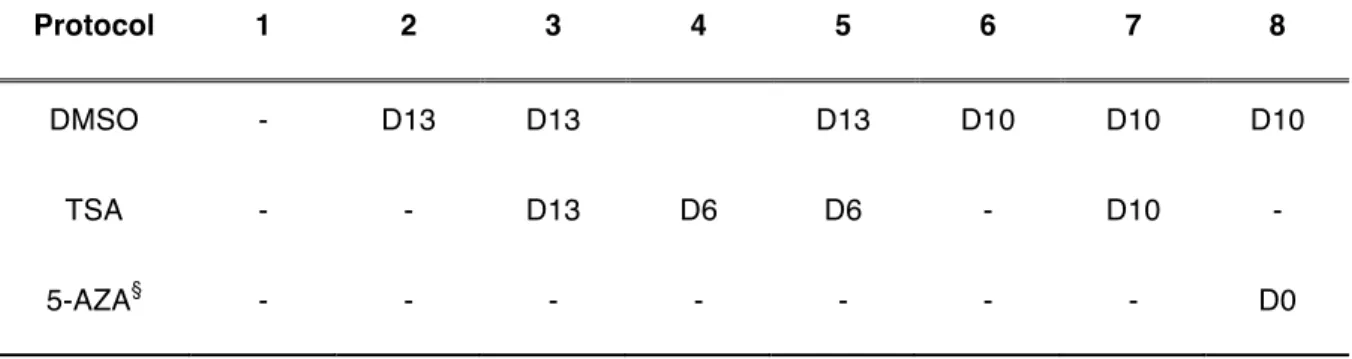
Hepatocyte differentiation of hnMSC ... 55
3.3.5
Gene expression ... 56
3.3.6
Histology ... 58
3.3.7
Albumin and urea production ... 59
3.3.8
Biotransformation activity ... 59
3.3.9
Protein quantification ... 60
3.3.10
Statistical analysis ... 60
3.4
Results ... 61
3.4.1
The endoderm marker HHEX is expressed in hnMSC and induced by FGF-2 61
3.4.2
Epigenetic modifiers improve hepatocyte phenotype on HLC ... 62
3.4.3
hnMSC-derived HLC present a partial hepatic differentiation at the transcriptional and functional levels at day 34 ... 65
3.5
Discussion ... 75
Chapter 4. Three-dimensional spheroid cultures of human neonatal mesenchymal stem cells ... 81
4.1
Abstract ... 82
4.2
Introduction ... 83
4.3
Material and Methods ... 85
4.3.2
Cell culture reagents ... 85
4.3.3
hnMSC isolation and culture ... 85
4.3.4
hnMSC viability evaluation ... 87
4.3.5
Immunofluorescence microscopy ... 88
4.3.6
Protein quantification ... 89
4.3.7
Statistical analysis ... 89
4.4
Results ... 89
4.4.1
Isolation and characterization of hnMSC cells ... 89
4.4.2
hnMSC form viable spheroids in spinner flask suspension cultures (SFSC) and ultra-low attachment (ULA) plates ... 89
4.4.3
hnMSC grown in 3D culture conditions maintain MSC antigen expression phenotype ... 93
4.4.4
hnMSC spheroid structures mimic the native environment ex vivo ... 93
4.5
Discussion ... 95
Chapter 5. Self-assembled 3D spheroids and hollow-fiber bioreactors improve MSC-derived hepatocyte-like cell maturation in vitro ... 99
5.1
Abstract ... 100
5.2
Introduction ... 101
5.3
Material and Methods ... 103
5.3.1
Reagents ... 103
5.3.2
Cell cultures ... 103
5.3.3
Human neonatal MSC cultivation and differentiation into HLCs ... 103
5.3.4
Cell viability assays ... 105
5.3.5
Determination of albumin secretion and urea synthesis ... 106
5.3.6
Biotransformation activity ... 106
5.3.7
Gene (transcript) expression ... 106
5.3.8
Histology ... 107
5.3.9
Statistical analysis ... 108
5.4.2
3D cultures allow the formation of viable tissue-like structures of HLCs ... 109
5.4.3
Hepatic-specific markers are upregulated in HLC 3D cultures ... 111
5.4.4
3D culturing improves cell metabolic activity ... 113
5.4.5
HLC Phase I and II enzyme activity is improved in 3D culture conditions ... 115
5.4.6
hnMSC-derived HLCs metabolize diclofenac and bupropion ... 118
5.4.7
HLCs show higher toxicity to diclofenac in spheroid suspension cultures ... 118
5.5
Discussion ... 120
5.6
Supplementary Material ... 126
Chapter 6. The integrated assessment of nevirapine biotransformation unveils the biocompetence of a 3D in vitro model of human hepatocyte-like cells for liver function
studies ... 127
6.1
Abstract ... 128
6.2
Introduction ... 129
6.3
Material and Methods ... 130
6.3.1
Reagents ... 130
6.3.2
Cell culture ... 131
6.3.3
Biotransformation activity ... 131
6.3.4
Gene expression ... 132
6.3.5
Quantification of nevirapine metabolites ... 133
6.3.6
Determination of the thiolomic profile ... 134
6.3.7
Statistical analysis ... 135
6.4
Results ... 135
6.4.1
NVP modulates key biotransformation enzymes in 2D and 3D cultures ... 135
6.4.2
3D cultures thiolomic profile is altered upon NVP exposure ... 140
6.5
Discussion ... 145
Chapter 7. Concluding remarks and future perspectives ... 149
LIST OF FIGURES
Figure 1.1 Multiscale liver tissue structures contribute to the diverse functional roles of the liver. ... 2
Figure 1.2 Distribution of extracellular matrix (ECM) in the liver acinus. A basement membrane is localized in the periportal and perivenous regions. ... 3
Figure 1.3 Elimination and Metabolism of the top 200 most prescribed drugs in 2002. ... 5
Figure 1.4 Elimination and Metabolism of drugs by the hepatocyte. ... 6
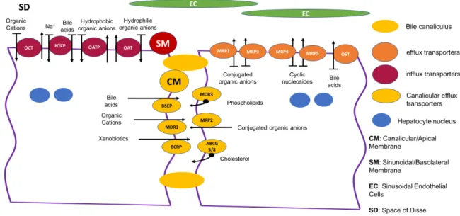
Figure 1.5 Schematic representation of the hepatic transporters in the sinusoidal/basolateral and in the canalicular/apical membrane of hepatocytes. ... 9
Figure 1.6 Major events in early mammalian endoderm development. ... 34
Figure 1.7. Epigenetic control of gene transcription. ... 38
Figure 3.1 Gene expression of HHEX on hnMSCs after exposure to different FGF cocktails in the differentiation Step 1 (D3). ... 61
Figure 3.2 Comparative characterization of hnMSC-derived HLC obtained with the differentiation protocols 1-8 (D24).. ... 63
Figure 3.3 Gene expression analyses of HLC-derived from hnMSCs upon exposure to protocols 1-3 and 6-8 (D24). ... 64
Figure 3.4 Functional capacity of HLC-derived from hnMSCs upon exposure to protocols 1-3 and 6-8 (D24) ... 65
Figure 3.5 hnMSCs-HLC morphology and glycogen storage ability upon sequential exposure to liver-specific factors and DMSO supplementation from day 10 onwards. ... 66
Figure 3.6 Effect of culture time on albumin and urea production in HLC-derived from hnMSCs up to day 34 in culture upon sequential exposure to liver-specific factors and DMSO supplementation from day 10 onwards (black bars) ... 67
Figure 3.7 Genome wide analyses of HLC-derived from hnMSCs at day 34 of differentiation. ... 68
Figure 3.8 Genome wide analyses of HLC-derived from hnMSCs at day 34 of differentiation.. ... 69
Figure 3.9 Gene expression analyses during hnMSCdifferentiation into HLCs upon sequential exposure to liver-specific factors and DMSO supplementation from day 10 onwards. ... 72
exposure to liver-specific factors and DMSO supplementation from day 10 onwards (D27).. ... 73
Figure 3.11 Effect of culture time on phase I and phase II activities in HLC-derived from hnMSCsup to day 34 in culture upon sequential exposure to liver-specific factors and DMSO supplementation from day 10 onwards. ... 91
Figure 4.2 3D spheroid cultures allow the extended maintenance of viable hnMSC without necrotic cores ... 92
Figure 4.3 Expression of extracellular matrix proteins by hnMSC spheroids. ... 94
Figure 5.1 Miniaturized 3D multi-compartment bioreactor for high-density liver cell perfusion ... 105
Figure 5.2 5-AZA triggers HLCs maturation.. ... 110
Figure 5.3 HLCs cell culture characterization in spheroids, bioreactors and in 2D cultures. ... 111
Figure 5.4 Gene expression analyses of the hepatic markers on HLCs (day 27) cultured in 2D culture, in spheroids and in bioreactors. ... 112
Figure 5.5 Immunohistochemical analysis of HLC-derived hnMSCs cultured in spheroids, in bioreactors and in 2D cultures (day 27). ... 114
Figure 5.6 Metabolic capacity of HLCs cultured in 3D cultures, bioreactor and spheroids, and in 2D cultures as a control from day 18 until day 27 ... 115
Figure 5.7 Phase I and Phase II metabolism on HLCs. ... 116
Figure 5.8 EROD activity on HLCs cultured in bioreactors, spheroids and 2D cultures at day 27 of the differentiation ... 117
Figure 5.9 Metabolic competence of HLCs evaluated by exposure to (a) diclofenac and (b) bupropion. ... 119
Figure 5.10 Comparison of diclofenac cytotoxicity in 3D spheroids and 2D monolayer cultures evaluated by MTS mitochondrial activity assay.. ... 119
Figure 5.11 Phase I and II metabolism on HLCs up to day 34. ... 123
Figure 6.1 Gene expression analyses of 3D- and 2D-HLCs after 3 (day 27) and 10 days (day 34) of NVP treatment.. ... 137
Figure 6.2. Enzymatic induction in 3D- and 2D-HLCs after 3 (day 27) and 10 days (day 34) of NVP treatment.. ... 138
Figure 6.3 Levels of NVP phase I and II metabolites in 3D- and 2D-HLCs after 3 (day 27) and 10 days (day 34) of NVP treatment. ... 139
Figure 6.4. Intracellular and extracellular thiolomic profile in non-NVP-treated 3D- and 2D-HLCs at day 27 and day 34. ... 141
Figure 6.5. Intracellular and extracellular thiolomic profile in 3D- and 2D-HLCs after 3 (day 27) and 10 (day 34) days of NVP treatment.. ... 142
Figure 6.6 NVP effect on glutathione oxidation (intracellular GSSG/GSH ratio) in 3D- and 2D-HLCs after 10 days of NVP treatment (day 34). ... 143
Table 1.1 Relative abundance of human CYP450 enzymes and their contribution to drug metabolism. ... 7
Table 1.2 Xenosensors target genes associated with xenobiotic metabolism and transport in humans and rodent animal models. ... 11
Table 1.3 Advantages and limitations of in vitro liver preparations. ... 16
Table 1.4 Advantages and limitations of spheroid cell cultures for in vitro toxicity testing applications. ... 20
Table 1.5 Advantages and limitations of complex in vitro hepatotoxicity cell model systems. ... 22
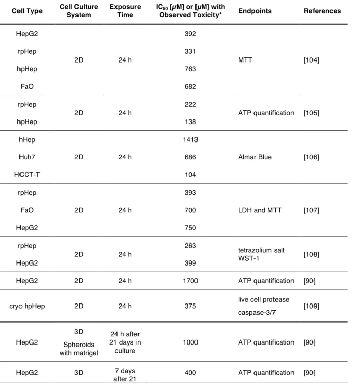
Table 1.6 Diclofenac cytotoxicity endpoints observed in different cell types and cell culture systems. ... 25
Table 1.7 Troglitazone cytotoxicity endpoints observed in different cell types and cell culture systems. ... 27
Table 3.1. Summary of the DMSO, TSA and 5-AZA supplementation times in the hepatic differentiation protocols. ... 55
Table 3.2 Primers used for qRT-PCR characterization of differentiated and undifferentiated hnMSCs, HepG2 and hpHeps. ... 57
Table 5.1 Albumin Production on the controls: undifferentiated cells (hnMSCS), HepG2 cell line, and primary hepatocytes, human (hpHep) and rat (rpHep) in 2D and in spheroids culture ... 126
Table 5.2 Urea Production on the controls: undifferentiated cells (hnMSCS), HepG2 cell line, and primary hepatocytes, human (hpHep) and rat (rpHep) in 2D and in spheroids culture 126
Table 6.1 Primers used for qRT-PCR characterization of differentiated HLCs, undifferentiated hnMSCs and HepG2 cell line ... 133
Table 6.2 Comparative summary of 3D- and 2D-HLC metabolic capacity upon 10 days of NVP treatment (day 34) ... 144
Table 6.3 Comparison of relative levels (expressed as percentage) of NVP metabolites in clinical, pre-clinical and in vitro studies. ... 146
ABREVIATIONS
2D Two dimensional
3-MC 3-methylcholantherene
3D Three dimensional
3 Rs Reduced, Replace and Refine
4-MU 4-methylumbelliferone
5-AZA 5-azacytidine
ABC ATP-binding cassette family
Ac-LDL Acetylated low-density lipoprotein
ADMET Absorption, Distribution, Metabolism, Excretion and Toxicity
AFP Alpha fetoprotein
AhR Aryl hydrocarbon receptor
ALB Albumin
AR Androgen receptor
ASC Adult stem cells
AT Alpha 1-Antitripsin
AT-MSC Adipose tissue MSC
BCA Bicinchoninic acid
BCRP Breast cancer resistance protein
BM-MSC Bone marrow mesenchymal stem cells
BMP Bone morphogenic proteins
BSA Bovine serum albumin
BSEP Bile salt export pump
c/EBPα CCAAT/enhancer binding protein
CAR Constitutive androstane receptor
cDNA Complementary deoxyribonucleic acid
CHMP Committee for Medicinal Products for Human Use
CK-18 Cytokeratin 18
CK-19 Cytokeratin 19
CYP450 Cytochrome P-450 superfamily
CYS Cysteine
CYS-GLY Cysteinylglycine
DAPI 4’,6-diamidino-2-phenylindole
DILI Drug-induced liver injury
DMSO Dimethyl sulfoxide
DNMTi DNA methyltransferase inhibitors
ECM Extracellular matrix
ECOD 7-Ethoxycoumarin-O-deethylase
ECVAM European Centre for the Validation of Alternative Methods
EMA European Medicines Agency
EPA Environmental Protection Agency
ER Estrogen receptor
EROD 7-Ethoxyresorufin-O-deethylase
ESC Embryonic stem cells
FBS Foetal bovine serum
FDA Food and Drug Administration
FDA/PI Flurescein diacetate/Propidium iodide
FGF-2 Fibroblast growth factor-2
FGF-4 Fibroblast growth factor-4
FMO flavin-containing monooxygenases
Foxa1-3 Forkhead box a1 to 3
FP7 7th Framework Programme for Research and Technological Development
(2007-2013)
G6P Glucose-6-phosphate
GAPDH glyceraldehyde-3-phosphate dehydrogenase
GATA 4-6 GATA binding factor 4 to 6
GCL Glutamate cysteine ligase
γGT Gama glutamyltranspeptidase GLU-CYS Glutamil-cysteine Gp130 Glycoprotein 130 GR Glucocorticoid receptor GSH Reduced glutatione GSSG Oxidized glutatione GST Glutathione transferases
H&E Hematoxylin-Eosin staining
H2020 Eurpean Uninion Framework Programme for Research and Innovation (2014-2020)
HbLC Hepatoblast-like cells
HDACi Histone deacetylase inhibitors
HepaRG Human hepatoma cell line
HepG2 Human hepatoma cell line
HepPar1 positive marker for hepatocellular differentiation
hERG Human Eher-a-go-go Related Gene
hESCs Human embryonic stem cells
HGF Hepatocyte growth factor
HHEX Hematopoietically-expressed homeobox protein
hiPSC Human induced Pluripotent Stem Cells
HL-60 Human promyelocytic leukemia cells
HLA-DR Human Leukocyte Antigen - antigen D Related
HLC Hepatocyte-like cells
hnMSC Human neonatal mesenchymal stromal cell
hpHep Human primary hepatocytes
HTS High Throughput Screening
hUCM-MSC Human umbilical cord matrix mesenchymal stromal cells
Huh7 Human hepatoma cell line
ICCVAM Interagency Coordinating Committee on the Validation of Alternative Methods
ICH International Conference on Harmonisation of Technical Requirements for Registration
of Pharmaceuticals for Human Use
IPA Ingenuity Pathway Analysis
ISCT International Society for Cellular Therapy
ISSCR International Society for Stem Cell Research
ITS Insulin–transferrin–sodium selenite
JAK/Stat3 Janus Kinase/Signal Transducer and Activator of Transcription 3
KLF4 Kruppel-like factor 4
LA Linoleic Acid
LDH Lactate dehydrogenase
LDL Low-density lipoprotein cholesterol
LMWT Low molecular weight thiol
MAO monoamine oxidases
MAPK Mitogen-activated protein kinases
MDR Multidrug resistance protein
MR Mineralocorticoid receptor
MRP Multidrug resistance-associated protein
MRP-2 Multidrug resistance protein 2
MSC Mesenchymal Stem Cells
MT Methytransferases
MTS 3-(4,5-dimethylthiazol-2-yl)-5-(3-carboxymethoxyphenyl)-2-(4-sulfophenyl)-2H-tetrazolium, inner salt
NADPH Nicotinamide adenine dinucleotide phosphate
NAT N-Acetytransferase
NPC non-parenchymal cells
NTCP Na+/bile acid cotransport protein
NVP Nevirapine
OAT Organic anion transporter family
OATP Organic anion transporter protein family
OATP-C Organic anion-transporting polypeptide C
OCT-4 Octomer-binding transcription factor-4
OCT Organic cation transporter family
OECD Organisation for Economic Cooperation and Development
OSM Oncostatin M
OST Organic solute transporter
P/S/A Peninsilin/streptomicin/ amphotericin B
PBS Phosphate Buffered Saline (pH=7,4)
PCA Principal Component Analysis
PDSC placenta derived stem cells
Pdx 1 Pancreatic and duodenal homeobox 1
PFA Paraformaldehyde
PPAR Peroxisome proliferator activated receptor
PR Progesterone receptor
Prox1 Prospero homeobox protein 1
PXR Pregnane X receptor
qPCR Quantitative real-time Polymerase Chain Reaction
REACH Registration, Evaluation, Authorization and Restriction of Chemicals
RNS/ROS Reactive nitrogen species / Reactive oxigen species
rpHep Rat primary hepatocytes
RXR Retinoid X receptor
SB Sodium butyrate
SC Stem cell
SCID Severe Combined Immunodeficiency
SD Standard deviation
SEM Standard error of the mean
SFSC Spinner flask suspension culture
Sox17 Sry-related HMG box 17
STM Septum transverse mesenchyme
SULT Sulfotransferases
TAT Tyrosine aminotransferase
TDO Tryptophan-2,3-dioxygenase
TGF-β Transforming growth factor β
TGZ Troglitazone
Tm Melting temperature
TSA Trichostatin A
UCX® ECBio’s proprietary Umbilical Cord eXpanded hnMSCs isolated from the Wharton’s Jelly
UGT uridine 5’-diphosphate glucuronosyltransferase
ULA Ultra-low attachment
US Tox21 Toxicity Testing in the 21st century: A vision and a strategy
VPA Valproic acid
1
Chapter 1. General Introduction
Part of the scientific content of the present chapter has been included in the following publications:
Cipriano M, Oliveira NG, Miranda JP. Revisiting 3D liver models for drug metabolism and
toxicology studies: A critical perspective. (submitted for publication)
Cipriano M, Miranda JP. Strategies for deriving stem cells into hepatocyte-like cells and its
Earlier and better understanding of drug mechanisms of action and toxicity is essential to improve the R&D process of a new drug [1, 2]. In fact, there is a relevant number of drug withdrawals for toxicological reasons, of which 21 % is due to hepatotoxicity [3]. The liver is the main organ responsible for xenobiotic metabolism, being particularly exposed to chemicals and its metabolites. Moreover, the hepatocyte is a particularly complex cell type that presents more individual functions than other ~200 terminally differentiated cell types in the human body [4].
1.1 Liver physiology and function
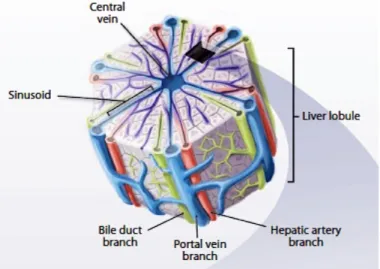
The liver is composed of 4 lobes, contains 5 cell types and distinct extracellular matrix (ECM) compositions. The liver parenchymal cells are the hepatocytes and the non-parenchymal cells are Kupffer cells, sinusoidal endothelial cells, bile duct epithelial cells and Ito cells (stellate cells). Hepatocytes represent 60 % of the liver’s cells, about 80 % of liver total cell mass and are responsible for the liver’s synthetic and metabolic functions. Hepatocytes are arranged in single-cell thick plates as represented in Figure 1.1.
Figure 1.1 Multiscale liver tissue structures contribute to the diverse functional roles of the liver. Hierarchical structure of the liver consisting of repeated functional tissue units, the liver lobules [5].
Regardless of hepatocytes’ morphological similarity, its functions vary per its location within the hepatic lobule, namely when located either closer to the periportal vein or to the central vein (Figure 1.2). This phenomenon is named hepatic zonation. Periportal hepatocytes are characterized by higher oxygen presence, glycogen synthesis from lactate and urea production since the blood that irrigates these hepatocytes flows from the whole circulation. On the other hand, the perivenus phenotype is exposed to lower oxygen pressure, and presents higher xenobiotic metabolism and glycogen synthesis from glucose given that these
cells are partially irrigated by blood flowing from the enterohepatic circulation [6, 7]. In the hepatic lobule, the blood flows toward the hepatic vein within the space of Disse contacting with the exposed surface areas of the hepatocyte plates, where toxins and nutrients within the blood stream are extracted by the hepatocytes. Kupffer cells are liver macrophages residing in the sinusoids. Their functions include phagocytosis of old red blood cells and bacteria as well as heme conversion into bilirubin. Sinusoidal endothelial cells are fenestrated allowing proteins to pass freely through the sinusoidal endothelium into the space of Disse, where they directly contact with hepatocytes, in a bi-directional fashion, meaning that proteins and other substances produced, stored or processed by the liver can also be transferred back into the blood. Bile duct epithelial cells line the interlobular bile ducts within the portal triads. Finally, Ito or stellate cells are found in the space of Disse and play an important role in liver fibrosis once these cells are responsible for collagen production [7, 8].
Figure 1.2 Distribution of extracellular matrix (ECM) in the liver acinus. A basement membrane is localized in the periportal and perivenous regions. Adapted from [7].
Besides the cellular component, the liver ECM composition and stiffness is of major importance for the maintenance of the parenchymal and non-parenchymal cell function. As represented in Figure 1.2, the hepatic parenchyma ECM is constituted by fibronectin and type I and III collagen, whereas the basal membrane, surrounding the blood vessels, is composed
by laminin, type IV collagen and perlecan. The endothelial sinusoids lack a basal lamina since the liver has no continuous barrier between epithelial cell surface and the plasma [7].
1.2 Role of liver in absorption, distribution, metabolism, excretion and toxicity
(ADMET)
The liver is a unique organ regarding the large number of vital functions. Its main functions are nutrient homeostasis, by storing glycogen through glucose metabolism; ammonia detoxification, by synthetizing urea; filtration of particulates; protein synthesis, such as albumin; biotransformation of endo and xenobiotics; cholesterol synthesis and homeostasis; formation of bilirrubin and billiary secretion [9]. It is, thus, strategically located to maintain the body’s metabolic homeostasis and it plays an essential role in ADMET (absorption, distribution, metabolism, excretion and toxicity) process for most drugs. In fact, the liver is one of the first organs contacting with oral xenobiotics after the process of absorption, through the portal vein. Within the liver, the distribution of molecules is facilitated by: (i) the capillary endothelium porosity, since the hepatic sinusoids present fenestrae that have 50 to 150 nm in diameter; (ii) the presence of specialized membrane transporters; (iii) accumulation in cell organelles and reversible intracellular binding. The hepatocytes’ high exposure to xenobiotics and its metabolic defence mechanisms contribute to molecules biotransformation and subsequent excretion. About 73 % of the 200 most prescribed drugs suffer biotransformation as primary elimination pathway, whereas, 22 % and 5 % suffer renal and biliary elimination, respectively (Figure 1.3.) [10]. The biotransformation mechanisms are classified as phase I, II and III reactions as schematically represented in Figures 1.3 and 1.4.
1.2.1 Phase I metabolism
Phase I reactions are responsible for the increase in compounds’ polarity by introducing or unmasking a functional group (e.g., -OH, -COOH, -NH2, or -SH) within a molecule to enhance
its hydrophilicity. It can occur through direct introduction of the functional group or by modifying existing functionalities. This might result in the reduction of ketones or aldehydes to alcohols, the oxidation of alcohols to acids, hydrolysis of ester and amides, the reduction of azo and nitro compounds or also the oxidative N-, O-, and S-dealkylation. Several enzymes are responsible for phase I reactions; those include the cytochrome P450 (CYP450) enzymes, flavin-containing monooxygenases (FMOs), monoamine oxidases (MAOs) and xanthine oxidase/aldehyde oxidase (XO/AO) [10, 11]. As represented in Figure 1.3, CYP450 enzymes are responsible for 75 % of the drug metabolism of the 200 most prescribed drugs. There is a
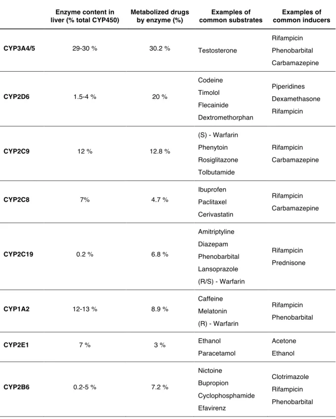
total of 18 human CYP450 gene families among which only three have a role in xenobiotic metabolism, whereas the remaining families are involved in cholesterol biosynthesis, vitamin D metabolism, bile acid metabolism and biosynthesis catabolism of steroids [7, 12]. CYP3A, CYP2C and CYP1A are the most important enzyme families involved in drug metabolism, being responsible for 70 % of drug biotransformation, representing 50 % of CYP450 enzymes in the liver. The overall CYP450 metabolism shows a different distribution of metabolized drugs by each CYP450 enzyme (Table 1.1) when compared to the data presented in Figure
1.3, that refers to the 200 most prescribed drugs. In fact, the relative amount of CYP450
enzymes in the liver is not directly correlated with the extension of drugs biotransformation and its activity is susceptible of specific induction, which results in an altered capacity to biotransform its substrates (Table 1.1).
Figure 1.3 Elimination and Metabolism of the top 200 most prescribed drugs in 2002. [10, 11]
1.2.2 Phase II metabolism
Phase II reactions are characterized by the addition of a functional group through conjugation reactions performed by UDP-Glucuronosyltransferases (UGT), Sulfotransferases (SULT), Glutathione transferases (GST), aminoacid transferases, N-Acetytransferase (NAT) or Methytransferases (MT). Biotransformation 73% Renal 22% Biliar 5%
Drugs Elimination Pathways Distribution
CYP1A 11% CYP3A 46% CYP2C9 15% CYP2C19 10% CYP2D6 15% CYP2B6 3%
CYP450 (Phase I) Enzymes Distribution
UGTs 35% GSTs 18% SULTs 17% NATs 14% Others 16%
Phase II Enzymes Distribution CYP450 (Phase I)
75% UGT (Phase II)
12% Esterases (Phase I)
10%
Others 3%
Figure 1.4 Elimination and Metabolism of drugs by the hepatocyte. Lipophilic xenobiotics (X) flow in sinusoidal blood and diffuse through the endothelial cell fenestrations into the space of Disse while still protein bound. Drugs may enter the hepatocyte by active transport or passive diffusion. Inside the cell, a drug will typically encounter a specific phase I enzyme, e.g. a CYP450 enzyme, capable of binding to the drug, and converting it to more water soluble and chemically reactive metabolites (X*). Typically, the resultant metabolite undergoes conjugation to a polar ligand in a reaction catalyzed by phase II enzymes. The resultant conjugated metabolite is then secreted by efflux transporters into the biliary canaliculus for excrection in bile, or secreted back into sinusoidal blood for ultimate excretion in urine, referred to as phase III metabolites. [7, 9, 13].
UGTs are responsible for 35 % of phase II drug metabolism being also important in bilirubin, steroid hormone, thyroid hormone, bile acid, and fat-soluble vitamin metabolism [14]. There are two broad families named UGT1 and UGT2, also known as the phenol/bilirubin family and the steroid/bile salt family, respectively. The liver is the major organ for glucuronidation, being UGT1A1 and UGT2B7 the most relevant enzymes for drug metabolism in the human liver [15]. SULTs are divided into four families, with the cytosolic SULTs being the most relevant enzymes for xenobiotic metabolism. In humans three SULT families, SULT1, SULT2, and SULT4, have been identified, containing at least 13 distinct members [16]. Within SULT1 family the isoform SULT1A1 was identified in a variety of species whereas SULT1A2, SULT1A3 and SULT1A4 were only identified in humans. In fact, SULT1A1 is the most expressed SULT in human liver [16]. SULT1A1 and SULT1A3 are abundantly expressed in the fetal liver whereas in adult liver SULT1A3 expression is much lower [16].
Table 1.1 Relative abundance of human CYP450 enzymes and their contribution to drug metabolism. Enzyme content in
liver (% total CYP450)
Metabolized drugs by enzyme (%) Examples of common substrates Examples of common inducers CYP3A4/5 29-30 % 30.2 % Testosterone Rifampicin Phenobarbital Carbamazepine CYP2D6 1.5-4 % 20 % Codeine Timolol Flecainide Dextromethorphan Piperidines Dexamethasone Rifampicin CYP2C9 12 % 12.8 % (S) - Warfarin Phenytoin Rosiglitazone Tolbutamide Rifampicin Carbamazepine CYP2C8 7% 4.7 % Ibuprofen Paclitaxel Cerivastatin Rifampicin Carbamazepine CYP2C19 0.2 % 6.8 % Amitriptyline Diazepam Phenobarbital Lansoprazole (R/S) - Warfarin Rifampicin Prednisone CYP1A2 12-13 % 8.9 % Caffeine Melatonin (R) - Warfarin Rifampicin Phenobarbital CYP2E1 7 % 3 % Ethanol Paracetamol Acetone Ethanol CYP2B6 0.2-5 % 7.2 % Nictoine Bupropion Cyclophosphamide Efavirenz Clotrimazole Rifampicin Phenobarbital Adapted from [17, 18]
GSTs are divided into two super-families comprising cytoplasmic and membrane bound enzymes. The cytoplasmic GSTs might also be present in nucleolus, mitochondria and peroxisomes and are responsible for 18 % of drug metabolism (Figure 1.3). This superfamily comprises five classes: a (A1-A4), µ (M1-M5), p (P1), k (K1) and q (T1, T2) [19]. In the liver,
GSTa is the most predominant class followed by GSTµ [20]. GSTs reduce the formation of hydroperoxides of fatty acids, phospholipids, cholesterol, and act against the redox-cycling of some quinone containing compounds, through glutathione conjugation. Thus, GST is part of the defence mechanisms against reactive oxygen species. GSTs have also an important role on the transport of a wide variety of both endogenous and exogenous compounds [21]. Glutathione (GSH) conjugation is not always enzymatic since hydrophobic molecules containing an electrophilic atom might react non-enzymatically with glutathione. This includes a variety of electrophilic xenobiotics resulting in glutathione conjugates that are thioethers, formed by nucleophilic attack of glutathione thiolate anion (GS−) with an electrophilic carbon atom in the xenobiotic containing electrophilic heteroatoms (O, N, or S) [9].
1.2.3 Phase III metabolism
Phase III reactions comprise the role of the transport of endo and xenobiotics and its metabolites. The hepatic transporters can be classified as (i) influx transporters, located in the sinusoidal/basolateral membrane, responsible for the uptake of molecules into the hepatocytes; and (ii) efflux transporters, that are mostly located in the canalicular/apical membranes, but may also be located in the sinusoidal/basolateral membrane as represented in Figure 1.5. The influx transporters include the Na+/bile acid cotransport protein (NTCP) and three families of organic ion exchanger transporters: OATP, the organic anion transporter protein family; OCT, the organic cation transporter family; and OAT, the organic anion transporter family [7]. The OATP family is also named as glycoprotein transporters, particularly important for drug transport and bilirubin uptake. OATP1B1 and OATP1B3 are liver specific, being OATPC1A2 and OATP2B1 also present in liver [22]. The efflux transporters include the ATP-binding cassette (ABC) family and the organic solute transporter a/b (OSTa / OSTb), responsible for bile acid transport. The canalicular/apical ABC family transporters comprise two functional classes: (i) the solute transporters such as the cation transporter multidrug resistance protein 1 (MDR1) also known as P-glycoprotein 1, the bile salt export pump (BSEP), the xenobiotic transporter breast cancer resistance protein (BCRP), and the multidrug resistance-associated protein 2 (MRP-2) also known as multi-specific organic anion transporter 2; and (ii) the translocators such as the phospholipid flippase MDR3, and the ABC half transporters involved in cholesterol transport ABCG5/8. On the sinusoidal/basolateral membrane there is the OSTa/OSTb transporter and the ABC transporters: MRP1 that transports various organic anions including GSH, glucuronide and sulfate conjugates of several drugs; MRP3 that is an organic anion transporter that primarily transports bile salts
![Figure 1.3 Elimination and Metabolism of the top 200 most prescribed drugs in 2002. [10, 11]](https://thumb-eu.123doks.com/thumbv2/123dok_br/19187383.948297/35.892.159.729.447.855/figure-elimination-metabolism-prescribed-drugs.webp)