Arq Neuropsiquiatr 2007;65(3-B):830-833
DEMENTIA PUGILISTICA
WITH CLINICAL
FEATURES OF ALZHEIMER’S DISEASE
Renata Areza-Fegyveres
1, Sergio Rosemberg
2, Rosa Maria R.P.S. Castro
3,
Claudia Sellitto Porto
1, Valéria Santoro Bahia
1, Paulo Caramelli
1, Ricardo Nitrini
1ABSTRACT - A 61-year-old ex-boxer presented with a three-year history of progressive memory decline. During a seven-year follow-up period, there was a continuous cognitive decline, very similar to that usual-ly observed in Alzheimer’s disease. Parkinsonian, pyramidal or cerebellar signs were conspicuoususual-ly absent. Neuropathological examination revealed the typical features of dementia pugilistica: cavum septi pellu-cidi with multiple fenestrations, numerous neurofibrillary tangles in the cerebral isocortex and hippocam-pus (and rare senile plaques). Immunohistochemistry disclosed a high number of tau protein deposits and scarce beta-amyloid staining. This case shows that dementia pugilistica may present with clinical features practically undistinguishable from Alzheimer’s disease.
KEY WORDS: dementia, dementia pugilistica, punch drunk, Alzheimer’s disease, chronic traumatic enceph-alopathy, parkinsonian/motor signs, neuropathology.
Dementia pugilistica com características clínicas de doença de Alzheimer
RESUMO - Um ex-boxeador de 61 anos apresentou-se com história de três anos de perda progressiva de memória e evoluiu com declínio cognitivo lentamente progressivo, sugestivo de doença de Alzheimer, rante seguimento de sete anos. Sinais parkinsonianos, piramidais ou cerebelares estiveram ausentes du-rante toda a evolução. Exame neuropatológico evidenciou características típicas de dementia pugilistica: cavum do septo pelúcido com múltiplas fenestrações, numerosos emaranhados neurofibrilares no isocór-tex cerebral e hipocampo (e raras placas senis). Imuno-histoquímica confirmou número elevado de depó-sitos de proteína tau e raros de beta-amilóide. Este caso demonstra que dementia pugilistica pode apre-sentar quadro clínico indistinguível daquele da doença de Alzheimer.
PALAVRAS-CHAVE: demência, dementia pugilistica, síndrome punch-drunk, doença de Alzheimer, encefa-lopatia traumática crônica, sinais motores/parkinsonianos, neuropatologia.
1
Behavioral and Cognitive Neurology Unit, Department of Neurology, University of São Paulo School of Medicine, São Paulo SP, Brazil; 2Department of Pathology, University of São Paulo School of Medicine, São Paulo SP, Brazil; 3Ludwig Institute for Cancer Research, Sao Paulo Branch, Sao Paulo SP, Brazil.
Received 18 January 2007, received in final form 8 May 2007. Accepted 15 June 2007.
Dra. Renata Areza-Fegyveres - Rua Oscar Freire 1702 / 44 - 05409-011 São Paulo SP - Brasil. E-mail: [email protected]
Martland, in 1928
1, first brought the expression
“
punch drunk syndrome
” to medical literature,
hith-erto used by the lay public and boxing fans to name
the condition that some boxers develop during or
af-ter their fighting career. The syndrome consisted of
extrapyramidal and cerebellar signs and symptoms,
associated or not, with cognitive and behavioral
ab-normalities. In 1937, the designation “
dementia
pu-gilistica
” was proposed
2. Critchley, in 1957
3, named
it “chronic progressive encephalopathy of the
box-er”, being more descriptive in that it represents the
long term cumulative effect of repetitive head
trau-ma
1,3-5. However, the label
dementia pugilistica
has
been more frequently used.
We report the clinical and neuropathological
find-ings of a case of
dementia pugilistica
presenting a
cognitive profile similar to Alzheimer’s disease (AD).
Informed consent to publish these data was given by
the patient’s wife and son.
CASE
Arq Neuropsiquiatr 2007;65(3-B) 831
no history of psychiatric disturbances, alcoholism or drug abuse and no family history of dementia. General physical examination revealed no abnormalities. On neurological examination, he scored 24 points in the Mini-Mental State Examination6. Muscle power, sensory system, cranial nerves, equilibrium, kinesis, muscle tonus, coordination, deep ten-don and superficial reflexes were all normal.
Neuropsychological evaluation showed predominant memory impairment. The total score on the Mattis Demen-tia Rating Scale (Mattis DRS)7 was 104 points out of 144, with greater impairment in the memory subscale.
A thorough laboratory investigation including cerebro-spinal fluid analysis was unremarkable. Apolipoprotein ε al-lele polymorphism detected the ε3/ε3 genotype. Comput-ed tomography (CT) scans revealComput-ed enlargement of the 3rd and lateral ventricles, cortico-subcortical atrophy and pres-ence of cavum septi pellucidi. Magnetic resonance imaging studies confirmed the CT findings. Single photon emission computed tomography showed mild hypoperfusion of the frontal lobes bilaterally.
This patient was followed with at least two medical evaluations a year for seven years. No cerebellar, pyrami-dal or extrapyramipyrami-dal signs were seen during the follow-up period. His cognitive impairment steadily worsened. Dur-ing the follow-up, the Mattis DRS total score decreased to 101 after six months, falling to 86 points after four years. With progression of the disease, predominant impairment in memory, attention and executive functions were con-firmed by poor performance in memory subtests from the Wechsler Memory Scale8, the Visual Retention Test: Multi-ple Choice9, the Block Design (Wechsler Adult Intelligence Scale - WAIS)10, the Hooper visual Organization Test11, the Trail Making Test12, the Maze Test13 and the category flu-ency test (animals per minute).
Rivastigmine was prescribed for approximately one year and gradually titrated to 6 mg BID with dubious results.
Death occurred seven years after the first consultation and was caused by gastric adenocarcinoma.
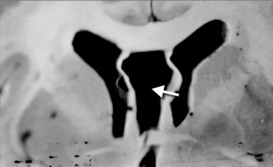
Macroscopic examination of the brain at autopsy showed moderate and symmetric atrophy, particularly of the frontal and temporal lobes, along with moderate en-largement of the lateral ventricles, especially at the level of the inferior horns. Presence of total cavum septi pellucidi, and a very thin septum with multiple fenestrations across almost its full extension was evident (Fig 1).
Slices were taken from the cingulate gyrus, frontal, pa-rietal, temporal, and occipital cortices, hippocampus, lentic-ular nucleus, thalamus, amygdala, midbrain, pons, medulla, cerebellar cortex, and dentate nucleus. All fragments were stained with H&E, Bielschowsky and Nissl stains, and Weil method for myelin. Immunohistochemistry was performed with the GFAP antibodies (Dako A/S, Denmark, M0761, clone 6F2, monoclonal mouse anti human glial fibrillary acidic protein); ubiquitin (Dako A/S, Denmark, Z0458, poly-clonal Ab-rabbit anti-human ubiquitin); beta-amiloid (Da-ko A/S, Denmark, M0872, clone 6F/3D, monoclonal mouse Ab- residue 8-17); tau protein (Dako A/S, Denmark, A0024, polyclonal Tau-P); GSK3 beta (Chemicon International Inc., USA, AB8687, polyclonal rabbit anti glycogen synthase ki-nase 3 beta); and alpha-synuclein (Chemicon International Inc., USA, AB5038, polyclonal rabbit anti synuclein alpha); using hydrolytic microwave antigenic retrieval and detec-tion with peroxidase Ab-Dako EnVision Labelled Polymer (Dako Corporation, USA).
Microscopic examination on H&E stain showed mild to moderate neuronal loss and reactive gliosis in all isocor-tical structures, predominantly at the frontal and tempo-ral lobes. Intense neuronal loss with reactive gliosis at the CA1 sector was evident, along with less intense loss in the CA4 sector of the hippocampus. Basal ganglia, thalami, brain stem and cerebellum were unremarkable, except for the presence of neurofibrillary degeneration of some
832 Arq Neuropsiquiatr 2007;65(3-B)
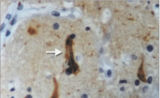
Fig 2. Imunohistochemistry of frontal cortex: neurofibrillary tangles. Bielschowsky method X 400.
Fig 3. Imunohistochemistry of frontal cortex: tau protein deposits: polyclonal antibody X 400.
rons of the substantia nigra and locus coeruleus. Howev-er, moderate reactive gliosis was detected at the putamen and thalamus on GFAP stain. Status spongiosus was ab-sent. Neurofibrillary tangles detected through Bielschowsky (Fig 2) and tau protein stains (Fig 3) were present in large amounts (5 to 6 per high power field-HPF) at the CA4, CA1 sectors of the hippocampus, at the pre-subicullum, subicul-lum and entorhinal cortex. They were present in moderate amounts (1 to 3 per HPF) at the frontal, cingulate, tempo-ral and parietal cortices, and absent at the occipital cor-tex. Rare beta-amyloid senile plaques were found only at the subicullum and entorhinal cortex. Thread-like depos-its were absent in all structures. Granulo-vacuolar
degen-eration was not detected. GSK3 beta and alpha synuclein stains gave negative results.
DISCUSSION
The exclusively cognitive impairment with
pre-dominant memory decline we are reporting in this
case differs from the typical case of
dementia
pugilis-tica
described in the literature. Furthermore, the
pat-tern of cognitive decline was also different from the
usual cases. In a broad review of this topic, Mendez
5Arq Neuropsiquiatr 2007;65(3-B) 833
which revealed difficulties in sequencing abilities,
complex attention tasks, frontal-executive functions,
memory, information processing speed and
finger-tapping speed. These cognitive abnormalities are
indicative of frontal-subcortical dysfunction. In the
case we are now describing, the cognitive decline
was more suggestive of AD, with memory
impair-ment followed by topographical disorientation.
A few cases of DP without motor signs have
al-ready been described, but in these reports either
neuropsychological evaluations were absent and/
or pathological study was not performed
1-3,14,15.
Ro-chon
14reported a case of an ex-boxer, who
present-ed memory impairment and aggressive behavior,
with no motor signs. The neuropsychological
evalu-ation showed mild anomia, disinhibition, difficulty
in maintaining an effective action strategy, slower
speed in the left hand, mental inflexibility and mild
perseveration, which are suggestive of
frontal-sub-cortical dysfunction. Naccache et al.
15also reported
a case of an ex-boxer with memory impairment and
highlighted the absence of motor signs. The subject’s
neuropsychological evaluation showed severe
mem-ory impairment and executive dysfunction. In
Bra-zil, Cordeiro-Junior and Oliveira reported a case of a
67-year-old ex-boxer that developed
neuropsychiat-ric manifestations (psychotic syndrome and memory
and spatial orientation deficits) after the end of his
sportive career possibly due to successive brain
inju-ries related to boxing
16. However, in the above
de-scribed cases, neuropathological examinations were
not performed.
The neuropathological findings of our case are
those usually described in
dementia pugilistica
, which
consist of
septum pellucidum
alterations (
cavum
and
fenestrations), cerebellar atrophy with Purkinje
neu-ronal loss; degeneration and loss of
substantia nigra
pigmented cells, presence of neurofibrillary tangles
(NFT) throughout the cortex and the brain stem,
es-pecially in the uncus, cortico-medial section of
amyg-daloid nucleus, fusiform and para-hippocampal gyri,
lateral temporal, insular and frontal cortices; paucity
or absence of senile plaques (SP)
17.
Dementia pugilistica
has been included among
the acquired tauopathies, and its pathophysiology
is still poorly understood
18. Geddes et al.
19examined
the brains of four young men, and a frontal
lobec-tomy specimen from a fifth individual, who all had
a history of traumatic brain injury in common, only
for different reasons (including 2 boxers). They
con-cluded that repetitive head injury in young adults is
initially associated with neocortical NFT formation in
the absence of A
b
deposition and that the
distribu-tion of tau pathology around the vessels in two of
the five brains examined suggested that the
patho-genesis of cytoskeletal abnormalities may involve
damage to blood vessels or perivascular elements.
In the case we are now reporting, the differential
diagnosis from AD proved very difficult when based
solely on clinical grounds, in spite of systematic
neu-rological and neuropsychological evaluations.
How-ever, the neuropathological findings were typical of
dementia pugilistica
.
REFERENCES
1. Martland HS. Punch drunk. J Am Med Assoc 1928;91:1103-1107. 2. Millspaugh JA. Dementia pugilistica. United States Naval Medicine
Bulletin 1937;35:297-303.
3. Critchley M. Medical aspects of boxing: particularly from a neurologi-cal standpoint. Br Med J 1957;1:357-362.
4. Roberts AH. Brain damage in boxers: a study of the prevalence of trau-matic encephalopathy among ex-professional boxers. London: Pitman Medical and Scientiic Publishing Company, Ltd., 1969.
5. Mendez MF. The neuropsychiatric aspects of boxing. Int J Psychiatry Med 1995;25:249-262.
6. Folstein MF, Folstein SE, McHugh PR. “Mini-mental state: a practical method for grading the cognitive state of patients for the clinician. J Psychiatr Res 1975;12:189-198.
7. Mattis S. Dementia rating scale: professional manual. Florida: Psycho-logical Assessment Resources, Inc, 1988.
8. Wechsler D. Wechsler Memory Scale - Revised. New York: The Psycho-logical Corporation, 1987.
9. Benton AL, Hamsher KS, Stone FB. Visual Retention Test: multiple choice I. Iowa: Division of Behavioral Neurology, Department of Neu -rology, University of Iowa Hospitals, 1977.
10. Wechsler D. Wechsler Adult Intelligence Scale. New York: Grune and Stratton, 1976.
11. Hooper HE. Hooper Visual Organization Test (HVOT): manual. Los Angeles: Western Psychological Services, 1983.
12. Spreen O, Strauss E. A compendium of neuropsychological tests: ad -ministration, norms and commentary, 2.Ed. New York: Oxford Univer -sity Press, 1998.
13. Lezak, MD. Neuropsychological assessment. 3.Ed. New York: Oxford University Press, 1995.
14. Rochon M. Présentation d’un cas: l’encephalopathie des boxeurs. Can J Psychiatry 1994;39:211-214.
15. Naccache L, Slachevsky A, Deweer B, Habert MO, Dubois B. “Démence pugilistique” sans signes moteurs. Press Med 1999;28:1352-1354. 16. Cordeiro-Junior Q, Oliveira AM. Parkinsonian, cerebellar, psychotic
and demential symptoms in ex-boxer: case report. Arq Neuropsiquiatr 2001;59:283-285.
17. Corsellis JAN, Bruton CJ, Freeman-Browne D. The aftermath of box -ing. Psychol Med 1973;270-303.
18. Lace GL, Wharton SB, Ince PG. A brief history of tau: the evolving view of the microtubule-associated protein tau in neurodegenerative diseas-es. Clin Neuropathol 2007;26:43-58.