ISSN Online: 2157-9458 ISSN Print: 2157-944X
Spirulina platensis
Alleviates the Liver, Brain
and Heart Oxidative Stress in Type 1 Diabetic
Rats
Francisca Adilfa de Oliveira Garcia
1,2*, Violet G. Yuen
1, Helioswilton Sales de Campos
3,
Eveline Turatti
4, Glauce Socorro de Barros Viana
2,5, Carlo José Freire Oliveira
3, John H. McNeill
11Faculty of Pharmaceutical Sciences, The University of British Columbia (UBC), Vancouver, Canada
2Departamento de Fisiologia, Faculdade de Medicina Estacio de Juazeiro do Norte (ESTACIO), Juazeiro do Norte, Brazil
3Departamento de Microbiologia, Imunologia e parasitologia, Universidade Federal do Triangulo Mineiro (UFTM), Minas Gerais, Brazil
4Faculdade de Odontologia, Universidade de Fortaleza (UNIFOR), Fortaleza, Brazil 5Departamento de Farmacologia, Universidade Federal do Ceará(UFC), Fortaleza, Brazil
Abstract
Spirulina platensis (SPI) is a microalga with a high content of functional compounds, such as phenolics, phycocyanins and polysaccharides that has been shown to have antioxidant, anti-inflammatory, hypoglycemic, neuroprotective and immunomodulatory effects. The objectives of the present work were to study the possible effects of SPI treatment on the glycemic-lipid profile, oxida-tive stress, lipid peroxidation and cardiac performance in diabetic rats. Diabetes was induced by streptozotocin (STZ) in male Wistar rats. In diabetic animals SPI, at a dose of 50 mg/kg/day, reduced lipid peroxidation, nitrite levels and lipids in plasma and tissues. SPI exhibited an effective improvement on +dP/dT and −dP/dT in non-diabetic rats. This study showed that SPI signifi-cantly suppressed nitrite generation and lipoperoxidation in the hearts of di-abetic animals, as well as an improvement in the cardiac function in control SPI-treated rats which is consistent with several studies that demonstrated the protective effect of antioxidants on oxidative stress-mediated injury caused by reactive oxygen species (ROS) produced in diabetic myocardial tissues.
Keywords
Spirulina platensis, Lipid Profile, Cardiac Function, Oxidative Stress, Diabetes
1. Introduction
Spirulina platensis is a multicellular and filamentous edible blue-green alga. Due How to cite this paper: de Oliveira Garcia,
F.A., Yuen, V.G., de Campos, H.S., Turatti, E., de Barros Viana, G.S., Oliveira, C.J.F. and McNeill, J.H. (2018) Spirulina platensis Alleviates the Liver, Brain and Heart Oxid-ative Stress in Type 1 Diabetic Rats. Food and Nutrition Sciences, 9, 735-750. https://doi.org/10.4236/fns.2018.96056
Received: May 19, 2018 Accepted: June 25, 2018 Published: June 28, 2018
Copyright © 2018 by authors and Scientific Research Publishing Inc. This work is licensed under the Creative Commons Attribution International License (CC BY 4.0).
to abnormally high levels of chlorophyll in SPI, it was initially placed in the plant kingdom but was later shifted to the bacterial kingdom based on new under-standing of SPI physiology, genetics and biochemical properties. SPI cells form long strands which look similar to a coiled spring; thus the name SPI, meaning “little spring” [1]. SPI is found naturally in alkaline lakes. It has also been cul-tured in a controlled environment for human consumption [1] [2].
SPI is used as a food supplement and the nutritional and therapeutic values have been well documented [2]. SPI contains 62% amino acids, is the world’s richest natural source of vitamin B12 and contains a whole spectrum of natural mixed carotenes and xanthophyll phytopigments. SPI is wrapped with a soft cell wall formed from complex sugars and proteins like rhamnose, xylose, glucose, galactose, and arabopyranose glucuronic acid [3] [4]. Algae of the genus Spiru-lina spp present approximately 15% of biliproteins (C-phycocyanin, allophyco-cyanin and phycoerythrin) [4], with C-phycocyanin being the major protein component of SPI.
More recently SPI has been found to have additional pharmacological properties through a variety of active constituents. SPI exhibits antioxidant, anti-inflammatory, neuroprotective, immunomodulatory, antihyperlipidemic, cardiovascular and anti-diabetic properties, making it a potential drug candidate in the therapeutic management of chronic disorders such as diabetes and hypertension [4]-[9] [2] [25]-[32] [44] [45].
Diabetes is a metabolic disorder characterised by chronic hyperglycaemia. The long-term effects of diabetes mellitus include cellular injury, inflammation and failure of various organs [10].
Diabetes mellitus is a well-known risk factor for the development of heart failure independent of coronary heart disease and hypertension and may cause a cardiomyopathy [11] [12] [13]. A significant number of diabetic patients exhibit diabetic cardiomyopathy (DCM), however, the development of DCM remains poorly understood and the underlying mechanisms have not yet been clearly elucidated. For many authors DCM is characterized by sustained hyperglycae-mia and hyperlipidehyperglycae-mia, oxidative stress, defective calcium handling, apoptotic and necrotic cell death, altered mitochondrial function, inflammation and myo-cardial fibrosis [11].
Clinical studies have showed that DCM increases the risk of death in the pa-tients [12]. Since there are no effective approaches to preventing the develop-ment and progression of diabetic cardiomyopathy in the clinic, the search for new therapeutic targets for the prevention or protection from DCM would be an important in therapeutic development [13].
malon-dialdehyde (MDA) which further damages the cells, the interaction of nitric oxide (NO) with ROS causes the production of several reactive nitrogen species (RNS ) such as nitrogen dioxide (NO2
−), peroxynitrite (OONO-), dinitrogen
tri-oxide (N2O3) and nitrous acid (HNO2) that potentiate cellular damage [14] [15]. Also, the variation in the levels of antioxidant enzymes, such as superoxide dis-mutase (SOD) and glutathione peroxidase (GSH-Px), tissue susceptible to oxida-tive stress leading to the development of diabetic complications such as DCM
[15].
It is well recognized that stress is closely associated with the pathogenesis of diabetes, and long-term exposure to oxidative stress in diabetes leads to chronic inflammation [16] [17]. It has been suggested that the ideal therapeutic agent to manage the multifactorial aspects of diabetes should act concomitantly, in the modulation of the molecular signaling where oxidative stress and inflammatory responses have been shown to cross-talk and closely interact with each other
[16].
Previous studies in our partner laboratory in Brazil focused on investigating the anti-inflammatory effects of SPI and showed that the oral administration of SPI for 5 days, at doses of 50 and 100 mg/kg/day significantly decreased paw edema volume in alloxan-induced diabetic mice and rats. The anti-inflammatory effect of SPI was further confirmed by a decrease in TNFα immunostaining in the inflamed paw and in the myeloperoxidase release from human neutrophils
[5].
The objectives of the present work were to study the possible effects of SPI treatment on the glycemic-lipid profile, oxidative stress, lipid peroxidation and cardiac performance in STZ-induced diabetic rats.
2. Materials and Methods
2.1. Cultivation and Collection of Botanical Material
The cultivation of SPI needs intense sunlight, high temperature and low rains, besides nutrients and a pH between 9 and 10. Initially, the innocula received from Antenna (Geneve, Switzerland) were cultivated in our laboratory of Feder-al University of Ceara, in Brazil, utilizing Zarrouk media [18]. After filtration, the material was weighed to determine the wet biomass, and submitted to desic-cation for 5 h (50˚C). The dried material was weighed to determine the dried biomass and the production per square meter (8 to 10 g/day/m2).
2.2. Experimental Diabetes
Animals and Induction of Diabetes
Vancou-ver, British Columbia, Canada. Care was given in accordance with the principles of the Canadian Council on Animal Care. The experimental protocol used in this study was reviewed and approved by the Animal Care Committee of the University of British Columbia. Diabetes was induced by a single intravenous tail vein injection (under halothane anaesthesia) of streptozotocin (STZ) (Sigma, St. Louis, MO. USA) (60 mg/kg). Immediately before use STZ was freshly dis-solved in 0.9% saline at a concentration of 60 mg/ml. An equivalent volume of saline was administrated to control animals. The rats were considered diabetic if blood glucose levels were greater than or equal to 15 mM 72 hours after STZ in-jection. The rats were divided into 4 treatment groups (n = 8 each): control (C), control plus SPI (CT), diabetic (D) and diabetic plus SPI (DT). Dried Spirulina prepared in distilled water was administered by oral gavage daily at a dose of 50 mg/kg for 6 weeks.
1) Isolated Working Heart Procedure
At termination animals were anaesthetized with a subcutaneous injection of pentobarbital, the chest cavity opened, the heart excised and the cardiac function was determined by the isolated working heart methodology used extensively in this laboratory [19] [20].
2) Biochemical Parameters
Plasma and tissues (liver, brain and heart) were collected, snap-frozen in liq-uid nitrogen, and stored at −80˚C for biochemical measurements. Plasma glu-cose was analyzed using a gluglu-cose analyzer II (Beckman instruments, USA). Plasma cholesterol and triglycerides levels, tissue malondialdehyde (MDA), ni-trite, superoxide dismutase (SOD) levels were analyzed using test kits from Cayman Chemical Co. (Ann Arbor, MI).
3) Effects of SPI on the Hepatic Tissue Morphology
At termination tissue samples of liver were promptly excised, rinsed with normal saline and fixed into 10% neutral formalin for histopathological exami-nations, by standard hematoxylin & eosin staining and examined under light microscope (Olympus BX51, Tokyo, Japan).
4) Statistical Analysis
Data were expressed as mean ± standard error of mean (SEM). Statistical ana-lyses were done with either one-way ANOVA or GLM repeated measures ANOVA as described in the figure legend followed by a post-hoc Newman-Keuls multiple comparisons test. Graph Pad prism program (version 7) and NCSS were used as analyzing software. A level of p ≤ 0.05 was taken as significant. The number of animals per group per study is as described in the figure legend.
3. Results
3.1. SPI 50 mg/kg/day Reduced Plasma Lipid Levels in Diabetic
Animals and Reduced Body Weight in Control Treated Animals
groups as compared to control. SPI, at a daily dose of 50 mg/kg/day, was not ef-fective in reducing hyperglycemia during 6 weeks of treatment (Figure 1(a)).
SPI 50 mg/kg/day decreased significantly the levels of plasma lipids. After 6 weeks of SPI treatment, plasma triglyceride levels were restored to normal levels in diabetic treated animals (Figure 1(b), Figure 1(c)).
SPI 50 mg/kg/day reduced body weight in control treated animals from week 3 of treatment through to termination. There was no effect of treatment on body weight in the diabetic group (Figure 1(d)).
Figure 1. Effects of 6 weeks of treatment with SPI on plasma glucose, cholesterol,
trigly-cerides levels and body weight in STZ-induced diabetic rats. The treatment groups were: control (C), control treated (CT), diabetic (D) and diabetic treated (DT). Data were ex-pressed as mean ± SEM and statistical analysis was performed using two-way ANOVA followed by Newman-Keuls multiple comparisons test, p < 0.05 taken as significant, n = 8/group. adifferent from all other groups and bdifferent from control untreated and con-trol treated groups.
3.2. SPI 50 mg/kg/day Reduced Lipid Accumulation in the Liver of
Treated Animals
cords. The diabetic untreated group showed an intra-cytoplasmic accumulation of triglyceride. Fat droplets displaced the centrally located nucleus forming fat vacuoles that are well delineated and optically “empty”. Many hepatocytes in the diabetic-untreated group possessed shrunken nuclei, ambiguous cell boundaries, granulated cytoplasm and dilated sinusoids (Figure 2).
Figure 2. Effects of 6 weeks of treatment with SPI on lipid accumulation in STZ-induced diabetic rats. The 4 treatment groups
were: control (C), control T (CT), diabetic (D) and diabetic T (DT). The control, control-treated and diabetic-treated groups showed normal histological features with hepatocytes arranged in single-cell thick plates. The diabetic-untreated group showed intra-cytoplasmic accumulation of triglyceride (neutral fats). Fat droplets displace the centrally located nucleus forming fat va-cuoles.
3.3. SPI 50 mg/kg /day Reduced Nitrite Levels in Diabetic Treated
Rats
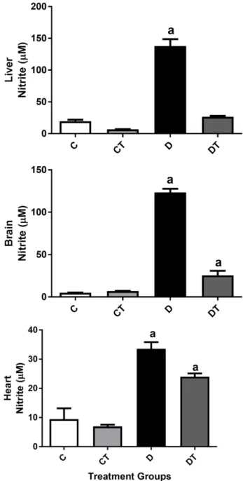
Nitrite and total superoxide dismutase, 2 markers of oxidative stress, were measured in the tissues of the liver, brain and heart. There was no effect of SPI treatment on total superoxide dismutase. Treatment of diabetic rats with SPI 50
mg/kg/day restored nitrite levels to control levels in liver tissue and significantly attenuated the increased nitrite content in both the heart and the brain (Figure 3).
3.4. SPI 50 mg/kg/day Inhibited Lipid Peroxidation in Diabetic
Treated Rats
Figure 3. Effects of 6 weeks of treatment with SPI on nitrite content in the liver, brain and heart of STZ-induced diabetic rats. The treatment groups were: control (C), control treated (CT), diabetic (D) and diabetic treated (DT). Data were expressed as mean ± SEM and statistical analysis was performed using two-way ANOVA followed by Newman-Keuls multiple comparisons test, p < 0.05 taken as significant, n = 8/group. adifferent from all other groups.
3.5. SPI 50 mg/kg/day Produced an Improvement in the Cardiac
Function in Control Treated Rats
Figure 4. Effects of 6 weeks of treatment with SPI on malondialdehyde (MDA) content in the liver, brain and heart of STZ-induced diabetic rats. The treatment groups were: con-trol (C), concon-trol treated (CT), diabetic (D) and diabetic treated (DT). Data were expressed as mean ± SEM and statistical analysis was performed using two-way ANOVA followed by Newman-Keuls multiple comparisons test, p < 0.05 taken as significant, n = 8/group a different from all other groups and b different from control group.
4. Discussion
(a)
(b)
(c)
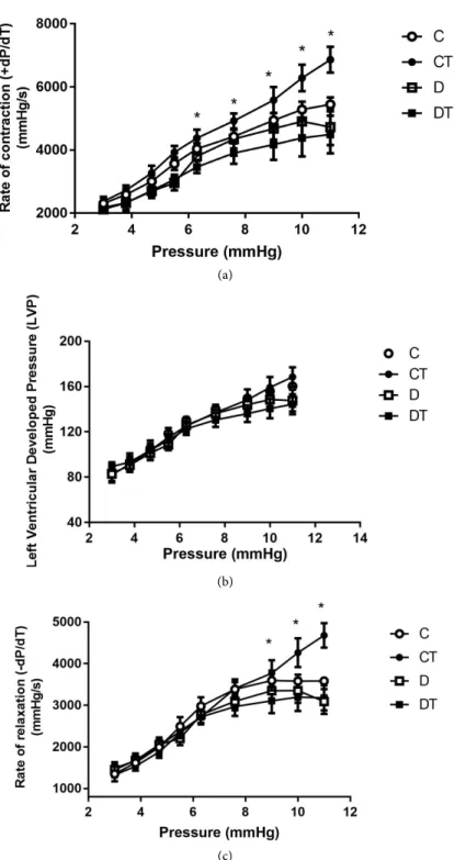
Figure 5. Effects of 6 weeks of treatment with SPI on rate of contraction (+dP/dT), on
rate of left ventricular developed pressure (LVP) and on rate of relaxation (−dP/dT) at different left atrial filling pressures in STZ-induced diabetic rats. The treatment groups were: control (C, n = 5), control treated (CT, n = 7), diabetic (D, n = 5) and diabetic treated (DT, n = 5). Data were expressed as mean ± SEM and statistical analysis was performed us-ing GLM repeated measures ANOVA followed by Newman-Keuls multiple comparisons test, p < 0.05 taken as significant, n = 8/group * different from all other groups.
SPI presents cardiovascular benefits which primarily result from its hypolipi-demic, antioxidant and anti-inflammatory activities, as demonstrated in a large number of preclinical studies and clinical trials [21]. In this experiment the in-termidate dose of SPI 50 mg/kg/day was used (based on preliminary studies done by our partner group in Brazil [5]) to study the effect of SPI on oxidative stress, glycemic and lipid profile in the STZ diabetic animal model. However af-ter 6 weeks of treatment the diabetic treated animals did not show a significant reduction in glycemic levels, perhaps which may be because the glycemic levels achieved in the STZ animals were highly elevated in this study. We hypothesized that the dose of SPI 50 mg/kg/day may have been too low in this animal model to reduce glycemic levels. However we did observe that SPI at this same dose significantly decreased triglyceride in diabetic treated animals and reduced plasma cholesterol levels in control treated animals.
Several animal and human studies have repeatedly reported the lipid-lowering effects of SPI [21] [22] [23] [24] [25]. However, the mechanisms of action of SPI on lipid metabolism are not well understood. Nagakoa and collaborators found that a concentrate of SPI inhibited jejunal cholesterol absorption and ileal bile acid reabsorption, proposing that C-phycocyanin is the molecule responsible for this effect [26]. Furthermore, it was found that phycocyanin inhibits pancreatic lipase [27]. These effects could explain the hypocholesterolemic and hypotria-cylglycerolemic effects of SPI [28].
We have also shown in this study that SPI 50 mg/kg/day (6 weeks) signifi-cantly reduced the weight of the control treated animals when compared to con-trol, while SPI did not change the weight of diabetic treated animals when com-pared to the diabetic control. Literature results of studies with SPI at higher does have shown that SPI succeeded inducing either an improvement in body weight or no weight reduction in human and animals [21]. However, more recent stu-dies have demonstrated that Spirulina platensis supplementation was effective in weight regulation, serum total cholesterol and appetite reduction in overweight patients [29] [30].
In this study we showed a normal morphological arrangement of the hepato-cytes in SPI treated diabetic animals as compared to hepatohepato-cytes from untreated diabetic animals in which fat droplets displaced the centrally located nucleus forming fat vacuoles. The observed fatty degeneration is linked to insulin defi-ciency and the dysregulation of mitochondrial β-oxidation of fatty acids. This leads to the esterification of fatty acids to triglyceride in the cytoplasm, which is characterized by the presence of multiple triglyceride droplets within the hepa-tocytes [31].
The concentration of nitric oxide (NO) (estimated as concentration of nitrite) was increased in all tissues of diabetic rats. NO levels were brought back to near normal values after the treatment with SPI 50 mg/kg/day. Generally, NO at phy-siological levels produces many benefits to the vascular system. However, in-creased oxidative stress and subsequent activation of the transcription factor NF-β enhanced the NO production, which is believed to be a mediator of dam-age to beta-cells [34].
The reduction of nitrite production in different tissues such as brain, heart and liver in our study could be considered an important factor of protection against oxidative damage to DNA, indicating its important protective effect against STZ-induced damage and these results are corroborated by numerous researchers who have studied the antioxidant and anti-inflammatory properties of SPI [35] [36] [37]. Many of the studies investigating the antioxidant effect of SPI have attributed this property to phycocyanin since this phycobilin pigment has radical scavenging properties, noting that some of these reports have also shown that phycocyanin directly inhibits NAD(P)H oxidase activity [38] [39]
and that NAD(P)H oxidases may be the main source of ROS in the tissues of di-abetic animals and patients [40] [41].
In addition, the level of malondialdehyde (MDA) was analyzed as a lipid pe-roxidation marker since it has an important role in the pathogenesis and the complications of diabetes. The results of the present analysis clearly highlight the efficacy of SPI as an MDA lowering agent. The MDA levels in diabetic rats treated with SPI were significantly lower in comparison to the diabetic control. (P < 0.05).
In this study we also evaluated the antioxidant biomarker enzyme superoxide dismutase (SOD), however there were no differences in total SOD activity among the groups studied.
Other researchers have tried to correlate the elevated lipid profile in diabetes to the development of cardiac dysfunction [19] [20] [42]. Lipid abnormalities associated with diabetes may lead to alterations in myocardial enzyme systems, subcellular organelles and myocardial fuel supply and eventually to cardiac dis-ease [19].
In this study hearts from control and diabetic rats were isolated and cardiac performance was evaluated under physiological and superphysiological filling pressures, simulated by increases in left atrial filling pressure from 2.5 to 11 mmHg. Paralleling the elevation of plasma glucose and triglycerides, the un-treated diabetic group exhibited decreased cardiac performance as assessed by their inability to respond to increases in left atria1 filling pressure. Treatment with SPI for 6 weeks did not restore the ability of the treated diabetic animals to respond to increases in filling pressure on the functional parameters studied. However, SPI in this same concentration produced an improvement in cardiac hemodynamic parameters such as dP/dTmax and dP/dTmin in control rats, suggesting a possible positive inotropic effect of Spirulina.
dys-function in diabetic rats may be due to the low dose used in this experiment, as some researchers reported evidence that both SPI and C-phycocyanin can have a preventive effect on drug-induced cardiac side effects as well as a protective ef-fect during heart attacks [43] [44] [45]. In the first two studies, they tested first SPI and then C-phycocyanin as protectants against the adverse cardiac side ef-fects of doxorubicin, a chemotherapy drug. They concluded that both SPI and C-phycocyanin significantly protected the mice from the cardiotoxic effects and was referred to as a “crucial role of the antioxidant nature of SPI and C-phycocyanin in cardioprotection” [44] [45]. Another study from the same group demonstrat-ed that pre-ischemic infusions of C-phycocyanin and SPI were cardioprotective against ischemia-reperfusion (I/R) injury, leading to enhanced recovery of con-tractile function, attenuation of infarct size, decreased apoptosis, and suppres-sion of oxidative stress in the post-ischemic reperfused heart. These results sug-gest that the underlying signaling mechanism(s) involved C-phycocyanin atten-uation of I/R-induced cardiac dysfunction through its antioxidant and anti-apoptotic actions and modulation of p38 MAPK and ERK1/2 [45].
5. Conclusion
Further studies will be needed to confirm an effect of SPI on diabetic cardi-omyopathy. However this study has shown that SPI significantly suppressed ni-trite generation and lipoperoxidation in the hearts of animals diabetics, as well as an improvement in the cardiac function SPI-treated control rats which is con-sistent with several studies that demonstrated that ROS produced in diabetic myocardial tissues causes oxidative stress-mediated injury that is protected by antioxidants.
Acknowledgements
The authors are thankful to the Science without Borders Project, CNPq (the Bra-zilian National Council for Scientific and Technological Development) for the financial support of this study.
References
[1] Tietze, H.W. (2004) Spirulina Micro Food Macro Blessing. 4th Edition, Harald W. Tietze Publishing, Australia.
[2] Kay, R.A (1991) Microalgae as Food and Supplement. Critical Reviews in Food Science and Nutrition, 30, 555-573. https://doi.org/10.1080/10408399109527556
[3] Piñero Estrada, J.E., Bermejo Bescós, P. and Villar del Fresno, A.M. (2001) Anti-oxidant Activity of Different Fractions of Spirulina Protean Extract. Farmaco, 56, 497-500. https://doi.org/10.1016/S0014-827X(01)01084-9
[4] Chen, J.C., Liu, K.S., Yang, T.J., Hwang, J.H., Chan, Y.C. and Lee, I.T. (2012) Spiru-lina and C-Phycocyanin Reduce Cytotoxicity and Inflammation-Related Genes Ex-pression of Microglial Cells. Nutritional Neuroscience, 15, 252-256.
https://doi.org/10.1179/1476830512Y.0000000020
S.A., et al. (2012) The Microalga SPI Presents Anti-Inflammatory Action as Well as Hypoglycemic and Hypolipidemic Properties in Diabetic Rats. Journal of Comple-mentary and Integrative Medicine, 9, 1553-3840.
https://doi.org/10.1515/1553-3840.1534
[6] Lima, F.A.V., Joventino, I.P., Joventino, F.P., de Almeida A.C., Neves. K.R.T., do Carmo, M.R., et al. (2017) Neuroprotective Activities of Spirulina platensis in the 6-OHDA Model of Parkinson’s Disease Are Related to Its Anti-Inflammatory Ef-fects. Neurochemical Research, 42, 3390-3400.
https://doi.org/10.1007/s11064-017-2379-5
[7] Pentón-Rol, G., Martínez-Sánchez, G., Cervantes-Llanos, M., Lagumersindez-Denis, N., Acosta-Medina, E.F., Falcón-Cama, V., et al. (2011) C-Phycocyanin Ameliorates Experimental Autoimmune Encephalomyelitis and Induces Regulatory T Cells. In-ternational Immunopharmacology, 11, 29-38.
https://doi.org/10.1016/j.intimp.2010.10.001
[8] Lee, J., Park, A., Kim, M.J., Lim, H.J., Rha, Y.A. and Kang, H.G. (2017) Spirulina Extract Enhanced a Protective Effect in Type 1 Diabetes by Anti-Apoptosis and An-ti-ROS Production. Nutrients, 9, E1363. https://doi.org/10.3390/nu9121363
[9] Farouk, K., El-Baz, F., Hanan Aly., El-Sayed A.B. and Amal, A. (2013) Mohamed. Role of Spirulina platensis in the Control of Glycemia in DM2 Rats. International Journal of Scientific & Engineering Research, 4, 1731-1740.
[10] American Diabetes Association (2012) Diagnosis and Classification of Diabetes Mellitus. Diabetes Care, 35, 564-571.
[11] Bugger, H. and Abel, E.D. (2014) Molecular Mechanisms of Diabetic Cardiomyo-pathy. Diabetologia, 57, 660-671. https://doi.org/10.1007/s00125-014-3171-6
[12] Secrest, A.M., Becker, D.J., Kelsey, S.F., LaPorte, R.E. and Orchard, T.J. (2010) All-Cause Mortality Trends in a Large Population-Based Cohort with Long-Standing Childhood-Onset Type 1 Diabetes: The Allegheny County Type 1 Diabetes Registry.
Diabetes Care, 33, 2573-2579. https://doi.org/10.2337/dc10-1170
[13] Dobrin, J.S. and Lebeche, D. (2010) Diabetic Cardiomyopathy: Signaling Defects and Therapeutic Approaches. Expert Review of Cardiovascular Therapy, 8, 373-391.
https://doi.org/10.1586/erc.10.17
[14] Cai, H. and Harrison, D.G. (2000) Endothelial Dysfunction in Cardiovascular Dis-eases: The Role of Oxidant Stress. Circulation Research, 87, 840-844.
https://doi.org/10.1161/01.RES.87.10.840
[15] Guzik T.J., Mussa, S., Gastaldi, D., Sadowski, J., Ratnatunga, C., Pillai, R. and Channon, K.M. (2002) Mechanisms of Increased Vascular Superoxide Production in Human Diabetes Mellitus: Role of NAD(P)H Oxidase and Endothelial Nitric Oxide Synthase. Circulation, 105, 1656-1662.
https://doi.org/10.1161/01.CIR.0000012748.58444.08
[16] Suzuki, H., Kayama, Y., Sakamoto, M., Iuchi, H., Shimizu, I., Yoshino, T., et al. (2015) Arachidonate 12/15-Lipoxygenase-Induced Inflammation and Oxidative Stress Are Involved in the Development of Diabetic Cardiomyopathy. Diabetes, 64, 618-630.https://doi.org/10.2337/db13-1896
[17] Kayama, Y., Raaz, U., Jagger, A., Adam, M., Schellinger, I.N, Sakamoto, M., et al. (2015) Diabetic Cardiovascular Disease Induced by Oxidative Stress. International Journal of Molecular Sciences, 16, 25234-25263.
https://doi.org/10.3390/ijms161025234
Maxima. PhD Thesis, Fac. Sci., University of Paris, Paris.
[19] Rodrigues, B., Xiang, H. and McNeill, J.H. (1988) Effect of Lcarnitine Treatment on Lipid Metabolism and Cardiac Performance in Chronically Diabetic Rats. Diabetes, 37, 1358-1364.https://doi.org/10.2337/diab.37.10.1358
[20] Rodrigues, B., Goyal, R.K. and McNeill, J.H. (1986) Effects of Hydralazine on STZ-Induced Diabetes Rats-Prevention of Hyperlipidemia and Improvement in Cardiac Function. The Journal of Pharmacology and Experimental Therapeutics, 237, 292-299.
[21] Deng, R. and Chow, T.J. (2010) Hypolipidemic, Antioxidant, and Antiinflammatory Activities of Microalgae Spirulina. Cardiovascular Therapeutics, 28, 33-45.
https://doi.org/10.1111/j.1755-5922.2010.00200.x
[22] Lee, E.H., Park, J.E., Choi, Y.J., Huh, K.B. and Kim, W.Y. (2008) A Randomized Study to Establish the Effects of Spirulina in Type 2 Diabetes Mellitus Patients. Nu-trition Research and Practice, 2, 295-300.https://doi.org/10.4162/nrp.2008.2.4.295
[23] Mani, U.V., Desai, U.V.S. and Iyer, U. (2000) Studies on the Long-Term Effect of Spirulina Supplementation on Serum Lipid Profile and Glycated Proteins in NIDDM Patients. Journal of Nutraceuticals, Functional and Medical Foods, 2, 25-32.https://doi.org/10.1300/J133v02n03_03
[24] Park, J.Y. and Kim, W.Y. (2003) The Effect of Spirulina on Lipid Metabolism, An-tioxidant Capacity and Immune Function in Korean Elderly. The Korean Journal of Nutrition, 36, 287-297.
[25] Parikh, P., Mani, U. and Iyer, U. (2001) Role of Spirulina in the Control of Glyce-mia and LipideGlyce-mia in Type 2 Diabetes Mellitus. Journal of Medicinal Food, 4, 193-199.https://doi.org/10.1089/10966200152744463
[26] Nagaoka, S., Shimizu, K., Kaneko, H., Shibayama, F., Morikawa, K., Kanamaru, Y.,
et al. (2005) A Novel Protein C-Phycocyanin Plays a Crucial Role in the Hypocho-lesterolemic Action of Spirulina platensis Concentrate in Rats. Journal of Nutrition, 135, 2425-2430.https://doi.org/10.1093/jn/135.10.2425
[27] Li-Kun, H., Dong-Xia, L., Lan, X., Xiao-Jie, G., Yasumasa, K., Isao, S. and Hiromi-chi, O. (2006) Isolation of Pancreatic Lipase Activity-Inhibitory Component of Spi-rulina platensis and It Reduce Postprandial Triacylglycerolemia. Yakugaku Zasshi, 126, 43-49.https://doi.org/10.1248/yakushi.126.43
[28] Iwata, K., Inayama, T. and Kato, T. (1990) Effects of Spirulina platensis on Plasma Lipoprotein Lipase Activity in Fructose-Induced Hyperlipidemic Rats. Journal of Nutritional Science and Vitaminology, 36, 165-171.
https://doi.org/10.3177/jnsv.36.165
[29] Miczke, A.A., SzulInska, M., Hansdorfer-Korzon, R., Kregielsk-Narozna, M., Suli-burska, J., Walkowiak, J., et al. (2016) Effects of Spirulina Consumption on Body Weight, Blood Pressure, and Endothelial Function in Overweight Hypertensive Caucasians: A Double Blind, Placebo-Controlled, Randomized Trial. European Re-view for Medical and Pharmacological Sciences, 20, 150-156.
[30] Zeinalian, R., Farhangi, M.A., Shariat, A. and Saghafi-Asl, M. (2017) The Effects of
Spirulina Platensis on Anthropometric Indices, Appetite, Lipid Profile and Serum Vascular Endothelial Growth Factor (VEGF) in Obese Individuals: A Randomized Double Blinded Placebo Controlled Trial. BMC Complementary and Alternative Medicine, 17, 225.https://doi.org/10.1186/s12906-017-1670-y
[31] Fromenty, B. and Pessayre, D. (1995) Inhibition of Mitochondrial Beta-Oxidation as a Mechanism of Hepatotoxicity. Pharmacology & Therapeutics, 67, 101-154.
[32] Bertolin, T.E., Pilatti, D., Giacomini, A.C.V.V., Bavaresco, C.S., Colla, L.M. and Costa, J.A.V. (2009) Effect of Microalga Spirulina platensis (Arthrospiraplatensis) on Hippocampus Lipoperoxidation and Lipid Profile in Rats with Induced Hyper-cholesterolemia. Brazilian Archives of Biology and Technology, 52, 1253-1259.
https://doi.org/10.1590/S1516-89132009000500024
[33] Dartsch, P.C. (2008) Antioxidant Potential of Selected Spirulina platensis Prepara-tions. Phytotherapy Research, 22, 627-633.https://doi.org/10.1002/ptr.2310
[34] Malik, Z.A., Tabassum, N. and Sharma, P.L. (2013) Attenuation of Experimentally Induced Diabetic Neuropathy in Association with Reduced Oxidative-Nitrosative Stress by Chronic Administration of Momordicacharantia. Advances in Bioscience and Biotechnology, 4, 356-363.https://doi.org/10.4236/abb.2013.43047
[35] Lee, J., Park, A., Kim, M.J., Lim, H.J., Rha, Y.A. and Kang, H.G. (2017) Spirulina Extract Enhanced a Protective Effect in Type 1 Diabetes by Anti-Apoptosis and An-ti-ROS Production. Nutrients, 9, 1363.https://doi.org/10.3390/nu9121363
[36] Romay, C., Delgado, R., Remirez, D., González, R. and Rojas, A. (2001) Effects of Phycocyanin Extract on Tumor Necrosis Factor-Alpha and Nitrite Levels in Serum of Mice Treated with Endotoxin. Arzneimittelforschung, 51, 733-736.
[37] Romay, C., González, R., Ledón, N., Remirez, D. and Rimbau, V. (2003) C-Phycocyanin: A Biliprotein with Antioxidant, Anti-Inflammatory and Neuropro-tective Effects. Current Protein & Peptide Science, 4, 207-216.
https://doi.org/10.2174/1389203033487216
[38] Riss, J., Décordé, K., Sutra, T., Delage, M., Baccou, J.C., Jouy, N., et al. (2007) Phy-cobiliprotein C-Phycocyanin from Spirulina platensis Is Powerfully Responsible for Reducing Oxidative Stress and NADPH Oxidase Expression Induced by an Athero-genic Diet in Hamsters. Journal of Agricultural and Food Chemistry, 55, 7962-7967.
https://doi.org/10.1021/jf070529g
[39] Lanone, S., Bloc, S., Foresti, R., Almolki, A., Taillé, C., Callebert, J., et al. (2005) Bi-lirubin Decreases nos2 Expression via Inhibition of NAD(P)H Oxidase: Implication for Protection against Endotoxic Shock in Rats. FASEB Journal, 19, 1890-1892.
https://doi.org/10.1096/fj.04-2368fje
[40] Inoguchi, T., Li, P., Umeda, F., Yu, H.Y., Kakimoto, M., Imamura, M., et al. (2000) High Glucose Level and Free Fatty Acid Stimulate Reactive Oxygen Species Produc-tion through Protein Kinase C-Dependent ActivaProduc-tion of NAD(P)H Oxidase in Cul-tured Vascular Cells. Diabetes, 49, 1939-1945.
https://doi.org/10.2337/diabetes.49.11.1939
[41] Inoguchi, T. and Nawata, H. (2005) NAD(P)H Oxidase Activation: A Potential Target Mechanism for Diabetic Vascular Complications, Progressive Beta-Cell Dysfunction and Metabolic Syndrome. Current Drug Targets, 6, 495-501.
https://doi.org/10.2174/1389450054021927
[42] Bayeva, M., Sawicki, K.T. and Ardehali, H. (2013) Taking Diabetes to Heart-Deregulation of Myocardial Lipid Metabolism in Diabetic Cardiomyopathy.
Journal of the American Heart Association, 2, e000433.
https://doi.org/10.1161/JAHA.113.000433
[43] Khan, M., Varadharaj, S., Ganesan, L.P., Shobha, J.C. and Naidu, M.U. (2005) C-Phycocyanin Protects against Ischemia-Reperfusion Injury of Heart through In-volvement of p38 MAPK and ERK Signaling. American Journal of Physiology.
Heart and Circulatory Physiology, 290, 2136-2145.
https://doi.org/10.1152/ajpheart.01072.2005
(2005) Protective Effect of SPI against Doxorubicin-Induced Cardiotoxicity. Phyto-therapy Research, 19, 1030-1037.https://doi.org/10.1002/ptr.1783
[45] Khan, M., Varadharaj, S., Shobba, J.C., Naidu, M.U., Parinandi, N.L., Kutala, V.K.,


