Subcortical ischemic vascular
disease and cognition
A systematic review
Gilberto Sousa Alves
1, Carlos Eduardo de Oliveira Alves
1, Maria Elisa Lanna
1,
Denise Madeira Moreira
2,3, Eliasz Engelhardt
2, Jerson Laks
1,4Abstract – Subcortical Ischemic Vascular Disease (SIVD) is underdiagnosed. This review investigates the re-lationship among SIVD severity, cognitive status and neuroimaging markers. Methods: Cohort, cross-sectional and case control studies were searched on ISI, Medline, Scielo, PsychoInfo and LILACS databases published between 1995 and 2006. Results: The most impaired cognitive domains were executive, attentional and memory retrieval mechanisms. These cognitive features were frequently associated to White Matter Lesions (WML).
Conclusions: WML is an independent factor in cognitive decline. However, the threshold for this impact is not yet clearly established .
Key words: neuropsychology, vascular dementia, cerebrovascular disorders, cognition.
Doença vascular isquêmica subcortical e cognição: uma revisão sistemática
Resumo – A doença vascular isquêmica subcortical (DVIS) é uma condição ainda subdiagnosticada. Esta revisão investiga a relação entre a gravidade da DVIS, o status cognitivo e marcadores de neuroimagem. Métodos: Estudos de coorte, seccionais e casos-controles foram pesquisados nas bases de dados ISI, Medline, Scielo, PsychoInfo e LILACS entre 1995 e 2006. Resultados: Os domínios cognitivos mais comprometidos foram as funções executivas, a atenção e a recuperação da memória. Estes achados cognitivos estiveram frequentemente associados à presença de lesões em substância branca (LSB). Conclusões: As LSB constituem um fator de risco independente para o declínio cognitivo. Contudo, o limiar de impacto desta variável ainda não pôde ser claramente estabelecido.
Palavras-chave: neuropsicologia, demência vascular, doença cerebrovascular, cognição.
1Institute of Psychiatry, Federal University of Rio de Janeiro, Brazil. 2Institute of Neurology, Federal University of Rio de Janeiro, Brazil. 3Radiology Service
of the Procardíaco Hospital, Brazil. 4State University of Rio de Janeiro, Brazil.
Gilberto Sousa Alves – Rua Otaviano Hudson, 16 / 608 - 22030-030 Rio de Janeiro RJ - Brazil. E-mail: [email protected] Received February 14, 2008. Accepted in final form May 6, 2008.
Elderly subjects commonly present with some degree of cerebrovascular lesion on brain Magnetic Resonance Imag-ing (MRI).1,2 Depending on the site, intensity and severity,
these lesions may either cause or contribute to further cog-nitive decline. Among the overall category of cerebrovas-cular disease (CVD), subcortical ischemic vascerebrovas-cular disease (SIVD) is particularly prevalent3,4 and encompasses three
basic pathological entities: small vessel disease, lacunar infarct and ischemic white matter lesions (WML). This categorization is depicted according to the primary type of brain lesions.5
Being a homogeneous construct, the diagnosis of SIVD allows better knowledge of the clinical picture as well as of the natural history, outcome and clinical response to
treat-ment strategies.5-8 Regardless of the causal pathological
factor of brain vascular lesions, a wide variety of complex mechanisms can be singled out as risk and intervening fac-tors for SIVD. These facfac-tors include interactions between vascular etiologies (hypoperfusion caused by arterial stiffness and its relationship to risk factors such as arterial hyperten-sion), microvascular changes in the brain (infarcts, WML, atrophy), host factors (age, education), hypotension, genetic inheritance (CADASIL), and cognitive characteristics.5,9
The main clinical manifestations of SIVD may be sum-marized as a “dysexecutive syndrome” due to the interrup-tion of prefrontal-subcortical loops10,11 affecting control,
goal-directed activities.12,13 The clinical picture also
in-cludes psychomotor slowness, forgetfulness, and changes in speech, affect and mood11,14. Despite the fact that
dys-executive syndrome and cognitive decline are frequently observed in SIVD, a debate remains over to what extent the vascular lesions have to be present in order to be respon-sible for the clinical and neuropsychological picture.15 This
review aimed to investigate the relationship between SIVD severity and cognitive status with possible neuroimaging markers, addressing two main questions: (1) how the clini-cal stage of subcorticlini-cal disease is related to impairment in specific cognitive domains and how it affects prognosis and functional status; (2) the impact of the presence, localiza-tion and extent of WML on cognilocaliza-tion.
Methods
A systematic review of the literature regarding SIVD was performed by searching data from ISI, Medline, Scielo, PsychoInfo, and LILACS web databases published between 1995 (January) and 2006 (July). The search strategy in-cluded key words aimed at investigating a broader spec-trum of primary vascular disorders affecting subcortical areas, mainly white-matter lesions: subcortical disease OR dementia, vascular dementia, small vessels disease, Binswanger disease, CADASIL, white-matter disease OR dementia, leukoencephalopathy, cerebrovascular disease or disorders, brain vascular disease OR disorder.
All abstracts were independently read by five authors (GSA; EE; CEOA; MEL; JL) and those studies which com-plied with the inclusion criteria were selected for further reading. A manual search was also performed to reach ar-ticles related to this subject found among the references of the selected studies. The articles which satisfied all the fol-lowing criteria were included for further reading and anal-ysis. They had (1) to be cohort, cross-sectional, or case con-trol studies with at least one criterion for vascular dementia (DSM-IV, or NINDS-AIREN, or ICD-10, or ADDTC); (2) to provide data on cognitively impaired patients ≥60 years of age, with or without clinical diagnosis of dementia; (3) to include a comprehensive neuropsychological assessment, as well as at least one neuroimaging exam (brain Com-puted Tomography or Magnetic Resonance). Studies that described reviews of the literature, case reports, samples with primary psychiatric disorders, exclusively cortical lesions (Alzheimer disease or cortical vascular disease), gray-matter, and cerebellar diseases were excluded from this review. The following items were chosen to describe the correlated variables: 1) methodological characteristics concerning the article; 2) cognitive outcomes and subcorti-cal disease; 3) neuroradiologic findings and cognitive-func-tional aspects.
To the best of our knowledge, only one other systematic review on the subject has sought to study the relationship between WML and cognition.16
Results
This search retrieved a total of 159 articles, only 32 of which remained eligible according to the inclusion and ex-clusion criteria. A further analysis excluded three other pa-pers, two because the majority of individuals in the sample were younger than 60 years and one because of absence of neuroimaging to ascertain the presence of SIVD. Tables 1 and 2 show the 29 articles included in this study, displaying two subgroups: firstly those that only studied the cognitive profile (Table 1), plus the studies that assessed the correla-tions with neuroimaging (Table 2).
Methodological characteristics of the articles
Twenty-four studies were cross-sectional and five longi-tudinal. The samples assessed in the articles were predomi-nantly outpatients (18), followed by inpatients (6 articles) and mixed samples (inpatients/outpatients) in one article.17
Seven articles involved samples from branches of multi-center projects. Three studies consisted of population based individuals,15,18-19 whereas four articles were from a study
involving outpatients.20-23 One article did not describe the
source of the patients.24
Cognitive manifestations and subcortical disease
The majority of the articles (n=28, 96.5 %) found a correlation between the clinical diagnosis of SIVD and cognitive alterations. The present analysis focused on ex-ecutive dysfunction and is complemented by data on other cognitive domains.
Executive dysfunction
A worse performance in executive functions was the main finding of the studies comparing SIVD patients with other causes of dementia, such as cortical stroke pa-tients,25,26 lacunar infarcts27 and AD.22
Executive dysfunction was considered a predictor of worse performance in complex activities of daily life in one study.28 In the Libon et al.22 study, the SIVD group
presented greater impairment in the executive control tests, such as the Boston Revision of the Wechsler Memory Scale-Mental Control Subtest (WMS-MC),29 in relation to AD
pa-tients. Impairment in tasks assessing the set shifting process was described in some studies, whether comparing SIVD to healthy controls30 or to other groups of patients such as
fronto temporal dementia21. Worse performance in
seen in a recent article.25 In the work by O’Sullivan et al.,30
a brief assessment of executive functions involving Trail Making and digit symbols was able to provide good sen-sitivity and specificity for distinguishing individuals with ischemic leukoaraiosis from subjects with healthy aging.
Impairment of executive functioning at early stages of the disease was investigated in two studies using outpatient samples diagnosed with CADASIL (autosomal dominant arteriopathy associated with subcortical infarcts and leu-koencephalopathy). One of these31 evaluated three distinct
clinical groups – patients with no previous history of ce-rebral infarct and two groups with recent stroke, with and without vascular dementia (VD). Using MRI, the authors observed impairment in executive functions in the group with mild radiological alterations of white matter. In the other article,32 impaired performance in executive tasks,
ev-idenced by a greater proportion of perseverative errors, was shown in the group with vascular cognitive impairment
(VCI). Accordingly, these findings should enhance the dif-ferential diagnosis between vascular and neurodegenerative diseases, since CADASIL has a clinical presentation with frontal executive impairment at very early stages.
Cognitive performance other than executive
Attentional deficits were also a frequently reported finding15,21,24,33-36 and were strongly correlated to subcortical
lesions. Lower scores in attention and concentration tests were seen in vascular groups compared to AD34 or healthy
controls.33 Slowing in processing speed, assessed by the Trail
Making Test,30,32-33 Block Design17 and Letter-Digit
substi-tution test15 was more associated to subcortical disease.
Distinct mechanisms of memory impairment differen-tiated AD from SIVD. SIVD patients tended to show better performance than the AD group on delayed recognition memory with or without the help of clues.22-23,34,37 Also, the
SIVD group performed better than patients with AD in
de-Table 1. Studies with only clinical and cognitive correlations.
Authors/year Sample (N) Design Cognition and diagnosis of SVI/SVD
Amberla et al.31 3 CADASIL groups CS Early impairment of EF and later deficits in mental speed and set-shifting
abilities Doody et al.34 AD (37)
IVD (18)
CS Lower results on visuoconstructional tasks in IVD group
Frisoni et al.35 MCIva (29)
MCIad (19) SVD (21)
L MCI: Poorer performance on frontal tests and more cognitive loss and de-terioration in ADL
Gainotti et al.38 AD (68)
VaD (40)
CS Vascular patients had better scores on selective and divided attention and fewer false alarm errors
Galluzzi et al.36 MCIam (14)
MCIva (29)
CS Verbal fluency was able to discriminate future conversion to SVD and AD
Mok et al.28 SVD
inpatients (75) controls (42)
L Patients with mild cognitive impairment had worse performance on cogni-tive tests than healthy controls
Moretti et al.20 SVD (144) L Executive function and planning behavior more impaired in SVD patients
Moretti et al.21 FLD (40)
SVD (40)
L VaD patients: lower executive functioning, lack of set-shifting, more rigid and apathetic
O’Sullivan et al.30 SIVD (32)
Controls (17)
CS Trail Making and Digit Symbol: assess set shifting and were the most dis-criminating tests
Peters et al.32 CADASIL (63)
controls (40)
L CADASIL group showed greater impairment in executive functions and processing speed test even in early stages of disease
Pohjasvaara et al.26 Post-stroke
inpatients (337)
CS SVD more impaired in executive functions
Tierney et al.37 AD (31)
SIVD (31)
CS Verbal recognition memory and verbal fluency distinguished AD from SIVD (with a double dissociation)
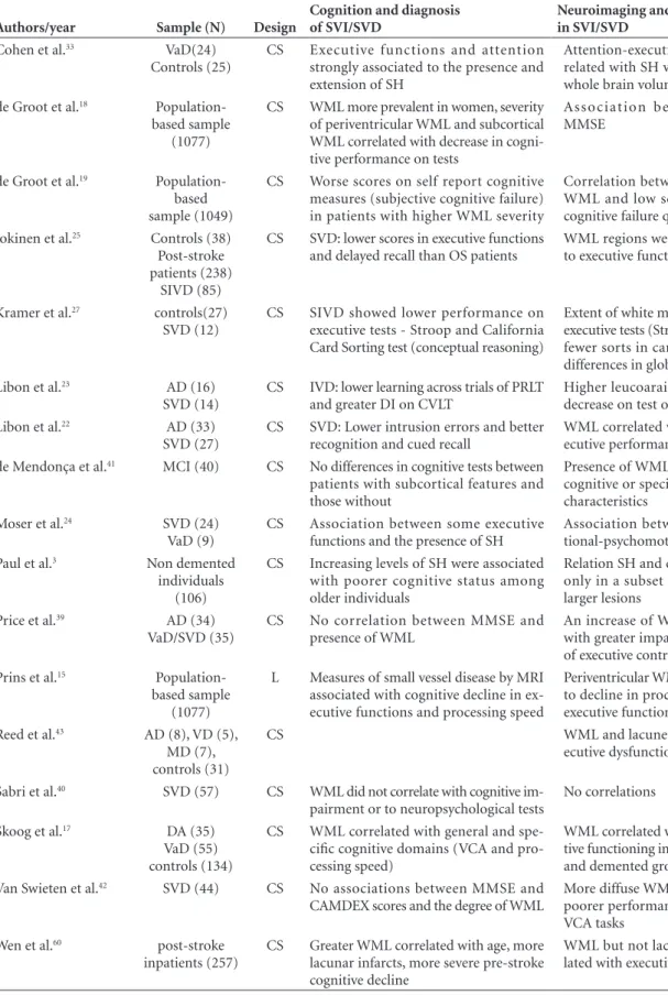
Table 2. Studies with correlations among clinical, cognitive and neuroimaging aspects.
Authors/year Sample (N) Design
Cognition and diagnosis of SVI/SVD
Neuroimaging and cognition in SVI/SVD
Cohen et al.33 VaD(24)
Controls (25)
CS Executive functions and attention strongly associated to the presence and extension of SH
Attention-executive dysfunction cor-related with SH volume but not with whole brain volume (WBV)
de Groot et al.18
Population-based sample (1077)
CS WML more prevalent in women, severity of periventricular WML and subcortical WML correlated with decrease in cogni-tive performance on tests
Association between WML and MMSE
de Groot et al.19
Population-based sample (1049)
CS Worse scores on self report cognitive measures (subjective cognitive failure) in patients with higher WML severity
Correlation between periventricular WML and low scores on subjective cognitive failure questionnaire Jokinen et al.25 Controls (38)
Post-stroke patients (238)
SIVD (85)
CS SVD: lower scores in executive functions and delayed recall than OS patients
WML regions were weakly correlated to executive functions
Kramer et al.27 controls(27)
SVD (12)
CS SIVD showed lower performance on executive tests - Stroop and California Card Sorting test (conceptual reasoning)
Extent of white matter correlated with executive tests (Stroop interference and fewer sorts in card sorting task). No differences in global cognitive abilities Libon et al.23 AD (16)
SVD (14)
CS IVD: lower learning across trials of PRLT and greater DI on CVLT
Higher leucoaraiosis correlated with decrease on test of psychomotor skills Libon et al.22 AD (33)
SVD (27)
CS SVD: Lower intrusion errors and better recognition and cued recall
WML correlated with decrease on ex-ecutive performance
de Mendonça et al.41 MCI (40) CS No differences in cognitive tests between
patients with subcortical features and those without
Presence of WML not associated with cognitive or specific neuropsychiatric characteristics
Moser et al.24 SVD (24)
VaD (9)
CS Association between some executive functions and the presence of SH
Association between SH and atten-tional-psychomotor speed tasks Paul et al.3 Non demented
individuals (106)
CS Increasing levels of SH were associated with poorer cognitive status among older individuals
Relation SH and cognition was found only in a subset of individuals with larger lesions
Price et al.39 AD (34)
VaD/SVD (35)
CS No correlation between MMSE and presence of WML
An increase of WMA was associated with greater impairment on measures of executive control and VCA
Prins et al.15
Population-based sample (1077)
L Measures of small vessel disease by MRI associated with cognitive decline in ex-ecutive functions and processing speed
Periventricular WM changes associated to decline in processing information/ executive functions
Reed et al.43 AD (8), VD (5),
MD (7), controls (31)
CS WML and lacunes correlated with
ex-ecutive dysfunction
Sabri et al.40 SVD (57) CS WML did not correlate with cognitive
im-pairment or to neuropsychological tests
No correlations
Skoog et al.17 DA (35)
VaD (55) controls (134)
CS WML correlated with general and spe-cific cognitive domains (VCA and pro-cessing speed)
WML correlated with impaired cogni-tive functioning in both non-demented and demented groups
Van Swieten et al.42 SVD (44) CS No associations between MMSE and
CAMDEX scores and the degree of WML
More diffuse WML were associated to poorer performance on executive and VCA tasks
Wen et al.60 post-stroke
inpatients (257)
CS Greater WML correlated with age, more lacunar infarcts, more severe pre-stroke cognitive decline
WML but not lacunar infarcts corre-lated with executive tests (in MDRS)
layed recall tasks,34 as wells as selective and divided attention
tasks,38 and in relation to the number of intrusive errors,
cued recall memory retrieval and visual recognition tests.22
Impairment of higher cognitive skills was discussed in addition to executive, memory and attention abnormali-ties. Visuo-spatial and visuo-construction abilities were assessed by specific tasks, such as the Block Design and Clock Drawing Test and were found to be more impaired in SIVD than in AD20,34,39 and healthy controls.17,31 One of
the studies observed greater impairment of such domains in mild stages of VaD.34 Language deficits, described as
se-mantic and phonological abnormalities, were seen in the vascular group in a two-year follow-up study.20 The
ac-curacy of verbal fluency (equal to 0.75 under the area of the Receiver Operating characteristic Curve, with a confi-dence interval of 95%) was considered by another study36
as one of the best parameters to discriminate individuals with amnestic complaints from those with mild subcorti-cal disease. Another article observed a double dissociation between AD and the SIVD group, with the latter showing a worse performance in verbal fluency and better scores in verbal recognition.37
Neuroimaging and cognitive aspects
Seventeen studies analyzed the association between WML and cognitive features. Sixteen of these articles (94.1%) found a relationship among neuroimaging, cog-nition and subcortical disease. The main findings are sum-marized under the topics below.
Presence of WML and cognitive performance in healthy individuals
Two studies described an association between WML and the presence of cognitive deficits in samples without a clinical diagnosis of dementia. One of these evaluated oldest old patients (aged ≥85 years) and observed an as-sociation between microangiopathy lesions and cognitive decline.17 Another article found a worse cognitive
perfor-mance (measured by ADAS-cog) and a greater intensity of small infarcts in a non-demented group (with Clinical Dementia Rating – CDR 0), in comparison with healthy individuals without radiological alterations.28
The presence of WML and cognitive alterations
There seems to be an association between the attention and executive function tests and the presence of subcorti-cal hyperintensities (SHI) in MRI, although the degree of global cortical atrophy was not related to the occurrence of SHI.33 Different studies found similar associations
be-tween the presence of subcortical lesions and impaired speed of thought and specific psychomotor skills. A
de-crease in cognitive processing speed was associated with the presence of WML17 and hyperintensities in MRI.24 The
presence of WML was also associated with lower scores in visuo-constructional,39 visuo-motor (Block Design), and
visuo-spatial skills (Clock-Drawing Test),17 attention and
concentration24,33 and the severity of the microangiopathic
lesions correlated to decreased learning of motor skills.23
Interestingly, other cognitive domains were not as-sociated with subcortical lesions. For instance, a greater intensity of WML neither correlated with a more serious impairment of memory and language39 nor to global
cogni-tive scores measured by the Mattis Dementia Rating Scale (MDRS).27 Two outpatient studies40-41 observed no
corre-lation between the severity of WML and performance in neuropsychological tests.
Localization and extent of WMLs and cognitive alterations
Six studies15,18-19,23,42-43 directly analyzed the correlation
between the presence and extension of WML and global cognitive performance and specific functions, with differ-ent findings.
Periventricular white matter lesions and cognition
A positive association between the degree of cogni-tive decline and the presence of periventricular WML (PWML) was observed in four studies.15,18-19,23 The
pres-ence of PWML, in comparison with diffuse subcortical lesions, was more associated with global cognitive impair-ment in two studies.18,19 A greater severity of PWML was
associated with more subjective complaints in the Cognitive Failure Questionnaire, a semi-structured instrument which evaluated the patient’s perception of their own cognition.19
PWML was associated with specific cognitive skills, execu-tive functions among them,15,42-43 and the processing of
in-formation.15 In one study the association with executive
dysfunction occurred independently to the presence of la-cunar infarcts.42 When compared with microangiopathy in
frontal or occipito-parietal areas, diffuse subcortical lesions were also associated with a worse performance in executive tests (Digit Symbol, Trail-Making B).42
Extent of WMLs and cognition
Only one article suggested a threshold of WML related to executive dysfunction. The authors observed that when 50% of white matter was compromised executive dysfunc-tion ensued.39
Discussion
number of prospective studies assessing the correlations of neuroimaging and cognitive features of SIVD may be explained partly by the high cost and the difficulties inher-ent to the recruitminher-ent and assessminher-ent of samples. Further-more, prospective studies of this disease are often faced with high rates of mortality-morbidity which impact the control of possible biases (attrition, survival or performance).16
The majority of the studies (n=15) found a positive clinical association between executive dysfunction and sub-cortical dementia. Loss of frontal executive control is a ma-jor component of cognitive disability11 and may also show
quantitative and qualitative differences between SIVD and other clinical groups (AD, cortical stroke patients), con-tributing as a method to validate the diagnostic criteria of subcortical disease, in addition to the assessment by brain imaging currently adopted.
The characteristic pattern of memory impairment pre-sented by SIVD in the selected articles dovetails with previ-ous literature and strengthens the hypothesis of possible different neurological pathways being involved in vascular and Alzheimer’s disease. While retrieval and evocation pro-cesses are highly impaired in SIVD because of disruption in executive circuits,44-46 recognition with the help of clues
(cued recall) is more preserved. On the other hand, encod-ing is severely impaired and cued recall tasks show a larger number of intrusions in AD.
In three studies,22,37,38 the cognitive assessment of
Al-zheimer and vascular patients demonstrated a pattern of double dissociation and reinforced previous literature outlining the frontal dysfunction as the most characteris-tic pattern of SIVD.47,48 The vascular group showed worse
performance in executive control and graphomotor tasks,22
whereas they performed better than the AD patients in the assessment of delayed recall tasks with or without the help of clues. Furthermore, a clear dissociation was observed in the early stages of VaD and in AD. The former group showed more impairment of sensory-motor and attention-al components whereas the latter presented worse perfor-mance on selective and divided attention.38
Studies on CADASIL, although few in number, offer an important diagnostic and prognostic model for the charac-terization of cognitive manifestations in subcortical vascular disease. They can also provide correlations with functional impairment and degree of cerebral lesion. Despite being rare, CADASIL merits interest as it can be considered a pure form of subcortical ischemic dementia. For this reason, longitudinal and multicentric studies could clarify the role of lacunes and WML in mood disorders and psychomotor and cognitive abnormalities in patients with this disease.49
Speed of psychomotor processing and some higher cor-tical functions such as language, spatial, and
visuo-constructional abilities were also frequently reported.24,33-36
It is possible that slowing of mental speed may account for lower scores on cognitive tests as an independent variable. Studies with timed and non-timed executive tests have shown that slow performance places greater demands on the retention of information in working memory and as-sociated neural circuits.30 As most neuropsychological
pro-tocols give emphasis to executive function,50 other aspects
of neuropsychological functioning may have been underes-timated and how language, visuo-spatial and visuo-motor skills can contribute in differentiating SIVD from other groups remains an open issue.
The importance of white matter disease as an isolated factor of cognitive impairment is frequently discussed,51
and some observations seem to suggest an effect which might be independent of large infarcts and degenerative lesions of AD.52 It is known that WML, like lacunar infarcts,
can interrupt subcortical pre-frontal loops, leading to im-paired functioning of the frontal lobe and to deficiency in the processing of information.11,46,53 There is evidence
that periventricular white-matter lesions and subcortical white-matter lesions affect cognitive functions in different ways54,55 where some articles have explored this hypothesis.
The main difference between these subcortical lesions is based on impaired neural pathways. While the periven-tricular WML damages long association fibers, in turn connected to distant cortical areas, lesions in short fibers result in the formation of more diffuse WML. Thus, WML abnormalities could compose disconnection syndromes, with diffuse WML more related to the interruption of local networks, whereas the periventricular lesions would lead to the impairment of functions which require coordination of multiple cortical areas distant from one another.12,56
One study found a threshold of WML for executive dysfunction,39 but the other twenty six drew no conclusion
on this issue. According to the author, important limita-tions were associated to this finding. Although neuroimag-ing has a leadneuroimag-ing role, any proposition of a more measur-able criterion for subcortical dementia has to balance not only the percentage of white matter lesions measured but also clinical and cognitive features, such as the influence of comorbid diseases (e.g., mixed dementia) and mea-sures of other cognitive functions (memory and language impairments).39
dysfunctions showed conflicting results. This limitation is probably due to methodological issues and to the difficul-ties in differentiating the cognitive effects of concurrent pathologies, such as cortical lesions in AD. The former is related to differences in the settings of the studies and in socio-demographic variables, the small size effect on cogni-tion of mild WML,57 the limited number of longitudinal
studies and the short period of follow-up,58 and finally,
dif-ferences in the criteria defining risk factors for cerebrovas-cular disease59 have hindered sound conclusions. In
addi-tion, inconsistent results have been collected because of the vast difference in methods of quantifying WML. Technical questions related to the acquisition and quality of cerebral images obtained by CT or MR, as well as the variations in the image contrast, resolutions and angles should also be noted.60 In relation to the latter aspect, post mortem studies
have evidenced that typical AD pathology was associated to executive deficits in mild stages of dementia and coexisted with small vessel disease.61 Thus, it is possible that in some
cases the impact of regional white matter lesions on cogni-tion could be overestimated and not consider a developing degenerative process.
Concluding, the concept of subcortical dementia, how-ever well-characterized in terms of neuropsychological al-terations, still awaits definition of concomitant factors and overlapping conditions, such as the presence of white mat-ter lesions, small armat-tery lesions (not always easily identifi-able by neuroimaging), cortical atrophy and degenerative lesions. In spite of this, the majority of the studies pointed to white matter lesions as an independent factor in cogni-tive decline. There appears to be a threshold for the impact of this effect and although this has not yet been clearly established, it seems certain that specific frontostriatal cir-cuits are impaired in the process.
Acknowledgments – The authors thank Ms. Luzinete
Alvarenga for her editorial assistance.
References
1. Almkvist O, Wahlund LO, Andersson-Lundman G, Basun H, Backman L. White-matter hyperintensity and neuropsycho-logical functions in dementia and healthy aging. Arch Neurol 1992;49:626-632.
2. Soderlund H, Nyberg L, Adolfsson R, Nilsson LG, Launer LJ. High prevalence of white matter hyperintensities in nor-mal aging: Relation to blood pressure and cognition. Cortex 2003;39:1093-1105.
3. Paul RH, Haque O, Gunstad J, et al. Subcortical hyperin-tensities impact cognitive function among a select subset of healthy elderly. Arch Clin Neuropsychol2005;20:697-704.
4. Chui H. Dementia due to subcortical ischaemic vascular dis-ease. Clinical Cornerstone 2001;3:40-51.
5. Erkinjuntti T. Subcortical ischemic vascular disease and de-mentia. Int Psychogeriatr 2003;15(suppl 1):23-26.
6. Erkinjuntti T, Inzitari D, Pantoni L, et al. Research criteria for subcortical vascular dementia in clinical trials. J Neural Transm Suppl 2000;59:23-30.
7. Erkinjuntti T, Inzitari D, Pantoni L, et al. Limitations of clini-cal criteria for the diagnosis of vascular dementia in cliniclini-cal trials. Is a focus on subcortical vascular dementia a solution? Ann NY Acad Sci 2000; 903: 262-72.
8. Pantoni L, Rossi R, Inzitari D, et al. Efficacy and safety of nimodipine in subcortical vascular dementia: A subgroup analysis of the Scandinavian multi-infarct dementia trial. J Neurol Sci 2000;175:124-134.
9. Brun A. Vascular burden of the white matter. Int Psychogeri-atr 2003;15(suppl 1):53-58.
10. Ishii N, Nishihara Y, Imamura T. Why do frontal lobe symp-toms predominate in vascular dementia with lacunes? Neu-rology 1986;36:340-345.
11. Román GC, Erkinjuntti T, Wallin A, Pantoni L, Chui HC. Subcortical ischaemic vascular dementia. Lancet Neurol 2002;1:426-436.
12. Wolfe N, Linn R, Babikian VL, Knoefel JE, Albert ML. Frontal systems impairment following multiple lacunar infarcts. Arch Neurol 1990;47:129-132.
13. Cummings JL. Frontal-subcortical circuits and human behav-ior. Arch Neurol 1993;50:873-880.
14. Román GC, Royall DR. Executive control function: a rational basis for the diagnosis of vascular dementia. Alzheimer Dis Assoc Disord 1999;13(suppl 3):69-80.
15. Prins ND, van Dijk EJ, den Heijer T, et al. Cerebral small-vessel disease and decline in information processing speed, executive function and memory. Brain 2005;128:2034-2041. 16. Ferro JM, Madureira S. Age-related white matter changes and
cognitive impairment. J Neurol Sci 2002;203-204:221-225. 17. Skoog I, Berg S, Johansson B, Palmertz B, Andreasson LA.
The influence of white matter lesions on neuropsychological functioning in demented and non-demented 85-year olds. Acta Neurol Scand 1996;93:142-148.
18. de Groot JC, de Leeuw FE, Oudkerk M, et al. Cerebral white matter lesions and cognitive function: the Rotterdam Scan Study. Ann Neurol 2000;47:145-151
19. de Groot JC, de Leeuw F-E, Achten E, et al. Prevalence of cerebral white matter lesions in elderly people: a population based magnetic resonance imaging study. The Rotterdam Scan Study. J Neurol Neurosurg Psychiatry 2001;70:9-14. 20. Moretti R, Torre P, Antonello RM, Cazzato G, Bava A, Manos
21. Moretti R, Torre P, Antonello RM, Cattaruzza T, Cazzato G, Bava A. Frontal lobe dementia and subcortical vascular dementia: a neuropsychological comparison. Psychol Rep 2005;96:141-151.
22. Libon DJ, Bogdanoff B, Bonavita J, et al. Dementia associ-ated with periventricular and deep white matter alterations: a subtype of subcortical dementia. Arch Clin Neuropsychol 1997;12:239-250.
23. Libon DJ, Bogdanoff B, Cloud BS, et al. Declarative and pro-cedural learning, quantitative measures of the hippocam-pus, and subcortical white alterations in Alzheimer’s disease and ischaemic vascular dementia. J Clin Exp Neuropsychol 1998;20:30-41.
24. Moser DJ, Cohen RA, Paul RH, et al. Executive function and magnetic resonance imaging subcortical hyperintensities in vascular dementia. Neuropsychiatry Neuropsychol Behav Neurol2001;14:89-92.
25. Jokinen H, Kalska H, Mantyla R, et al. White matter hyper-intensities as a predictor of neuropsychological deficits post-stroke. J Neurol Neurosurg Psychiatry 2005;76:1229-1233. 26. Pohjasvaara T, Mantyla R, Ylikoski R, Kaste M, Erkinjuntti T.
Clinical features of MRI-defined subcortical vascular disease. Alzheimer Dis Assoc Disord 2003;17:236-242.
27. Kramer JH, Reed BR, Mungas D, Weiner MW, Chui HC. Ex-ecutive dysfunction in subcortical ischaemic vascular disease. J Neurol Neurosurg Psychiatry 2002;72:217-220.
28. Mok VC, Wong A, Lam WWM, et al. Cognitive impairment and functional outcome after stroke associated with small ves-sel disease. J Neurol Neurosurg Psychiatry 2004;75:560-566. 29. Cloud BS, Swenson R, Malamut BL, et al. The Boston Revi-sion of the Wechsler Memory Scale-Mental Control subtest. Presented at the 22nd annual meeting of the International Neuropsychological Society, Cincinnati, OH, 1994.
30. O’Sullivan M, Morris RG, Markus HS. Brief cognitive assess-ment for patients with cerebral small vessel disease. J Neurol Neurosurg Psychiatry 2005;76:1140-1145.
31. Amberla K, Waljas M, Tuominen S, et al. Insidious cognitive decline in CADASIL. Stroke 2004;35:1598-1602.
32. Peters N, Opherk C, Danek A, Ballard C, Herzog J, Dichgans M. The pattern of cognitive performance in CADASIL: a monogenic condition leading to subcortical ischemic vascular dementia. Am J Psychiatry 2005;162:2078-2085.
33. Cohen RA, Paul RH, Ott BR, et al. The relationship of sub-cortical MRI hyperintensities and brain volume to cogni-tive function in vascular dementia. J Int Neuropsychol Soc
2002;8:743-752.
34. Doddy RS, Massman PJ, Mawad M, Nance M. Cognitive con-sequences of subcortical magnetic resonance imaging changes in Alzheimer’s disease: comparison to small vessel ischemic vascular dementia. Neuropsychiatry Neuropsychol Behav Neurol 1998;11:191-199.
35. Frisoni GB, Galluzzi S, Bresciani L, Zanetti O, Geroldi C. Mild cognitive impairment with subcortical vascular fea-tures: clinical characteristics and outcome. J Neurol 2002;249: 1423-1432.
36. Galluzzi S, Sheu C-F, Zanetti O, Frisoni GB. Distinctive clinical features of mild cognitive impairment with subcor-tical cerebrovascular disease. Dement Geriatr Cogn Disord 2005;19:196-203.
37. Tierney MC, Black SE, Szalai JP, et al. Recognition memory and verbal fluency differentiate probable Alzheimer disease from subcortical ischemic vascular Dementia. Arch Neurol 2001;58:1654-1659.
38. Gainotti G, Marra C, Villa G. A double dissociation be-tween accuracy and time of execution on attentional tasks in Alzheimer’s disease and multi-infarct dementia. Brain 2001;124:731-738.
39. Price CC, Jefferson AL, Merino JG, Heilman KM, Libon DJ. Subcortical vascular dementia: integrating neuropsychologi-cal and neuroradiologic data. Neurology 2005;65:376-382. 40. Sabri O, Ringelstein EB, Hellwig D, et al. Neuropsychological
impairment correlates with hypoperfusion and hypometabo-lism but not with severity of white matter lesions on MRI in patients with cerebral microangiopathy. Stroke 1999;30: 556-566.
41. de Mendonça A, Ribeiro F, Guerreiro M, Palma T, Garcia C. Clinical significance of subcortical vascular disease in pa-tients with mild cognitive impairment. Eur J Neurol 2005;12: 125-130.
42. van Swieten JC, Staal S, Kappelle LJ, Derix MM, van Gijn J. Are white matter lesions directly associated with cogni-tive impairment in patients with lacunar infarcts? J Neurol 1996;243:196-200.
43. Reed BR, Eberling JL, Mungas D, Weiner M, Kramer JH, Jag-ust WJ. Effects of white matter lesions and lacunes on cortical function. Arch Neurol 2004;61:1545-1550.
44. Alexander GE, DeLong MR, Strick PL. Parallel organization of functionally segregated circuits linking basal ganglia and cortex. Ann Rev Neurosci 1986;9:357-381.
45. Takashima Y, Yao H, Koga H, et al. Frontal lobe dysfunction caused by multiple lacunar infarction in community dwelling elderly subjects. J Neurol Sci 2003;214:37-41.
46. Tekin S, Cummings JL. Frontal-subcortical neuronal circuits and clinical neuropsychiatry: an update. J Psychosom Res 2002;53:647-654.
47. Perry RJ, Hodges JR. Attention and executive deficits in Alzheimer´s disease. A critical review. Brain 1999;122:383-404. 48. Perry RJ, Watson P, Hodges JR. The nature and staging of at-tention dysfunction in early (minimal and mild) Alzheimer´s disease: relation to episodic and semantic memory impair-ment. Neuropsychologia 2000;38:252-271.
cerebral white matter changes on the transition to disability - the LADIS study: rationale, design and methodology. Neu-roepidemiology 2005;24:51-62.
50. Looi JCL, Sachdev OS. Vascular dementia as a frontal subcor-tical system dysfunction. Psychol Med 2000;30:997-1003. 51. Wolf H, Ecke GM, Bettin S, Dietrich J, Gertz HJ. Do white
matter changes contribute to the subsequent development of dementia in patients with mild cognitive impairment? A longitudinal study. Int J Geriatr Psychiatry 2000;15:803-812. 52. Esiri MM. Which vascular lesions are of importance in
vas-cular dementia? Ann NY Acad Sci 2000;903:239-243. 53. Cummings JL. Frontal-subcortical circuits and human
behav-ior. J Psychosom Res 1998;44:627-628.
54. Filley CM. The behavioral neurology of cereral white matter. Neurology 1998;50:1535-1540.
55. Fazekas F, Kleinert R, Offenbacher H, et al. Pathological cor-relates of incidental MRI white matter signal hyperintensities. Neurology 1993;43:1683-1689.
56. Mesulam MM. Large-scale neurocognitive networks and dis-tributed processing for attention, language and memory. Ann Neurol 1990;28:597-613.
57. Ross ED, Hansel SL, Orbelo DM, Monnot M. Relationship of leukoaraiosis to cognitive decline and cognitive aging. Cogn Behav Neurol 2005;18:89-97.
58. Garde E, Mortensen EL, Krabbe K, Rostrup E, Larsson HBW. Relation between age-related decline in intelligence and cere-bral white-matter hyperintensities in healthy octogenarians: A longitudinal study. Lancet 2000;356:628-634.
59. Pantoni L. Pathophysiology of age-related cerebral white mat-ter changes. Cerebrovasc Dis 2002;13(suppl 2):7-10. 60. Wen HM, Mok VC, Fan YH, et al. Effect of white matter
changes on cognitive impairment in patients with lacunar infarcts. Stroke 2004;35:1826-1830.