Willian J. G. Guirro*, Karina Maria Salvatore de Freitas**, Marcos Roberto de Freitas***,
José Fernando Castanha Henriques***, Guilherme Janson****, Luiz Filiphe Gonçalves Canuto*****
Relapse of maxillary anterior crowding in
Class I and Class II malocclusion treated
orthodontically without extractions
Objective: The present study aimed to retrospectively compare the postretention stability of maxillary anterior incisors alignment in patients with Class I and Class II malocclusions. Methods: Sample comprised 38 patients of both genders, treated with no extraction and Edgewise mechanics, divided into two groups: Group 1 comprised 19 patients, at a mean age of 13.06 years, with Class I malocclusion and initial maxillary anterior crowding greater than 3 mm. Group 2 comprised 19 patients, at a mean age of 12.54 years, with Class II malocclusion, and also with an initial maxillary anterior crowding greater than 3 mm. In the dental casts of pretreatment, post-treatment and postretention, the Little irregularity index, intercanine width and width between first and second premolars, intermolar width and maxillary arch length were measured. For intragroup comparison among the three evaluation times the one-way ANOVA was used followed by Tukey test. Intergroup comparison was performed by independent t test. To verify the presence of correlation, the Pearson correlation test was used. Results: Results evidenced greater stability of treatment in Group 2 (Class II), because during the postretention period it was observed a smaller relapse of maxillary anterior crowding in Group 2 (0.80 mm) than in Group 1 (1.67 mm). Conclusion: It was concluded that treatment of maxillary anterior crowding is more stable in Class II malocclusion than in Class I malocclusion.
Abstract
Keywords: Relapse. Maxillary anterior crowding. Post-treatment stability.
* MSc in Orthodontics, Bauru Dental School, University of São Paulo.
** MSc and PhD in Orthodontics, Bauru Dental School, University of São Paulo. Coordinator of the Masters in Dentistry, area of concentration: Orthodon-tics, Ingá University, Maringá-PR.
*** Head Professor, Department of Orthodontics, Bauru Dental School, University of São Paulo. **** Head Professor, Department of Orthodontics, Bauru Dental School, University of São Paulo. ***** MSc and PhD in Orthodontics, Bauru Dental School, University of São Paulo.
How to cite this article: Guirro WJG, Freitas KMS, Freitas MR, Henriques
JFC, Janson G, Canuto LFG. Relapse of maxillary anterior crowding in Class I and Class II malocclusion treated orthodontically without extractions. Dental Press J Orthod. 2011 Sept-Oct;16(5):43.e1-16.
intROduCtiOn
The orthodontic treatment has as main ob-jective the correction of malocclusions, howev-er, these corrections present considerable varia-tion in relavaria-tion to the postretenvaria-tion stability. Al-though there is a consensus in the orthodontic literature that some occlusal changes will inevi-tably occur after the end of orthodontic treat-ment,19,30 it was evidenced that the stability of teeth alignment is highly variable and widely unpredictable.2,20 In relation to the scientific ap-proach, despite numerous studies regarding the etiology of the relapse in orthodontic correction of mandibular anterior crowding,3,4,11,18 a small number of studies was conducted focusing the post-treatment changes in the maxillary ante-rior region and the possible factors associated to the magnitude of this relapse.9,14,23,27,29
LitERAtuRE REViEW
The stability of orthodontically aligned teeth was found to be highly variable.11 Little17 and other authors20,28,32 concluded that long-term de-creases in arch perimeter and length are usual in extraction and non-extraction cases or even in patients who did not undergo orthodontic treat-ment. Little,17 in 1990, stated that no clinical finding, dental cast or cephalometric parameter, before or after treatment, seemed to predict the relapse. However, as well as in most studies relat-ed to crowding relapse, only the mandibular arch was evaluated. Thus, extrapolation of these find-ings to the maxillary arch should be restricted.
Maxillary anterior crowding relapse is less prevalent when compared to the relapse in mandibular incisors alignment.26,31,32
After many decades of research, there is a consensus that stability of the aligned teeth is variable and largely unpredictable, particularly in the mandibular anterior segment.11 Surbeck et al29 noted that less than 7% of the patients had severe irregularity on the maxillary ante-rior teeth in the long-term out of retention.
However, Kahl-Nieke et al15 found that mean irregularity index of maxillary incisors increases in 23% from post-treatment to postretention.
According to Little,16 postretention crowd-ing of mandibular incisors is the first evidence of the progressive instability of orthodontic treatment outcomes. Regardless of the relapse etiology, irregularity of the mandibular incisors seems to be the precursor of maxillary crowd-ing, overbite and deterioration of treatment.
In 1994, Sadowsky et al27 studied orth-odontic cases previously treated, aiming to evaluate maxillary and mandibular long-term alignment stability. All cases were treated non-extraction with fixed Edgewise appliances and stood without retainers for a minimum of 5 years. Data were obtained from dental casts. The average retention time with a mandibular fixed lingual retainer was 8.4 years. The pre-treatment irregularity index was 8.0 mm in the maxillary arch and 5.2 mm in the mandibular arch; at the end of treatment it was 0.9 mm and 1.0 mm, respectively, and at the postretention stage it was 2.0 mm and 2.4 mm, respectively. The treatment was accomplished without in-cisor advancement or distal movement of the mandibular molars; however, both arches were transversely expanded. During the postreten-tion stage all variables showed relapse except for the intercanine and interpremolar width in the expanded maxillary arch.
Vaden et al,32 in 1997, quantified changes in tooth relationships in a series of extraction cases at 6 years and again at 15 years postre-tention. The authors32 concluded that maxil-lary and mandibular arches became shorter and narrower with age. After 15 years, most (96%) of the maxillary incisors irregularity correction was maintained. In general, 90% of the patients in this study were better off 15 years after treat-ment than they were before treattreat-ment.
alignment in the pre- (T1) and post-treatment (T2) stages, and also in the postretention stage, aiming to verify the influence of initial crowd-ing amount at the postretention relapse. As a sample selection criterion, it was used the pres-ence of all anterior maxillary teeth in the case of orthodontically treated patients, with or without tooth extraction. The sample was divided into 3 groups, according to the postretention dental casts configuration: One with significant spacing (1); one with significant irregularity (2); and one with perfect alignment (3). Logistic regression analyses revealed that irregularity was associated with greater anatomic contact displacement and with greater incisor rotation both at T1 and T2. Correlation analyses revealed that the pattern of pretreatment rotational displacement has a strong tendency to repeat itself after retention.
Huang and Artun14 reported that previous studies had suggested a poor association be-tween initial and postretention pattern of inci-sor irregularity. One explanation may be that the incisor movements are limited by the boundar-ies provided by the incisors in the opposite arch. If so, postretention displacement of the maxil-lary and mandibular incisors may be related to the forces exerted by the lips. According to the authors, the positioning of mandibular incisors and lip function could have a greater role at relapse in the buccolingual direction of ante-rior maxillary teeth than movements performed during orthodontic mechanics. They suggested that the positioning of the mandibular anterior teeth influences the positioning of the maxil-lary teeth and vice versa and, thus, relapse of the anterior teeth in one of the arches could be associated with the relapse of teeth alignment on the opposite arch.
To test this hypothesis, long-term postre-tention dental casts of 96 patients, most Class II malocclusion subjects, with acceptable oc-clusion at the time of appliance removal were examined. Postretention period ranged from 4 to
25 years. Statistical analyses demonstrated a sig-nificant association between the overall irregu-larity of the maxillary and mandibular incisors. The amount and direction of displacement of antagonistic pairs of maxillary and mandibular central incisors were also associated.
statistically significant relapse occurred for both groups. The treatment resulted in statistically significant improvement in the incisors crowd-ing – both maxillary and mandibular – in both groups, and the mandibular incisors showed a significant relapse of this crowding, being, re-spectively, 0.97 mm and 0.99 mm in groups with and without extractions. Maxillary incisor irregularity relapse was smaller than mandibu-lar incisor relapse for both groups. Clinically ac-ceptable stability was obtained, accordingly to Little.16 No statistically significant differences were recorded between the extraction and non-extraction groups regarding incisor alignment postretention stability.
Canuto,5 compared the long-term stability of maxillary incisors alignment in cases treat-ed with or without rapid maxillary expansion (RME) during orthodontic treatment. The sample comprised 48 subjects presenting Class I and Class II malocclusions, treated non-extrac-tion and with Edgewise fixed appliances. The sample was divided into two groups accord-ing to the treatment protocol: Group 1 (with RME) comprised 25 patients at a mean initial age of 13.53 years, who underwent rapid max-illary expansion during orthodontic treatment. Group 2 (without RME) comprised 23 patients at a mean initial age of 13.36 years, treated with fixed appliances and no rapid maxillary expan-sion. Dental casts measurements were obtained at three evaluation times (pretreatment, post-treatment and postretention) and the variables assessed were Little irregularity index, interca-nine, interpremolar and intermolar widths, and maxillary arch length and perimeter. The results evidenced significant transversal increases in the group treated with RME (Group 1), however, during the postretention period, no significant differences were observed between the groups in the amount of maxillary incisors alignment relapse (+1.52 mm in both groups), as well as in most of the variables evaluated. Therefore,
it was concluded that the RME procedure did not influence the long-term maxillary anterior crowding relapse.
Martins21 evaluated the influence of RME on maxillary anterior alignment stability in pa-tients treated with premolar extraction. The sample comprised 60 patients of both genders, with Class I and Class II malocclusions, treated with extraction of 4 premolars and Edgewise mechanics. The sample was divided into two groups according to the treatment protocol. Group 1 comprised 30 patients, with initial mean age of 13.55 of years, orthodontically treated by extraction of four premolars. Group 2 also comprised 30 patients, with initial mean age of 13.98 years, orthodontically treated by rapid maxillary expansion followed by correc-tive mechanics with extraction of four premo-lars or two maxillary premopremo-lars. Dental casts obtained from all patients at initial (T1), final (T2) and postretention stages (T3) were as-sessed by measurements of the Little irregular-ity index, intercanine, interpremolar and inter-molar widths, maxillary arch length and perim-eter. The results demonstrated that the Little ir-regularity index presented 9.40% of relapse for Group 1 and 13.57% for Group 2. There was no statistically significant difference between groups regarding the relapse in intercanine, interpremolar and intermolar widths, length and perimeter of the maxillary arch. However, Group 2 exhibited a greater amount of relapse in the maxillary anterior crowding. Thus, rapid maxillary expansion influenced negatively the maxillary incisors alignment stability.
PROPOSitiOn
The objective of this retrospective study was to evaluate the relapse of the maxillary ante-rior crowding in cases treated orthodontically without extractions, using the Little irregularity index, aiming to:
crowding between the Angle Class I and Class II malocclusions.
» Correlate the Little irregularity index, the intercanine, interpremolar and inter-molar widths, as well as the arch length at the initial and final stages and postre-tention period.
MAtERiAL And MEtHOdS Material
The sample used in this retrospective study consisted of 38 orthodontic records of patients treated at the Postgraduate Course in Ortho-dontics, University of São Paulo – Bauru Den-tal School, which showed, initially, Class I or Class II malocclusion and orthodontic treat-ment without extractions.
The criteria for sample selection also in-cluded the presence of all permanent teeth erupted at the beginning of orthodontic treat-ment (up to the first molars) and the absence of dental anomalies of shape and/or number. All patients were treated with fixed appliances and Edgewise mechanics and had complete orthodontic records, including dental casts at the initial, final and postretention stages.
The sample was divided into two groups by the classification of malocclusion according to Angle.
Thus, the groups were distributed as follows: » Group 1: Patients with Angle Class I — consisting of 19 patients who had maxillary an-terior crowding at the beginning of orthodontic treatment.
» Group 2: Patients with Angle Class II — com-prising 19 patients with maxillary anterior crowd-ing at the beginncrowd-ing of orthodontic treatment.
All patients used as retention, at the end of ac-tive orthodontic treatment, a removable Hawley retainer in the maxillary arch and a bonded lin-gual retainer from canine to canine in the man-dibular arch. The Hawley was used for an average of one year, while the bonded lingual retainer for a mean period of 3 years.
Angle Class I group: Group 1
The Angle Class I group had 19 Caucasian patients (12 females and 7 males), with initial mean age of 13.06 years (SD = 1.27). The mean time of orthodontic treatment was 2.15 years (SD = ± 0.89). After treatment, all patients had a satisfactory finishing. In this final phase, pa-tients had a mean age of 15.19 years (SD = ± 1.24). Patients belonging to Group 1 were eval-uated after a mean postretention period of 8.60 years (SD = ± 1.83).
Regarding the initial malocclusion, Group 1 had 19 patients with Class I malocclusion, with a maxillary anterior irregularity according to Little16 greater or equal to 3 mm.
Angle Class II group: Group 2
Group 2 comprised patients who had an ini-tial Angle Class II malocclusion, with 19 Cau-casians patients (14 female and 5 male) with a mean age of 12.54 years (SD = ± 1.37) at the beginning of orthodontic therapy. The mean treatment time was 2.32 years (SD = ± 0.73). After treatment, all patients, as well as patients belonging to Group 1, had a satisfactory finish-ing. In this phase, patients had a mean age of 14.93 years (SD = ± 1.50) and were reassessed after a mean postretention period of 8.04 years (SD = ± 2.11).
Regarding the initial malocclusion, Group 1 had 19 patients with Class I malocclusion, with a maxillary anterior irregularity according to Little16 greater or equal to 3 mm.
Methods
post-graduate students of specialization course (Latu sensu) and MSc/PhD (Strictu sensu) courses in Orthodontics at that institution. After the registration of all selected cases, those whose dental casts presented with artifacts of technique, absence of one or more follow-up stages (initial, final and postretention), or even badly damaged as to make it impossible their use were discarded.
The orthodontic records of the selected sam-ple were used to obtain some relevant data to conduct this research. The personal information form was used to record the full patients names, gender and birth date. The clinical procedure re-cords were examined as for beginning and end of treatment stages and completion of post-treat-ment controls. The time of retention removal was also noted. These data, together with the patient’s date of birth, allowed accurate determi-nation of the total treatment time, postretention time and patient ages in the studied phases.
When factors that might interfere with the sample standardization were noted, such as fail-ures in the maintenance of records and models, inconsistencies in relation to the severity and type of malocclusion or inappropriate postre-tention evaluation period, the case was imme-diately excluded from the sample.
Dental casts’ evaluation
Dental casts at the beginning of treatment (T1-initial), end of treatment (T2-final) and postretention (T3-postretention) were evalu-ated. The post-treatment dental casts were ob-tained at least 5 years after the end of treat-ment. All the measurements performed were obtained using a digital caliper (Mitutoyo Sul Americana Ltda., São Paulo, Brazil, model/code 500-143B), with a capacity of 150 mm, with precision of 0.01 mm.
The variables studied in the maxillary dental casts were:
Little Irregularity Index (modified) (LITTLE):
The irregularity index proposed by Little16 was
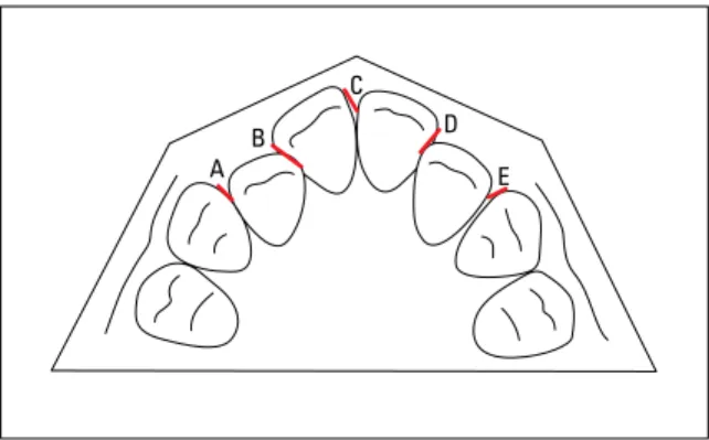
ideally elaborated for the evaluation of the den-tal crowding in the mandibular anterior seg-ment. However, due to its great reproducibility and precision, the same methodology to eval-uate the dental displacement was used in the present study for the evaluation of the maxillary anterior crowding. Little index was calculated in the maxillary dental casts in the three stud-ied phases (LITTLE1, LITTLE2 and LITTLE3). For this measurement, a digital caliper was used positioned parallel to the occlusal plane. The ir-regularity index was measured in this manner and characterized by the sum of the linear dis-tance among the anatomic contact points of the maxillary anterior teeth (canines and incisors). This measure represents the distance in which the contact points should be moved to attain alignment. According to Little,16 even though the contact points may vary in the vertical di-rection, the correction of these discrepancies will not affect significantly the anterior length of the arch, this way, changes in the vertical di-rection were not considered (Fig 1).
Intercanine width (INTERC): Distance mea-sured in milimeters, from cusp to cusp of the right and left maxillary canines. In cases where canines presented wear surfaces, the cusp was estimated.
Interpremolar width (INTERPB and INTERPB’):
Distances measured in milimeters, between the mesial cavity of the right and left first maxillary premolars (B) and of the second maxillary pre-molars (B’), respectively.
Intermolar width (INTERM): Distance mea-sured in milimeters, from mesiobuccal cusps of the right and left first maxillary molars. In cases where molars presented wear surfaces, the cusp was estimated.
Error of the method
The intra-examiner error was evaluated by new measurements of the studied variables per-formed on the initial, final and postretention casts of 10 randomly selected patients belonging to both groups. The reassessed variables (LITTLE, INTERC, INTERPB, INTERPB’, INTERM and ALENGTH) were also randomly selected. The first and second measurements were performed with a one month time difference. The formula proposed by Dahlberg6 (Se2 = ∑d2/2n) was used to estimate the order of magnitude of the casual errors, while the systematic errors were analyzed by paired t-tests, according to Houston.13
Statistical methods
Descriptive statistics was performed (mean, standard deviation and number) for Groups 1 (Class I) and 2 (Class II) for the measurements obtained by Little irregularity index, interca-nine, interpremolar and intermolar widths and arch length, in the initial (T1), final (T2) and postretention (T3) phases. Descriptive statistic was also performed for the difference of the measures obtained from the dental casts be-tween initial and final phases (T2-T1), charac-terizing the correction achieved with treatment,
and for the difference between the postreten-tion and final phases (T3-T2), characterizing the changes during postretention period, and for the difference between postretention and initial phases (T3-T1), characterizing the changes dur-ing the whole observation period.
For compatibility evaluation between Groups 1 and 2 regarding gender distribution and in rela-tion to the initial malocclusion severity, the Chi-square test was used. Aiming to verify compatibil-ity between groups regarding the amount of ini-tial crowding (LITTLE), iniini-tial age (Age T1), final age (Age T2) and age at postretention (Age T3), treatment time, retention time and postretention evaluation, independent t-test was used.
For intragroup comparison among the three evaluation times (Initial – T1; Final – T2; Postretention – T3), the dependent ANOVA test was used and, in case of a significant re-sult, the Tukey test. The test was applied for the evaluation of the variables measured in dental casts from Groups 1 and 2 together, in the three evaluated periods.
For the intergroup comparison of the values obtained for variables evaluated in the dental casts of initial, final and postretention phases, as well as the changes of these variables during treatment (T2-T1), postretention (T3-T2) and total changes (T3-T1), the independent t-test was used.
Finally, to verify the presence of correlation between relapse of maxillary anterior crowd-ing and relapse of the variables intercanine, interpremolar and intermolar widths and arch length, the Pearson’s correlation test was used.
All tests were performed by STATISTICA for Windows software (Release 6.0 – StatSoft, Inc. 2001), adopting a significance level of 5%.
RESuLtS
Table 1 shows the results of random and systematic errors, performed by the Dahlberg’s formula6 and paired t-tests, respectively, applied
FIGURE 1 - Little irregularity index (modified) = A+B+C+D+E. A
B C
D
to the variables LITTLE, INTERC, INTERPB, INTERPB’, INTERM and ALENGTH, and mea-sured on dental casts within a one-month interval. Group compatibility regarding gender dis-tribution was evaluated by the Chi-square test (Table 2). There was no statistically significant difference between the groups regarding gender distribution.
The independent t-test was used to as-sess the compatibility of groups regarding the amount of initial maxillary anterior crowd-ing (LITTLE1), initial age (Age T1), final age (Age T2), age at postretention stage (Age T3), treatment time (TREATTIME) and postreten-tion time (POSTTREATTIME). There was no significant difference between groups on these variables (Table 3).
The results of the ANOVA for the variables measured on dental casts of Groups 1 and 2, respectively, in the three studied periods (T1, T2 and T3) can be verified in Tables 4 and 5. In the presence of a significant result, the Tukey test was performed. The results for Group 1 showed a significant reduction in maxillary an-terior crowding and statistically significant di-mensional increments between the initial (T1) and final phases (T2), except for the intercanine width (INTERC) (Table 4).
It was observed that these changes tended to be stable in the postretention period, except for the interpremolar width (INTERPB), and arch length (ALENGTH), which had a statistically significant decrease in postretention (T3) (Ta-ble 4). The results of the ANOVA for Group 2 show that Little’s index had significant changes during treatment and during the postretention period (Table 5). However, there were no statis-tically significant changes for most of the other variables during these phases, except for the intercanine (INTERC) and intermolar widths (INTERM) which showed a significant decrease between the final (T2) and postretention stages (T3) (Table 5).
Variables
1st Measure-ment N=10
2nd
Measure-ment N=10 Dahlberg P
Mean SD Mean SD
LITTLE1 7.02 3.08 7.05 3.13 0.05 0.397
LITTLE2 0.10 0.33 0.11 0.34 0.01 0.343
LITTLE3 1.47 0.81 1.49 0.80 0.02 0.422
INTERC1 34.21 3.06 33.25 4.58 0.25 0.322
INTERC2 34.38 2.28 34.44 2.25 0.07 0.135
INTERC3 34.60 2.33 34.57 2.34 0.05 0.527
INTERPB1 32.35 2.49 32.63 2.48 0.35 0.220
INTERPB2 36.03 2.09 35.82 2.23 0.26 0.246
INTERPB3 35.30 2.18 35.39 2.55 0.22 0.564
INTERPB’1 37.04 3.41 37.10 3.64 0.28 0.755
INTERPB’2 41.13 2.56 41.21 2.59 0.15 0.454
INTERPB’3 40.40 2.39 40.48 2.64 0.20 0.564
INTERM1 49.21 3.94 49.29 3.86 0.10 0.207
INTERM2 52.19 3.85 52.33 3.80 0.13 0.092
INTERM3 52.38 3.50 52.48 3.71 0.15 0.303
ALENGTH1 67.96 5.22 68.21 5.51 0.25 0.121
ALENGTH2 72.59 4.59 72.58 4.75 0.23 0.990
ALENGTH3 70.86 4.06 71.36 4.71 0.65 0.249
Variables
Group 1 - Class I (N=19)
Group 2 - Class II
(N=19) P
Mean SD Mean SD
LITTLE1 7.83 3.14 6.35 2.67 0.126
Age T1 13.06 1.27 12.54 1.37 0.233
Age T2 15.19 1.24 14.93 1.50 0.552
Age T3 21.67 2.52 20.62 2.41 0.201
TREATTIME 2.15 0.89 2.32 0.73 0.534
POSTTREATTIME 8.60 1.83 8.04 2.11 0.388
TABLE 1 - Results of t test and Dahlberg’s formula,6 applied to the
evaluated variables to estimate systematic and casual errors, re-spectively.
TABLE 3 - Results of independent t test, applied to the variables initial Little index; initial, final and postretention age; treatment time and time of postretention evaluation for Groups 1 and 2, for evaluation of the in-tergroup compatibility.
TABLE 2 - Results of the Chi-square test for evaluation of compatibility of the Groups 1 and 2 regarding gender distribution.
Female Male Total
Group 1 - Class I 12 7 19
Group 2 - Class II 14 5 19
Total 26 12 38
Variables
Initial T1
Final T2
Postretention
T3 p
Mean (SD) Mean (SD) Mean (SD)
LITTLE 7.83 (3.14)A 0.34 (0.68)B 2.01 (1.87)B 0.000*
INTERC 33.79 (2.36)A 34.46 (1.48)A 34.29 (1.47)A 0.306
INTERPB 32.62 (1.91)A 35.91 (1.63)B 34.66 (1.54)C 0.000*
INTERPB’ 37.91 (2.94)A 40.90 (2.19)B 40.02 (2.04)B 0.000*
INTERM 49.49 (3.16)A 51.53 (2.86)B 51.34 (2.69)B 0.000*
ALENGTH 68.33 (4.72)A 71.01 (3.45)B 69.48 (3.38)A 0.000*
Variables
Initial T1
Final T2
Postretention
T3 p
Mean (SD) Mean (SD) Mean (SD)
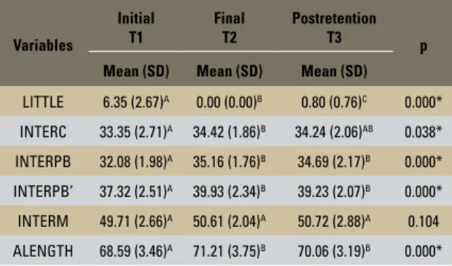
LITTLE 6.35 (2.67)A 0.00 (0.00)B 0.80 (0.76)C 0.000*
INTERC 33.35 (2.71)A 34.42 (1.86)B 34.24 (2.06)AB 0.038*
INTERPB 32.08 (1.98)A 35.16 (1.76)B 34.69 (2.17)B 0.000*
INTERPB’ 37.32 (2.51)A 39.93 (2.34)B 39.23 (2.07)B 0.000*
INTERM 49.71 (2.66)A 50.61 (2.04)A 50.72 (2.88)A 0.104
ALENGTH 68.59 (3.46)A 71.21 (3.75)B 70.06 (3.19)B 0.000*
TABLE 4 - Results of ANOVA for the variables from dental casts, of Group 1 (N=19), in the three studied phases (T1, T2 and T3). In the presence of a significant result, the Tukey test was performed (different letters show sig-nificant difference between the measurements).
TABLE 5 - Results of ANOVA for the variables from dental casts, of Group 2 (N=19), in the three studied phases (T1, T2 and T3). In the presence of a significant result, the Tukey test was performed (different letters show significant difference between the measurements).
*Statistically significant for p<0.05. *Statistically significant for p<0.05.
Tables 6, 7 and 8 show results of the inde-pendent t-test for intergroup comparison of the variables studied in the initial (T1), final (T2) and postretention stages (T3), respectively. In the initial phase, there was no statistically signif-icant difference for all variables between Groups 1 and 2 (Table 6). In the final and postreten-tion stages there were significant differences be-tween Groups 1 and 2 for the amount of maxil-lary anterior crowding (LITTLE2 and LITTLE3, Tables 7 and 8, respectively).
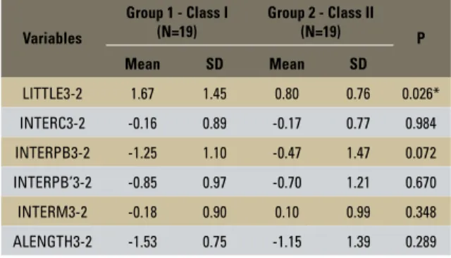
Changes in variables measured in dental casts during treatment (T2-T1), during postretention period (T3-T2) and total changes (T3-T1) in both groups, were compared by t-tests (Tables 9, 10 and 11, respectively). In relation to changes that occurred during treatment (T2-T1), there were no significant differences between Groups 1 and 2 (Table 9). During the postretention
period (T3-T2), only the amount of maxillary anterior crowding presented statistically sig-nificant difference between groups (Table 10). In relation to the total changes (T3-T1), there were no statistically significant differences be-tween Groups 1 and 2 (Table 11).
Table 12 shows the results of the Pearson cor-relation test. There was a negative corcor-relation, sta-tistically significant, between the amount of max-illary anterior crowding relapse and the amount of reduction in the intermolar width (Table 12).
diSCuSSiOn
stability,8,9,14,23,29 probably because maxillary an-terior crowding relapse is less prevalent when compared to the mandibular one.27,31 Despite of that, the search for predictive factors that might
TABLE 6 - Results of independent t test, applied to the studied vari-ables, to verify the differences in the initial stage (T1), between the Groups 1 and 2.
TABLE 7 - Results of independent t test, applied to the studied variables, to verify the differences in the final stage (T2), between Groups 1 and 2.
Variables
Group 1 - Class I (N=19)
Group 2 - Class II
(N=19) P
Mean SD Mean SD
LITTLE1 7.83 3.14 6.35 2.67 0.126
INTERC1 33.79 2.36 33.35 2.71 0.603
INTERPB1 32.62 1.91 32.08 1.98 0.401
INTERPB’1 37.91 2.94 37.32 2.51 0.514
INTERM1 49.49 3.16 49.71 2.66 0.820
ALENGTH1 68.33 4.72 68.59 3.46 0.849
*Statistically significant for p<0.05.
Variables
Group 1 - Class I (N=19)
Group 2 - Class II
(N=19) P
Mean SD Mean SD
LITTLE2 0.34 0.68 0.00 0.00 0.034*
INTERC2 34.46 1.48 34.42 1.86 0.943
INTERPB2 35.91 1.63 35.16 1.76 0.182
INTERPB’2 40.90 2.19 39.93 2.34 0.198
INTERM2 51.53 2.86 50.61 3.04 0.347
ALENGTH2 71.01 3.45 71.21 3.75 0.868
TABLE 8 - Results of independent t test, applied to the studied variables, to verify the differences in the postretention stage (T3), between Groups 1 and 2.
*Statistically significant for p<0.05.
Variables
Group 1 - Class I (N=19)
Group 2 - Class II
(N=19) P
Mean SD Mean SD
LITTLE3 2.01 1.87 0.80 0.76 0.012*
INTERC3 34.29 1.47 34.24 2.06 0.939
INTERPB3 34.66 1.54 34.69 2.17 0.958
INTERPB’3 40.02 2.04 39.23 2.07 0.246
INTERM3 51.34 2.69 50.72 2.88 0.498
ALENGTH3 69.48 3.38 70.06 3.19 0.591
TABLE 11 - Results of independent t test, applied to the studied variables, to verify the differences of changes in the whole period of observation (T3-T1), between Groups 1 and 2.
TABLE 9 - Results of independent t test, applied to the studied variables, to verify the differences in the treatment period (T2-T1), between Groups 1 and 2.
Variables
Group 1 - Class I (N=19)
Group 2 - Class II
(N=19) P
Mean SD Mean SD
LITTLE2-1 -7.48 3.24 -6.35 2.67 0.247
INTERC2-1 0.56 2.67 1.06 2.07 0.525
INTERPB2-1 3.29 1.78 3.08 1.88 0.723
INTERPB’2-1 2.98 2.27 2.60 2.69 0.641
INTERM2-1 2.03 2.13 0.90 2.78 0.169
ALENGTH2-1 2.68 3.18 2.61 3.01 0.951
TABLE 10 - Results of independent t test, applied to the studied variables, to verify the differences in the post-treatment period (T3-T2), between Groups 1 and 2.
*Statistically significant for p<0.05.
Variables
Group 1 - Class I (N=19)
Group 2 - Class II
(N=19) P
Mean SD Mean SD
LITTLE3-2 1.67 1.45 0.80 0.76 0.026*
INTERC3-2 -0.16 0.89 -0.17 0.77 0.984
INTERPB3-2 -1.25 1.10 -0.47 1.47 0.072
INTERPB’3-2 -0.85 0.97 -0.70 1.21 0.670
INTERM3-2 -0.18 0.90 0.10 0.99 0.348
ALENGTH3-2 -1.53 0.75 -1.15 1.39 0.289
Variables
Group 1 - Class I (N=19)
Group 2 - Class II
(N=19) P
Mean SD Mean SD
LITTLE3-1 -5.81 3.94 -5.55 2.34 0.804
INTERC3-1 0.50 2.03 0.89 2.34 0.588
INTERPB3-1 2.04 1.56 2.61 2.24 0.370
INTERPB’3-1 2.10 2.20 1.90 2.13 0.775
INTERM3-1 1.84 2.34 1.01 2.42 0.287
ALENGTH3-1 1.14 3.10 1.46 2.29 0.718
maxillary crowding tends to become more vis-ible and, consequently, promote more esthetic problems than mandibular incisor irregularity. However, depending on patient’s smile height, it may also not occur.
Sample selection included dental cast evalu-ation. Group 1 comprised patients who exhibit-ed Class I molar relationship and Group 2 con-sisted of patients presenting at least ¾ of a Class II molar relationship. Both groups were treated without extractions. Another sample selection criterion was that all patients had been treated with fixed Edgewise appliances, in both arches.
During sample selection, one of the con-cerns was to eliminate possible factors that might influence the results. One of the main objectives during this study development was to obtain compatible groups which would fa-cilitate comparison and, consequently, would favor interpretation and reliability of the re-sults. For this, the characteristics were homog-enized in the beginning and end of the orth-odontic treatment. The groups were compat-ible regarding to treatment protocol, the kind of orthodontic accessory used, sex distribu-tions and initial malocclusion. Besides, groups
had other compatible characteristics, such as: Initial and final ages, treatment time, postre-tention evaluation time. Thus, the changes that occurred in the postretention period could be analyzed safely.
Regarding stability, there are considerable controversies in the literature about long-term post retention maxillary crowding relapse in different types of malocclusion (Class I and Class II subjects).25,31 Some studies reported that the greater the initial malocclusion se-verity, the greater the long-term relapse.10,18 Therefore, relapse of maxillary anterior crowd-ing was evaluated in two groups that presented similar pretreatment incisors irregularity. The intergroup compatibility evaluation regard-ing pretreatment incisor irregularity was per-formed by t-test. No significant differences were observed between groups regarding ini-tial irregularity (Table 3).
Intragroup comparison
The results for ANOVA and Tukey’s test in Group 1 (Table 4) showed statistically signifi-cant changes in Little’s irregularity index be-tween initial and final or post-treatment phas-es. No statistically significant differences were noted between final and post-treatment phases. It may be inferred by interpreting these results, that orthodontic treatment resulted in signifi-cant maxillary crowding correction. During post-treatment period, there was no significant maxillary crowding relapse.
Intercanine width showed no significant changes during the three treatment stages (Ta-ble 4). These results could support the concept that stable results can be gained only when in-tercanine width is maintained.12,28 However, it is difficult to distinguish between intercanine width relapse and the normal decrease of this measure that occurs over the years in normal occlusion development, as others studies have reported.18,28
Variables r P
LITTLE1 x LITTLE3 0.101 0.545
LITTLE1 x LITTLE3-2 0.104 0.533
LITTLE2-1 x LITTLE3-2 -0.021 0.899
LITTLE3-2 x INTERC3-2 0.128 0.441
LITTLE3-2 x INTERPB3-2 -0.296 0.071
LITTLE3-2 x INTERPB’3-2 -0.177 0.286
LITTLE3-2 x INTERM3-2 -0.342 0.035*
LITTLE3-2 x ALENGTH3-2 -0.301 0.065
TABLE 12 - Results of the Pearson’s correlation test.
Regarding changes in maxillary arch dimen-sions during treatment, significant transversal increases were noted (variables INTERPB, IN-TERPB’ INTERM). Mean arch length increase was also significant. Only interfirstpremolar width decreased significantly during postre-tention, but not enough to reach initial values. The arch length width also showed a significant change during post-treatment, reaching a mean value close to the initial one.
Results for ANOVA and Tukey’s test in Group 2 (Table 5) showed statistically sig-nificant differences in Little’s irregularity in-dex in the three stages evaluated. Thus, it was observed that the changes in variable LITTLE were not similar to Group 1. Significant re-duction in maxillary crowding occurred dur-ing treatment. However, durdur-ing post-treatment period, there was significant maxillary anterior irregularity relapse, that did not reach pre-treatment mean value.
The variables INTERC, INTERPB, INTER-PB’ and ALENGTH showed statistically sig-nificant increases during treatment and did not exhibit a significant relapse when evaluating the final and postretention stages. The intermolar width showed no statistically significant chang-es in any of the studied phaschang-es.
Intergroup comparison
When comparing Groups 1 and 2 (Class I and Class II malocclusion subjects, respectively) in pretreatment, it was observed that none of the evaluated variables showed significant dif-ferences between groups (Table 6). Some stud-ies that evaluated crowding relapse during long-term post-treatment mention that pretreatment irregularity is directly related to the amount of relapse,1 although other authors have not ob-served this correlation.20
Regarding the maxillary arch dimensions, it was noted that Groups 1 and 2 exhibited similar transverse dimensions (Table 6). McNamara22
commented that, generally, transpalatal widths from 36 to 39 mm may accommodate an aver-age size permanent dentition, without crowd-ing or spaccrowd-ing.
As previously mentioned, the initial maxil-lary crowding severity, and other pretreatment variables were similar between the groups at T1, allowing a reliable comparative evaluation of the long-term post-treatment changes.
When comparing Groups 1 and 2 at post-treatment, the only variable that differed sig-nificantly between the groups was Little’s irregularity index. There was more incisor ir-regularity in Class I subjects, indicating that Group 2 patients exhibited more quality in maxillary incisor alignment at T2 (Table 7). Although statistically significant, difference in irregularity between the groups was only 0.34 mm, which may not be considered clini-cally significant. Accordingly to Little,18 dental arch irregularity values between 0 and 1 mm consist in ideal alignments.
During postretention, the only variable that showed a significant intergroup difference was Little’s irregularity index (LITTLE3), while other variables as INTERC3, INTERPB3, IN-TERPB’3, INTERM3 and ALENGTH3 showed no statistically significant differences (Table 8). These results suggested a similar behavior of the groups during postretention regarding dimen-sional changes.
Intergroup comparison of treatment chang-es revealed no significant difference between groups in the amount of maxillary crowding correction (LITTLE2-1, Table 9). This result was expected since there were no statistically significant differences between groups in the initial and final maxillary incisor irregularities.
during treatment because the sample exhibited less pretreatment incisor irregularity.8
The fact is that all these studies aimed at eliminating the incisors irregularity during treatment. Thus, variation in the amount of crowding correction is often due to variation in initial crowding severity.
The amount of maxillary incisors crowding relapse (LITTLE3-2) was statistically differ-ent between groups. Group 1 (Angle Class I subjects) exhibited a mean crowding relapse of 1.67 mm (SD = 1.45 mm). Group 2 (Angle Class II subjects) showed a mean crowding relapse of 0.80 mm (SD = 0.76 mm). Thus, there was greater treatment stability in Group 2 (Table 10).
This significant difference between the groups regarding incisors alignment stabil-ity may be due to orthodontic mechanics per-formed in patients of each group. In Class II subjects (Group 2), treated without extraction, there was, necessarily, distalization of maxillary molars. Therefore, more space could be gained for teeth alignment and this fact might have fa-vored on stability. In Group 1 (Class I malocclu-sion), the molars remained stable in their initial positions during treatment. The crowding was corrected by dental protrusion and maxillary arch expansion, perhaps contributing to maxil-lary anterior crowding relapse.
Sadowsky et al27, while evaluating the long-term stability of non-extraction cases, observed a similar amount of relapse (1.1 mm) five years postretention. However, Moussa, O’Reilly and Close23 observed more favorable results 8-10 years postretention. Vaden et al32 found that 96% of maxillary crowding correction was main-tained 15 years post-treatment. The amount of crowding increased from 1.5 mm (post-treat-ment) to 1.8 mm (postretention). Ferris et al9 also evaluated the relapse of maxillary anterior crowding in cases treated without extractions and observed an increase in maxillary incisors
irregularity of only 0.47 (SD = 1.19) during postretention (7.9 years). The greater maxillary incisors alignment stability of these studies may be due to the prolonged use of retention.3,23,27 In Sadowsky et al27 study, the mean retention pe-riod was 8.4 years. Moussa, O’Reilly and Close23 study adopted a mean retention period of 6.6 years in the mandibular arch (fixed retention) and 2 years for the maxillary arch (Hawley re-tainer). In the research conducted by Vaden et al,32 patients used Hawley retainers in mandibu-lar and maxilmandibu-lary arches or Hawley retainer in the maxillary arch and bonded lingual retainer in the lower arch. The first long-term post-treat-ment evaluation was carried out only 6 years after treatment. In Ferris et al9 study, patients were submitted to a retention protocol that in-cluded the use of maxillary removable retainers for at least 3 years (one year of continuous us-age) and a bonded lingual retainer or a Hawley retainer in mandibular arch during a mean pe-riod of 3 years. In the present study, all patients used a Hawley retainer in the maxillary arch during 1 year and a mandibular bonded lingual retainer for a mean period of 3 years.
Surbeck et al29 observed a direct influence of pretreatment maxillary irregularity sever-ity on amount of postretention relapse. The authors29 suggested the adoption of individual retention protocols and that the orthodontist should explain to patients the probability of post-treatment relapse, accordingly to the ini-tial irregularity severity.
However, analyzing the results of other au-thors and ours, a positive correlation between the amount of pretreatment crowding and the amount of long-term post-treatment relapse seems unlikely. For example, in the present study, Groups 1 and 2 presented 7.83 mm (SD = 3.14) and 6.35 mm (SD = 2.67) of pretreatment maxillary irregularity, respec-tively. A mean maxillary irregularity relapses of 1.67 mm (SD = 1.45 mm) for Group 1 and 0.80 mm (SD = 0.76 mm) for Group 2 was observed. The amount of irregularity relapse in the present study was greater than the crowd-ing relapse observed by Ferris et al,9 Sadowsky et al27 and Vaden et al.32 However, the sam-ple in these studies showed greater maxillary pretreatment irregularity: 10.45 mm, 8.0 mm and 7.9 mm, respectively. Thus, even showing slightly greater amounts of initial crowding than the present study, maxillary incisors alignment in those studies was more stable during postre-tention (0.47 mm, 1.1 mm, 0.3 mm of long-term post-treatment relapse, respectively).
When evaluating overall changes (T3-T1), it was observed that maxillary anterior irregular-ity decreased 5.81 mm (SD = 3.94) and 5.55 mm (SD = 2.34) for Groups 1 and 2, respec-tively, and there were no statistically significant differences between groups (Table 11).
Correlation
Correlation tests were performed in the total sample to obtain the Pearson’s correlation co-efficients. Results showed a significant correla-tion between pretreatment and post-treatment
incisors irregularity (LITTLE1 and LITTLE3), between initial crowding (LITTLE1) and post-treatment crowding relapse (LITTLE3-2), and between crowding correction (LITTLE2-1) and postretention crowding relapse (LITTLE3-2). We also attempted to determine a possible cor-relation between maxillary crowding relapse (LITTLE3-2) and postretention changes in maxillary arch dimensions (INTERC3-2, IN-TERPB3-2, INTERPB’3-2, INTERM3-2 and ALENGTH3-2). Results are shown in Table 12.
Most results of the correlation tests were not significant. It was observed that pretreatment maxillary crowding severity did not influence the postretention crowding relapse, as described in previous studies.2,3,20 Surbeck et al,29 however, reported a positive correlation between pretreat-ment crowding severity and the amount of maxil-lary anterior crowding relapse. Accordingly to the authors,29 the odds of maxillary anterior relapse increase 2.3 times for every 0.2 mm displacement of incisors anatomic contact points relative to the dental arch, and 2.7 times for every 4° of incisor rotation. The authors also pointed out that incom-plete alignment during active treatment is a sig-nificant risk factor for relapse.
COnCLuSiOnS
According to the sample and methodology used and based on the presented and discussed results, it was concluded that:
» Class I malocclusion subjects treated non-extraction exhibited greater maxillary anterior crowding relapse than Class II subjects treated with the same protocol.
Contact address
Karina Maria Salvatore de Freitas
Rua Jamil Gebara, 1-25, apto. 111, Jd. Paulista CEP: 17.017-150 – Bauru/SP, Brazil
E-mail: [email protected]
1. Artun J, Garol JD, Little RM. Long-term stability of mandibular incisors following successful treatment of Class II, division 1, malocclusions. Angle Orthod. 1996;66(3):229-38.
2. Artun J, Krogstad O, Little RM. Stability of mandibular incisors following excessive proclination: a study in adults with surgically treated mandibular prognathism. Angle Orthod. 1990;60(2):99-106.
3. Azizi M, Shrout MK, Haas AJ, Russell CM, Hamilton EH Jr. A retrospective study of Angle Class I malocclusions treated orthodontically without extractions using two palatal expansion methods. Am J Orthod Dentofacial Orthop. 1999;116(1):101-7.
4. Blake M, Bibby K. Retention and stability: a review of the literature. Am J Orthod Dentofacial Orthop. 1998;114(3):299-306.
5. Canuto LFG. Avaliação da inluência da realização da expansão rápida da maxila sobre a recidiva do apinhamento ântero-superior, em casos tratados ortodonticamente sem extrações [dissertação]. Bauru (SP): Universidade de São Paulo; 2006.
6. Dahlberg G. Statistical methods for medical and biological students. New York: Interscience; 1940.
7. Destang DL, Kerr WJ. Maxillary retention: is longer better? Eur J Orthod. 2003 Feb;25(1):65-9.
8. Erdinc AE, Nanda RS, Isiksal E. Relapse of anterior crowding in patients treated with extraction and nonextraction of premolars. Am J Orthod Dentofacial Orthop. 2006;129(6):775-84.
9. Ferris T, Alexander RG, Boley J, Buschang PH. Long-term stability of combined rapid palatal expansion-lip bumper therapy followed by full ixed appliances. Am J Orthod Dentofacial Orthop. 2005 Sep;128(3):310-25.
10. Freitas KM, Janson G, Freitas MR, Pinzan A, Henriques JF, Pinzan-Vercelino CR. Inluence of the quality of the inished occlusion on postretention occlusal relapse. Am J Orthod Dentofacial Orthop. 2007 Oct;132(4):428.e9-14. 11. Freitas KM, de Freitas MR, Henriques JF. Postretention
relapse of mandibular anterior crowding in patients treated without mandibular premolar extraction. Am J Orthod Dentofacial Orthop. 2004;125(4):480-7.
12. Glenn G, Sinclair PM, Alexander RG. Nonextraction orthodontic therapy: posttreatment dental and skeletal stability. Am J Orthod Dentofacial Orthop. 1987 Oct;92(4):321-8.
13. Houston WJB. The analysis of errors in orthodontic measurements. Am J Orthod. 1983 May;83(5):382-90. 14. Huang L, Artun J. Is the postretention relapse of maxillary
and mandibular incisor alignment related? Am J Orthod Dentofacial Orthop. 2001;120(1):9-19.
15. Kahl-Nieke B, Fischbach H, Schwarze CW. Post-retention crowding and incisor irregularity: a long-term follow-up evaluation of stability and relapse. Br J Orthod. 1995;22(3):249-57.
REfEREnCES
16. Little RM. The irregularity index: a quantitative score of mandibular anterior alignment. Am J Orthod. 1975;68(5):554-63.
17. Little RM. Stability and relapse of dental arch alignment. Br J Orthod. 1990;17(3):235-41.
18. Little RM. Stability and relapse of mandibular anterior alignment: University of Washington studies. Semin Orthod. 1999;5(3):191-204.
19. Little RM, Riedel RA, Stein A. Mandibular arch length increase during the mixed dentition: postretention evaluation of stability and relapse. Am J Orthod Dentofacial Orthop. 1990;97(5):393-404.
20. Little RM, Wallen TR, Riedel RA. Stability and relapse of mandibular anterior alignment — irst premolar extraction cases treated by traditional Edgewise orthodontics. Am J Orthod. 1981 Oct;80(4):349-65.
21. Martins PP. Avaliação da inluência da expansão rápida da maxila sobre a recidiva do apinhamento ântero-superior em casos tratados ortodonticamente com extrações de pré-molares [dissertação]. Bauru (SP): Universidade de São Paulo; 2007. 22. McNamara JA. Maxillary transverse deiciency. Am J Orthod
Dentofacial Orthop. 2000;117(5):567-70.
23. Moussa R, O’Reilly MT, Close JM. Long-term stability of rapid palatal expander treatment and Edgewise mechanotherapy. Am J Orthod Dentofacial Orthop. 1995;108(5):478-88.
24. Naraghi S, Andrén A, Kjellberg H, Mohlin BO. Relapse tendency after orthodontic correction of upper front teeth retained with a bonded retainer. Angle Orthod. 2006;76(4):570-6.
25. Ormiston JP, Huang GJ, Little RM, Decker JD, Seuk GD. Retrospective analysis of long-term stable and unstable orthodontic treatment outcomes. Am J Orthod Dentofacial Orthop. 2005;128(5):568-74; quiz 669.
26. Sadowsky C, Sakols EI. Long-term assessment of orthodontic relapse. Am J Orthod. 1982;82(6):456-63.
27. Sadowsky C, Schneider BJ, BeGole EA, Tahir E. Long-term stability after orthodontic treatment: nonextraction with prolonged retention. Am J Orthod Dentofacial Orthop. 1994;106(3):243-9.
28. Sinclair PM, Little RM. Maturation of untreated normal occlusions. Am J Orthod. 1983;83(2):114-23.
29. Surbeck BT, Artun J, Hawkins NR, Leroux B. Associations between initial, posttreatment, and postretention alignment of maxillary anterior teeth. Am J Orthod Dentofacial Orthop. 1998;113(2):186-95.
30. Thilander B. Orthodontic relapse versus natural development. Am J Orthod Dentofacial Orthop. 2000;117(5):562-3.
31. Uhde MD, Sadowsky C, BeGole EA. Long-term stability of dental relationships after orthodontic treatment. Angle Orthod. 1983;53(3):240-52.
32. Vaden JL, Harris EF, Gardner RL. Relapse revisited. Am J Orthod Dentofacial Orthop. 1997;111(5):543-53.
Submitted: June 4, 2009